

Subcellular localization of intercellular adhesion molecule-5 (telencephalin) in the visual cortex is not developmentally regulated in the absence of matrix metalloproteinase-9
- PMID: 23897576
- PMCID: PMC3877171
- DOI: 10.1002/cne.23440
Subcellular localization of intercellular adhesion molecule-5 (telencephalin) in the visual cortex is not developmentally regulated in the absence of matrix metalloproteinase-9
Abstract
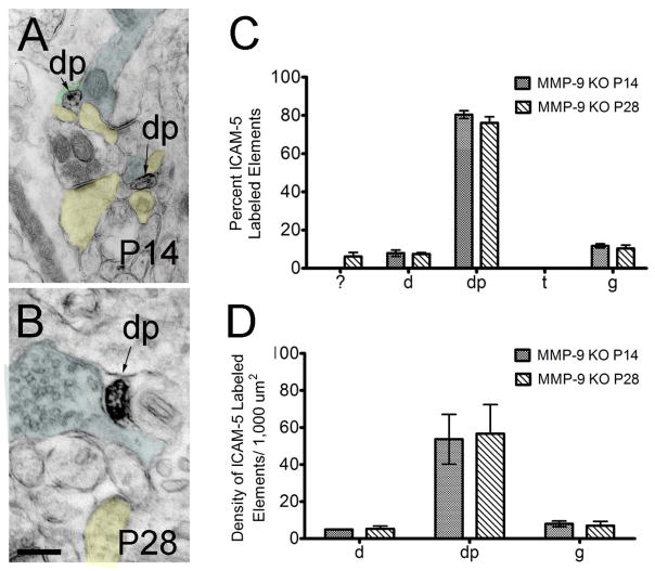
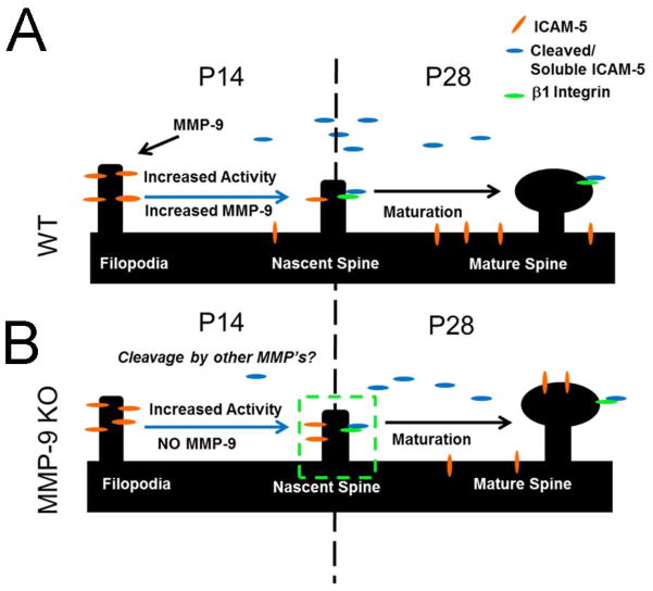
The telencephalon-associated intercellular adhesion molecule-5 (telencephalin; ICAM-5) regulates dendritic morphology in the developing brain. In vitro studies have shown that ICAM-5 is found predominantly within dendrites and immature dendritic protrusions, with reduced expression in mushroom spines, suggesting that ICAM-5 downregulation is critical for the maturation of synaptic structures. However, developmental expression of ICAM-5 has not been explored in depth at the ultrastructural level in intact brain tissue. To investigate the ultrastructural localization of ICAM-5 with transmission electron microscopy, we performed immunoperoxidase histochemistry for ICAM-5 in mouse visual cortex at postnatal day (P)14, a period of intense synaptogenesis, and at P28, when synapses mature. We observed the expected ICAM-5 expression in dendritic protrusions and shafts at both P14 and P28. ICAM-5 expression in these dendritic protrusions decreased in prevalence with developmental age to become localized predominantly to dendritic shafts by P28. To understand better the relationship between ICAM-5 and the endopeptidase metalloproteinase-9 (MMP-9), which mediates ICAM-5 cleavage following glutamate activation during postnatal development, we also explored ICAM-5 expression in MMP-9 null animals. This analysis revealed a similar expression of ICAM-5 in dendritic elements at P14 and P28; however, an increased prevalence of ICAM-5 was noted in dendritic protrusions at P28 in the MMP-9 null animals, indicating that, in the absence of MMP-9, there is no developmental shift in ICAM-5 subcellular localization. Our ultrastructural observations shed light on possible functions mediated by ICAM-5 and their regulation by extracellular proteases.
Keywords: ICAM-5; MMP-9; dendrite; electron microscopy; plasticity; telencephalin.
Copyright © 2013 Wiley Periodicals, Inc.
Conflict of interest statement
The authors have no conflict of interests to report.
Figures

Similar articles
-
Interactions between intercellular adhesion molecule-5 positive elements and their surroundings in the rodent visual cortex.Commun Integr Biol. 2013 Nov 1;6(6):e27315. doi: 10.4161/cib.27315. Epub 2013 Dec 10. Commun Integr Biol. 2013. PMID: 24753788 Free PMC article.
-
Activation of NMDA receptors promotes dendritic spine development through MMP-mediated ICAM-5 cleavage.J Cell Biol. 2007 Aug 13;178(4):687-700. doi: 10.1083/jcb.200612097. Epub 2007 Aug 6. J Cell Biol. 2007. PMID: 17682049 Free PMC article.
-
Interaction between telencephalin and ERM family proteins mediates dendritic filopodia formation.J Neurosci. 2007 Aug 15;27(33):8866-76. doi: 10.1523/JNEUROSCI.1047-07.2007. J Neurosci. 2007. PMID: 17699668 Free PMC article.
-
ICAM-5: a neuronal dendritic adhesion molecule involved in immune and neuronal functions.Adv Neurobiol. 2014;8:117-32. doi: 10.1007/978-1-4614-8090-7_6. Adv Neurobiol. 2014. PMID: 25300135 Review.
-
Telencephalin: a neuronal area code molecule?Neurosci Res. 1994 Dec;21(2):119-24. doi: 10.1016/0168-0102(94)90153-8. Neurosci Res. 1994. PMID: 7724062 Review.
Cited by
-
Proteolytic regulation of synaptic plasticity in the mouse primary visual cortex: analysis of matrix metalloproteinase 9 deficient mice.Front Cell Neurosci. 2015 Sep 22;9:369. doi: 10.3389/fncel.2015.00369. eCollection 2015. Front Cell Neurosci. 2015. PMID: 26441540 Free PMC article.
-
Effects of Mephedrone and Amphetamine Exposure during Adolescence on Spatial Memory in Adulthood: Behavioral and Neurochemical Analysis.Int J Mol Sci. 2021 Jan 8;22(2):589. doi: 10.3390/ijms22020589. Int J Mol Sci. 2021. PMID: 33435576 Free PMC article.
-
Calsyntenin-1 Negatively Regulates ICAM5 Accumulation in Postsynaptic Membrane and Influences Dendritic Spine Maturation in a Mouse Model of Fragile X Syndrome.Front Neurosci. 2019 Oct 18;13:1098. doi: 10.3389/fnins.2019.01098. eCollection 2019. Front Neurosci. 2019. PMID: 31680833 Free PMC article.
-
Extracellular matrix control of dendritic spine and synapse structure and plasticity in adulthood.Front Neuroanat. 2014 Oct 20;8:116. doi: 10.3389/fnana.2014.00116. eCollection 2014. Front Neuroanat. 2014. PMID: 25368556 Free PMC article. Review.
-
Ultrastructure of geniculocortical synaptic connections in the tree shrew striate cortex.J Comp Neurol. 2016 Apr 15;524(6):1292-306. doi: 10.1002/cne.23907. Epub 2015 Oct 13. J Comp Neurol. 2016. PMID: 26399201 Free PMC article.
References
-
- Benson DL, Yoshihara Y, Mori K. Polarized distribution and cell type-specific localization of telencephalin, an intercellular adhesion molecule. J Neurosci Res. 1998;52(1):43–53. - PubMed
-
- Bouvier D, Corera AT, Tremblay ME, Riad M, Chagnon M, Murai KK, Pasquale EB, Fon EA, Doucet G. Pre-synaptic and post-synaptic localization of EphA4 and EphB2 in adult mouse forebrain. Journal of neurochemistry. 2008;106(2):682–695. - PubMed
-
- Bouvier D, Tremblay ME, Riad M, Corera AT, Gingras D, Horn KE, Fotouhi M, Girard M, Murai KK, Kennedy TE, McPherson PS, Pasquale EB, Fon EA, Doucet G. EphA4 is localized in clathrin-coated and synaptic vesicles in adult mouse brain. Journal of neurochemistry. 2010;113(1):153–165. - PubMed
Publication types
MeSH terms
Substances
Grants and funding
LinkOut - more resources
Full Text Sources
Other Literature Sources
Molecular Biology Databases
Research Materials
Miscellaneous
