. 2013 Oct;81(10):3855-64.
doi: 10.1128/IAI.00525-13. Epub 2013 Jul 29.

Combined stimulation of Toll-like receptor 5 and NOD1 strongly potentiates activity of NF-κB, resulting in enhanced innate immune reactions and resistance to Salmonella enterica serovar Typhimurium infection

Affiliations

Combined stimulation of Toll-like receptor 5 and NOD1 strongly potentiates activity of NF-κB, resulting in enhanced innate immune reactions and resistance to Salmonella enterica serovar Typhimurium infection

Amir I Tukhvatulin et al. Infect Immun. 2013 Oct.

Abstract

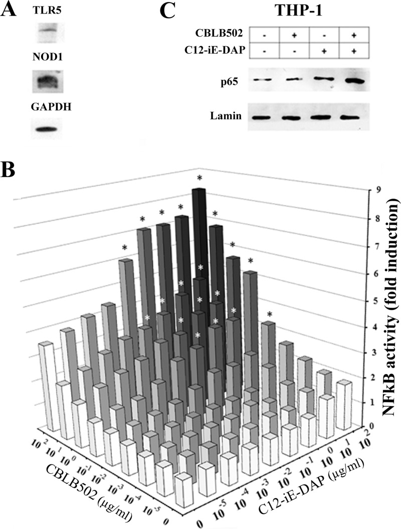
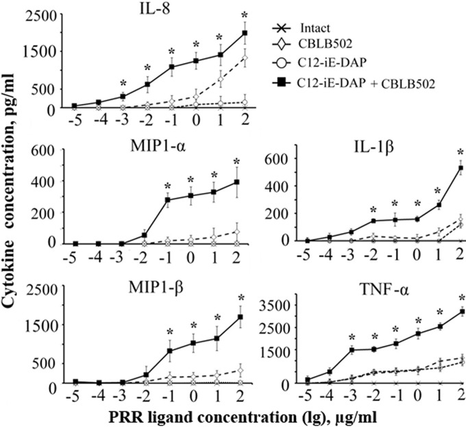
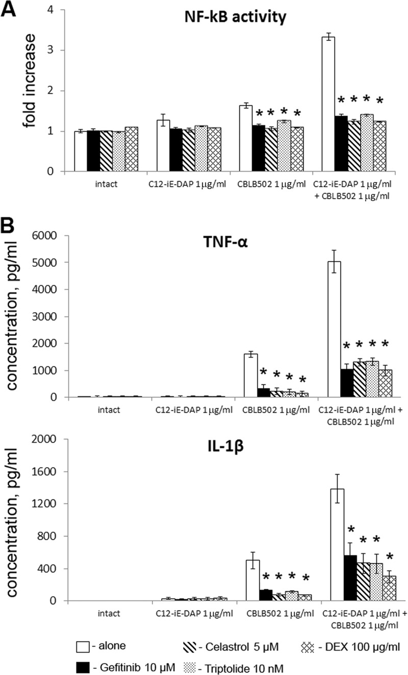
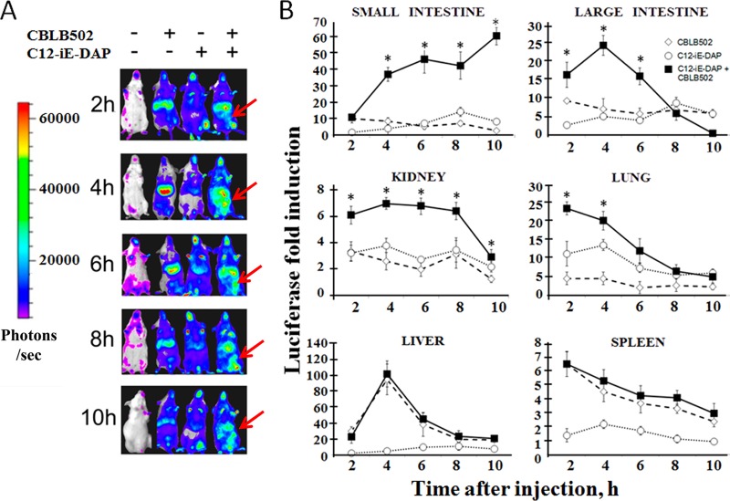
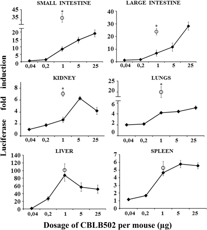
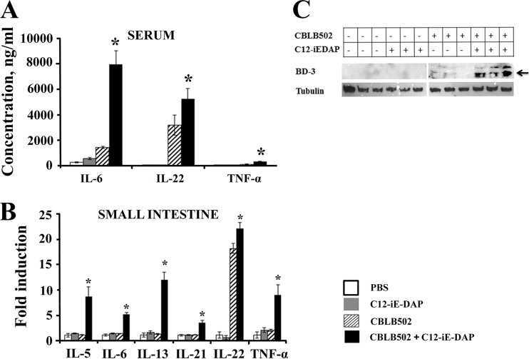
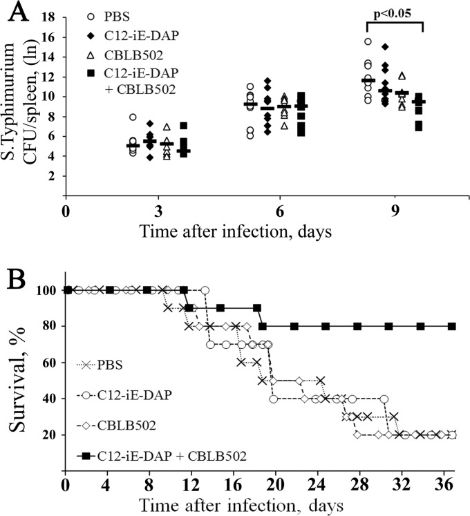
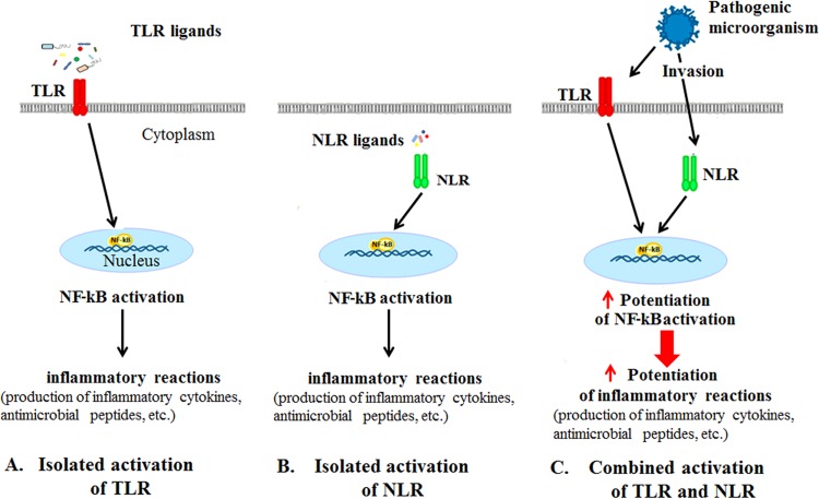
Pathogen recognition receptors (PRRs) are essential components of host innate immune systems that detect specific conserved pathogen-associated molecular patterns (PAMPs) presented by microorganisms. Members of two families of PRRs, transmembrane Toll-like receptors (TLRs 1, 2, 4, 5, and 6) and cytosolic NOD receptors (NOD1 and NOD2), are stimulated upon recognition of various bacterial PAMPs. Such stimulation leads to induction of a number of immune defense reactions, mainly triggered via activation of the transcription factor NF-κB. While coordination of responses initiated via different PRRs sensing multiple PAMPS present during an infection makes clear biological sense for the host, such interactions have not been fully characterized. Here, we demonstrate that combined stimulation of NOD1 and TLR5 (as well as other NOD and TLR family members) strongly potentiates activity of NF-κB and induces enhanced levels of innate immune reactions (e.g., cytokine production) both in vitro and in vivo. Moreover, we show that an increased level of NF-κB activity plays a critical role in formation of downstream responses. In live mice, synergy between these receptors resulting in potentiation of NF-κB activity was organ specific, being most prominent in the gastrointestinal tract. Coordinated activity of NOD1 and TLR5 significantly increased protection of mice against enteroinvasive Salmonella infection. Obtained results suggest that cooperation of NOD and TLR receptors is important for effective responses to microbial infection in vivo.


Figures

Fig 1
Fig 1
Combined stimulation of NOD1 and TLR5 receptors leads to enhanced NF-κB activity in THP1-XBlue-CD14 cells. (A) Western blot analysis of NOD1, TLR5, and GAPDH expression in THP1-XBlue-CD14 cells. (B) NF-κB activity (NF-κB-dependent SEAP reporter gene expression) in THP1-XBlue-CD14 cells treated for 18 h with the indicated doses (μg/ml) of C12-iE-DAP and CBLB502 (alone or in combination). Results are expressed as the fold increase in NF-κB activity relative to intact (untreated) cells (values are mean from three independent experiments, each performed in duplicate). Means were compared using the Student t test. Asterisks indicate significant differences (P < 0.05) in NF-κB activity between treatment with combined agonists and treatment with CBLB502 or C12-iE-DAP alone. (C) Detection of p65 nuclear translocation as an indicator of NF-κB activation in THP1-XBlue-CD14 cells left untreated or treated with C12-iE-DAP (100 ng/ml) or CBLB502 (10 ng/ml) alone or in combination. Thirty minutes after the addition of PRR agonists, nuclear protein extracts were prepared and assessed by Western blotting using anti-p65 and anti-lamin (loading control) antibodies.
Fig 2
Fig 2
Combined stimulation of NOD1 and TLR5 receptors leads to enhanced cytokine production in THP1-XBlue-CD14 cells. Concentrations of IL-1β, IL-8, MIP-1α, MIP-1β, and TNF-α in culture supernatants of THP1-XBlue-CD14 cells treated with C12-iE-DAP, CBLB502, and their combination, as indicated, for 18 h. Intact cells (no treatment) were used as controls. For combined activation of NOD1 and TLR5, C12-iE-DAP was used at a fixed concentration of 1 μg/ml in combination with the concentrations of CBLB502 indicated on the x axis of the figure. Asterisks indicate significant differences (P < 0.05) in cytokine production levels between treatment with combined agonists and treatment with CBLB502 or C12-iE-DAP alone.
Fig 3
Fig 3
Role of NF-κB in enhanced cytokine production after combined stimulation of NOD1 and TLR5 receptors in THP1-XBlue-CD14 cells. (A) NF-κB activity (SEAP reporter expression) in THP1-XBlue-CD14 cells treated for 18 h with C12-iE-DAP (1 μg/ml), CBLB502 (1 μg/ml), or their combination without (alone) or with the inhibitor gefitinib (10 μM), celastrol (5 μM), triptolide (10 nM), or DEX (100 μg/ml), as indicated. Data are presented as the mean fold change relative to untreated cells (no PRR agonists and inhibitors) in three independent experiments, each performed with duplicates. Error bars indicated standard deviations (SD). Asterisks indicate significant differences (P < 0.05) in NF-κB responses between inhibitor-treated and untreated cells. (B) Concentrations of IL-1β and TNF-α in the culture supernatants of THP1-XBlue-CD14 cells treated with C12-iE-DAP (1 μg/ml), CBLB502 (1 μg/ml), or their combination without (alone) or with the inhibitor gefitinib (10 μM), celastrol (5 μM), triptolide (10 nM), or DEX (100 μg/ml), as indicated. Data are presented as the means ± SD (error bars) from three independent experiments, each performed with duplicates. Asterisks indicate significant differences (P < 0.05) in cytokine production levels between inhibitor-treated and untreated cells.
Fig 4
Fig 4
Kinetics of NF-κB-luciferase activity in NF-κB-Luc transgenic mice exposed to TLR5 and NOD1 ligands. (A) In vivo imaging of NF-κB-luciferase activity in live NF-κB-Luc transgenic mice at 2, 4, 6, 8, or 10 h after s.c. injection of PBS or of CBLB502 (1 μg/mouse), C12-iE-DAP (200 μg/mouse), or their combination. Mice were anesthetized and injected i.p. with d-luciferin prior to imaging. Pseudocolored images reflect the intensity of bioluminescence (i.e., NF-κB activity) according to the scale shown on the left side of the figure, with red indicating the most intense light emission and purple indicating the weakest signal. Three additional experiments gave results similar to those shown here. Red arrows indicate enhancement of the NF-κB-dependent luciferase reporter signal in the abdominal region of mice that received CBLB502 and C12-iE-DAP in combination. (B) NF-κB activity in tissue homogenates prepared from NF-κB-Luc transgenic mice at the indicated time points after injection of 200 μg/mouse C12-iE-DAP alone, 1 μg/mouse CBLB502 alone, or their combination. Control mice were injected with PBS. Results are expressed as the fold increase in luciferase activity relative to PBS-treated control animals. Each point represents the mean of three mice per group ± SD (error bars). Data shown are pooled from two independent experiments. Asterisks indicate significant differences (P < 0.05) in NF-κB activity levels between combined agonists administration and CBLB502 or C12-iE-DAP alone.
Fig 5
Fig 5
NF-κB-dependent luciferase reporter activity in NF-κB-Luc transgenic mice injected with different doses of CBLB502 versus combined injection of CBLB502 and C12-iE-DAP. Luciferase levels were measured in tissue homogenates prepared 4 h after injection of NF-κB-Luc reporter mice with 0.04 μg, 0.2 μg, 1 μg, 5 μg, or 25 μg of CBLB502 per mouse as indicated on the x axis. Results are expressed as the mean fold change in luciferase activity relative to the mean in PBS-treated control mice (n = 5 mice per group). Open circles represent the fold induction of luciferase 4 h after combined injection of CBLB502 (1 μg/mouse) and C12-iE-DAP (200 μg/mouse). Error bars indicate SD. Asterisks indicate significant differences (P < 0.05) in NF-κB activity levels between administration of combined agonists and of CBLB502 alone.
Fig 6
Fig 6
Combined stimulation of NOD1 and TLR5 receptors results in potentiation of cytokine production in mouse serum and small intestine homogenates. (A and B) Mice (n = 5 per group) were injected s.c. with PBS or with CBLB502 (1 μg/mouse), C12-iE-DAP (200 μg/mouse), or their combination. Blood and small intestine samples were collected 2 h after PRR ligand administration. (A) Concentrations of IL-6, IL-22, and TNF-α were measured in blood serum. The mean absolute cytokine concentration ± SD is shown. Asterisks indicate significant differences (P < 0.05) in NF-κB activity levels between administration of combined agonists and of CBLB502 alone. (B) Concentrations of IL-5, IL-6, IL-13, IL-21, IL-22, and TNF-α were measured in small intestine homogenates. The mean fold change in cytokine concentration relative to the mean concentration in PBS-treated animals is shown. Error bars indicate SD. Asterisks indicate significant differences (P < 0.05) in NF-κB activity levels between administration of combined agonists and of CBLB502 alone. (C) Mice (n = 3 per group) were injected s.c. with PBS or with CBLB502 (1 μg/mouse), C12-iE-DAP (200 μg/mouse), or their combination. Homogenates were prepared from small intestine samples collected 16 h after PRR ligand administration and used for Western blot analysis of beta-defensin-3 (BD3) and tubulin (loading control). The arrow indicates beta-defensin-3. The experiment was repeated three times.
Fig 7
Fig 7
Combined stimulation of TLR5 and NOD1 results in improved immunity against Salmonella serovar Typhimurium in vivo. (A) S. Typhimurium titers in the spleens (CFU/organ) of mice injected s.c. with PBS or with CBLB502 (1 μg/mouse), C12-iE-DAP (200 μg/mouse), or their combination 9 h prior to oral infection with a lethal dose (5 × 10⁶ CFU) of Salmonella serovar Typhimurium. Bacterial loads were monitored in spleens isolated at 3, 6, and 9 days after infection by spleen homogenates on LB medium. Symbols represent individual mice, and the horizontal bars indicate per-group means (n = 10 mice/group). P values were calculated with Student's t test. Bacterial loads were statistically significant (P < 0.05) between administration of combined agonists and administration of PBS. (B) Kaplan-Meier survival curves for groups of mice treated with PBS, CBLB502, C12-iE-DAP, or CBLB502 plus C12-iE-DAP (n = 10/group) as described for panel A. P values were calculated with a log rank test. Survival of the mice administered a combination of CBLB502 and C12-iE-DAP versus administration of PBS, CBLB502, or C12-iE-DAP was determined to be significantly different (P < 0.05).
Fig 8
Fig 8
Schematic illustration of the synergistic activity of TLR and NOD receptors. Isolated activation of TLR or NOD receptors by specific PAMPs leads to induction of NF-κB-dependent immune responses. Pathogens present multiple PAMPs, which leads to combined stimulation of multiple PRRs, such as TLR and NOD receptors. Activation of multiple PRR-dependent signaling pathways results in enhanced NF-κB activation and enhanced NF-κB-regulated immune responses.

Comment in

  • Arginine cools the inflamed gut.
    Fritz JH. Fritz JH. Infect Immun. 2013 Oct;81(10):3500-2. doi: 10.1128/IAI.00789-13. Epub 2013 Jul 29. Infect Immun. 2013. PMID: 23897606 Free PMC article. No abstract available.

Similar articles

Cited by

References

    1. Kawai T, Akira S. 2009. The roles of TLRs, RLRs and NLRs in pathogen recognition. Int. Immunol. 21:317–337 - PMC - PubMed
    1. Reiling N, Hölscher C, Fehrenbach A, Kröger S, Kirschning CJ, Goyert S, Ehlers S. 2002. Cutting edge: Toll-like receptor (TLR)2- and TLR4-mediated pathogen recognition in resistance to airborne infection with Mycobacterium tuberculosis. J. Immunol. 169:3480–3484 - PubMed
    1. Takeuchi O, Hoshino K, Akira S. 2000. Cutting edge: TLR2-deficient and MyD88-deficient mice are highly susceptible to Staphylococcus aureus infection. J. Immunol. 165:5392–5396 - PubMed
    1. Silva GK, Gutierrez FR, Guedes PM, Horta CV, Cunha LD, Mineo TW, Santiago-Silva J, Kobayashi KS, Flavell RA, Silva JS, Zamboni DS. 2010. Cutting edge: nucleotide-binding oligomerization domain 1-dependent responses account for murine resistance against Trypanosoma cruzi infection. J. Immunol. 184:1148–1152 - PubMed
    1. Kobayashi KS, Chamaillard M, Ogura Y, Henegariu O, Inohara N, Nuñez G, Flavell RA. 2005. Nod2-dependent regulation of innate and adaptive immunity in the intestinal tract. Science 307:731–734 - PubMed

Publication types

MeSH terms