Review
. 2013 Nov-Dec;5(6):657-76.
doi: 10.1002/wsbm.1236. Epub 2013 Jul 29.

Reprogramming cells and tissue patterning via bioelectrical pathways: molecular mechanisms and biomedical opportunities

Affiliations
Review

Reprogramming cells and tissue patterning via bioelectrical pathways: molecular mechanisms and biomedical opportunities

Michael Levin. Wiley Interdiscip Rev Syst Biol Med. 2013 Nov-Dec.

Abstract

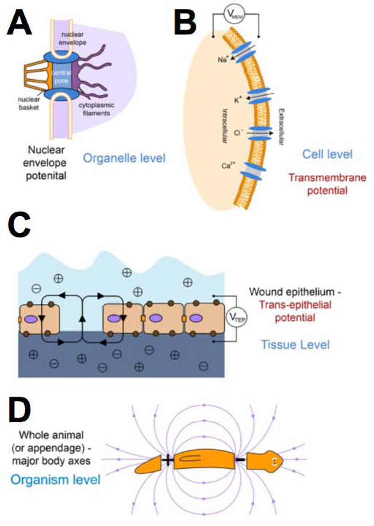
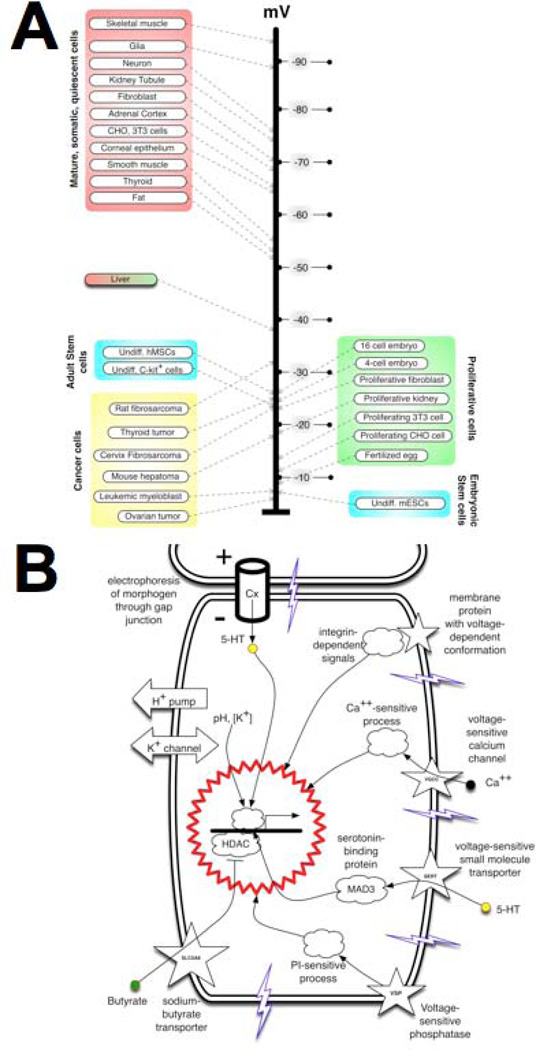
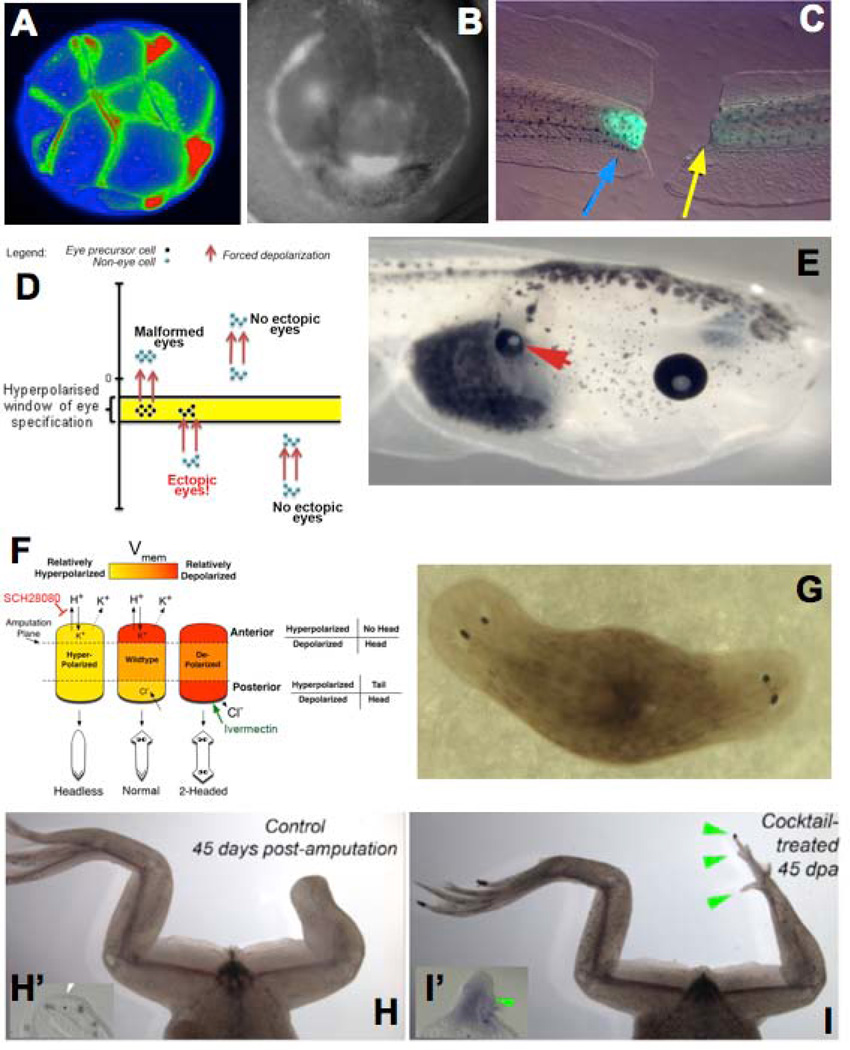
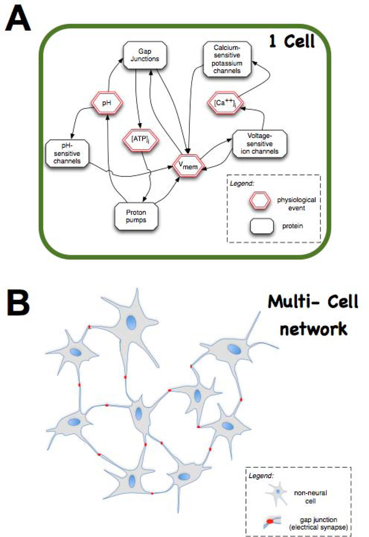
Transformative impact in regenerative medicine requires more than the reprogramming of individual cells: advances in repair strategies for birth defects or injuries, tumor normalization, and the construction of bioengineered organs and tissues all require the ability to control large-scale anatomical shape. Much recent work has focused on the transcriptional and biochemical regulation of cell behavior and morphogenesis. However, exciting new data reveal that bioelectrical properties of cells and their microenvironment exert a profound influence on cell differentiation, proliferation, and migration. Ion channels and pumps expressed in all cells, not just excitable nerve and muscle, establish resting potentials that vary across tissues and change with significant developmental events. Most importantly, the spatiotemporal gradients of these endogenous transmembrane voltage potentials (Vmem ) serve as instructive patterning cues for large-scale anatomy, providing organ identity, positional information, and prepattern template cues for morphogenesis. New genetic and pharmacological techniques for molecular modulation of bioelectric gradients in vivo have revealed the ability to initiate complex organogenesis, change tissue identity, and trigger regeneration of whole vertebrate appendages. A large segment of the spatial information processing that orchestrates individual cells' programs toward the anatomical needs of the host organism is electrical; this blurs the line between memory and decision-making in neural networks and morphogenesis in nonneural tissues. Advances in cracking this bioelectric code will enable the rational reprogramming of shape in whole tissues and organs, revolutionizing regenerative medicine, developmental biology, and synthetic bioengineering.


Conflict of interest statement

No conflict of interest.

Figures

Figure 1
Figure 1. the morphogenetic field
(A) Cell activity is guided by a complex, spatially-distributed set of signals from the host organism mediated by diffusing chemical, extracellular matrix, tension/pressure, and bioelectrical properties. (B) This morphogenetic field orchestrates cell behaviour towards large-scale anatomical programs during development and regeneration; its influence is subverted during oncogenic transformation and aging. Mastery of the information stored in this field, and of the mechanisms by which cells interact with it, will result in the ability to reprogram large-scale tissue and organ shape, with transformative implications for the fields of birth defects, regenerative medicine, cancer, and synthetic bioengineering.
Figure 2
Figure 2. bioelectric gradients exist at multiple scales of size and levels of biological organization
Organelles (A) and whole cells (B) are bound by membranes containing ion channel, pump, and transporter proteins. The activity of these ion translocators give rise to differences in resting potential (Vmem) across the membrane. Stacked in parallel, cells also give rise to a trans-epithelial potential (C), and electric fields have been characterized that correspond to appendages (limbs) or entire body axes (D). This overlapping set of cues provides positional information, organ identity, and other cues for cell behaviour and morphogenesis. This figure was drawn by Maria Lobikin, and is used with permission.
Figure 3
Figure 3. Vmem at the level of single cells: its transduction impacts cell states
(A) A sample survey of many cell types (modified and updated after), and recent functional data, , , –, reveals that at the level of single cells, Vmem determines cell plasticity and proliferation potential. Depolarized cells tend to be rapidly proliferating and undifferentiated (e.g., embryonic, stem, or tumor cells) while terminally-differentiated somatic cells tend to be highly polarized. Importantly, cell state can be functionally altered (switched between these two classes, in either direction) by artificial change of Vmem. This panel is modified after Fig. 1 of. (B) A range of mechanisms have now been characterized that transduce alterations of Vmem into downstream effector cascades (transcriptional changes). These include signalling proteins with a voltage-sensitive conformation (e.g., integrins and voltage-sensitive phosphatases) and transporters of small signalling molecules whose activity is regulated by Vmem (such as gap junctions, voltage-gated calcium channels, and solute carriers, which allow Vmem changes to signal via serotonin, Ca++, butyrate, and likely many other yet-to-be-discovered compounds). This figure is modified after Fig. 1B of.
Figure 4
Figure 4. Large-scale tissue and organ reprogramming by Vmem gradients
Voltage-sensitive fluorescent dyes reveal spatio-temporal patterns of bioelectrical gradients in vivo. Examples of gradients in the Xenopus laevis (frog) model include cleavage stages (A), craniofacial patterning (B, showing the hyperpolarizations in tissues that will become eye, branchial arches, and cement gland), and tail regeneration (C, regenerating tail on the left, and one that is prevented from regeneration by inhibition of V-ATPase activity on the right; green fluorescence signal indicates the normal repolarization of the wound (blue arrow), and when the repolarization is experimentally prevented (yellow arrow), regeneration is blocked). Functional data reveal a model in which a narrow range of Vmem (D) forces somatic cells, such as gut cells, to form a whole complete eye (E, arrowhead). A similar bioelectric circuit model describes the pathway regulating head vs. tail identity of regenerating tissue in planaria (F); experimental control of Vmem in this model results in 2-head animals after amputation (G), revealing the ability to control the shape of organs constructed by adult stem cells by bioelectric signalling. Brief treatment with sodium ionophore cocktail induces froglet hindlegs, which normally do not regenerate (H, showing wound region and lack of expression of the blastema gene MSX1 in inset panel H’) to regenerate legs with toes and toenails (I, showing blastema and induction of MSX1 expression in inset panel I’). This figure is modified after figures from references, , , .
Figure 5
Figure 5. Bioelectric signals enable non-neural cell fields to function as a computational medium
(A) At the level of single cells, elements such as voltage-sensitive ion channels result in feedback loops between the activity of ion translocator proteins and physiological parameters such as Vmem. These feedback loops ensure that physiological networks have non-obvious (emergent) behaviour dynamics, which can display hysteresis and multiple attractor states – thus able to store information encoded in stable Vmem states (e.g., depolarized = 1, hyperpolarized = 0) that would be invisible to genetic or proteomic profiling. (B) Even more interestingly, multiple (non-neural) cells communicating electrically via gap junctions (electrical synapses) could potentially store information and make decisions in the same way as do neural networks. The testing of this speculative hypothesis (using paradigms well-developed in computational neuroscience) may reveal entirely novel ways to understand and manipulate tissue-wide information that directs morphogenesis, and new approaches for the development of new (biologically-embedded) computational platforms. This figure is modified after figure 5 of reference.

Similar articles

Cited by

References

    1. Levin M. Morphogenetic fields in embryogenesis, regeneration, and cancer: non-local control of complex patterning. Bio Systems. 2012;109:243–261. - PMC - PubMed
    1. Rubin H. Cancer as a dynamic developmental disorder. Cancer Res. 1985;45:2935–2942. - PubMed
    1. Smithers DW. An attack on cytologism. Lancet. 1962;1:493–499. - PubMed
    1. Waddington CH. Cancer and the theory of organisers. Nature. 1935;135:606–608.
    1. Gentile L, Cebria F, Bartscherer K. The planarian flatworm: an in vivo model for stem cell biology and nervous system regeneration. Dis Model Mech. 2011;4:12–19. - PMC - PubMed

Publication types

LinkOut - more resources