. 2013 Oct;31(10):2116-26.
doi: 10.1002/stem.1481.

Transplantation of human umbilical mesenchymal stem cells cures the corneal defects of mucopolysaccharidosis VII mice

Affiliations

Transplantation of human umbilical mesenchymal stem cells cures the corneal defects of mucopolysaccharidosis VII mice

Vivien Jane Coulson-Thomas et al. Stem Cells. 2013 Oct.

Abstract

Mucopolysaccharidosis (MPS) are a family of related disorders caused by a mutation in one of the lysosomal exoglycosidases which leads to the accumulation of glycosaminoglycans (GAGs). MPS VII, caused by a mutation in β-glucuronidase, manifests hepatomegaly, skeletal dysplasia, short stature, corneal clouding, and developmental delay. Current treatment regimens for MPS are not effective for treating corneal clouding and impaired mental development. We hypothesized that human umbilical mesenchymal stem cells (UMSCs) transplanted into the corneal stroma could participate in the catabolism of GAGs providing a means of cell therapy for MPS. For such treatment, human UMSCs were intrastromally transplanted into corneas of MPS VII mice. UMSC transplantation restored the dendritic and hexagonal morphology of host keratocytes and endothelial cells, respectively, and in vivo confocal microscopy (HRT-II) revealed reduced corneal haze. Immunohistochemistry using antibodies against heparan sulfate and chondroitin sulfate chains as well as lysosomal-associated membrane protein 2 revealed a decrease in GAG content and both lysosomal number and size in the treated corneas. Labeling UMSC intracellular compartments prior to transplantation revealed the distribution of UMSC vesicles throughout the corneal stroma and endothelium. An in vitro coculture assay between skin fibroblasts isolated from MPS VII mice and UMSC demonstrated that neutral vesicles released by the UMSC are taken up by the fibroblasts and proceed to fuse with the acidic lysosomes. Therefore, transplanted UMSCs participate both in extracellular GAG turnover and enable host keratocytes to catabolize accumulated GAG products, suggesting that UMSC could be a novel alternative for treating corneal defects associated with MPS and other congenital metabolic disorders.

Keywords: Cornea; Exosomes; Glycosaminoglycans; Mucopolysaccharidosis; Umbilical cord mesenchymal stem cells.


Conflict of interest statement

Disclosure of Potential conflicts of interest

The authors indicate no conflict of interest.

Figures
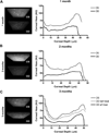
Figure 1
Figure 1
Corneal haze by In vivo confocal microscopy. Analysis of stromal haze was performed using Heidelberg Retinal Tomograph-HRTII Rostock Cornea Module (HRT-II, Heidelberg Engineering Inc., Germany). A series of images was collected to cover the whole stromal thickness as a continuous z-axis scan through the entire corneal stroma at 2 µm increments starting from the basal layer of the corneal epithelium and ending at the corneal endothelium. 3D reconstitution and histograms of light scattering were performed of images captured from (A) animals treated at 1 month, (B) animals treated at 2 months, and (C) animals treated at 3 months. Histograms produced using Fiji reveal significant corneal haze in untreated MPS VII corneas (OS) at 3.5 months (black graphs) and in the stroma of mice treated at 3 months prior to UMSC administration (C before treat). A significant decrease in haze can be observed in treated corneas at all time frames (OD) (grey graph).
Figure 2
Figure 2
Corneal morphology by in vivo confocal microscopy. Analysis of stromal morphology was performed using in vivo confocal microscopy. A representative image at the stromal depth of approximately 8 µm is shown for (A) littermate control wild-type mouse; (B) untreated MPSVII mice at 3 months; (C) untreated MPSVII mice at 3.5 months; (D) animals treated at 1 month at the conclusion of treatment; (E) animals treated at 2 months at the conclusion of treatment; (F) animals treated at 3 months at the conclusion of treatment. Amoeboid keratocytes are present throughout the stroma of untreated MPS VII corneas, however, a significant decrease in amoeboid keratocytes is observed in the stroma of mice treated at all time frames. Scale bar 50 µm.
Figure 3
Figure 3
Phalloidin staining after UMSC treatment. Corneas excised from the eyeballs were stained with phalloidin (green) in order to analyze the integrity of stromal keratocytes and endothelium (A and B). Phalloidin staining revealed significantly improved keratocyte morphology in treated corneas compared to the untreated corneas. (A) Untreated corneas (OS) display strong phalloidin staining revealing small rounded keratocytes as well as keratocytes with an amoeboid shape while treated corneas (OD) present dendritic host keratocytes. (B) DiI labeled UMSC (red) were present throughout the stroma of treated corneas. DiI positive cells (arrow) can be observed in the stroma establishing cell-cell contacts with host keratocytes (Asterisk). DiI speckles can be observed in host keratocytes surrounding DiI positive UMSC. Scale bar 20 µm.
Figure 4
Figure 4
Effectiveness of stem cell treatment by analysis of glycosaminoglycan and lysosomal content in MPS VII corneas treated with UMSC. (A) Immunocytochemistry was performed with mouse monoclonal anti-CS native sulfation motif (clones 4C3 and 6C3) and anti-HS (10E4 epitope). Corneas from 3.5 month-old MPS VII (OS) present a significant increase in both CS (red) and HS (green) accumulation with strong staining along the corneal lamellae, dense staining within the keratocytes and staining evidencing accumulation in clumps throughout the interfibrillar space. A drastic decrease in CS and HS staining was observed in the MPS VII corneas that received UMSC treatment (OD). (B and C) The total GAG content was measured using DMB. (B) Treated corneas (OD) at 1 month (black circle), 2 months (triangle) and 3 months (square) presented a significant reduction of at least 20% in overall GAG content when compared to the contralateral untreated corneas (OS). (C) A mean 30% reduction in GAG content was observed in treated corneas (OD) when compared to the untreated corneas (OS). The level of GAGs in the animals treated at 1 and 2 months (black circle and triangle, respectively) decreased to the level of the littermate wild-type controls (white circle), whereas the level of GAGs in the animals treated at 3 months (square) decreased significantly compared to the untreated corneas. (D) An overall decrease in lysosomal content (green) was observed in keratocytes throughout the stroma of animals treated at all-time points. A representative image at an approximate stromal depth of 10 µm is shown for LAMP2 staining in host keratocytes of animals treated at 2 months demonstrates the drastic decrease in the number and size of lysosomes in DiI negative cells. Nuclei were stained with DAPI (blue). Scale bar 20 µm. *p<0.05.
Figure 5
Figure 5
Lysosomal content and cell-cell trafficking between UMSC and MPS VII keratocytes and fibroblasts. (A) UMSC (asterisk) were labeled with FM 1-43FX (red) prior to intrastromal administration and eyeballs were excised after 3 days and corneas processed for whole mount. (B) MPS VII skin fibroblasts exposed to UMSC that had been previously labeled with LysoSensor™ Yellow/Blue DND-160 and seeded in transwell inserts with 0.44 µm pores for 15, 30 and 60 minutes. The live cells were analyzed using a Zeiss LSM710 confocal microscope. The nuclei of the UMSC and fibroblasts were labeled with SYTO® 59 (red). Neutral organelles are observed as blue and acid organelles as yellow/green. Scale bar 20µm. (C) MPS VII fibroblasts placed in co-culture with UMSC for 15 minutes or 6 hours and labeled with LAMP2 (green) in order to evaluate the lysosomal content. Naïve fibroblasts were used as the control. Nuclei were stained with DAPI (blue). Scale bar 20 µm. (D) The average number of pixels in 12 images for each experimental condition was calculated and the results displayed in a bar graph evidencing a statistically significant decrease in lysosome content in MPS VII fibroblasts exposed to UMSC. *p<0.05.
Figure 6
Figure 6
A schematic representation of the mechanism by which UMSC (red) aid corneal GAG turnover in MPSVII mice. (i) Endocytosis of accumulated extracellular GAGs and PGs by UMSC; (ii) UMSC catabolize the GAGs; (iii) intercellular trafficking active β-glucuronidase between UMSC and host keratocytes; (iv) sequential degradation of accumulated GAGs in host keratocyte.

References

    1. Freeze HH. Genetic Disorders of Glycan Degradation. 2009 - PubMed
    1. Cantz M, Gehler J. The mucopolysaccharidoses: inborn errors of glycosaminoglycan catabolism. Hum Genet. 1976;32:233–255. - PubMed
    1. Bernsen PL, Wevers RA, Gabreels FJ, et al. Phenotypic expression in mucopolysaccharidosis VII. J Neurol Neurosurg Psychiatry. 1987;50:699–703. - PMC - PubMed
    1. Neufeld EF, d'Azzo A. Biosynthesis of normal and mutant beta-hexosaminidases. Adv Genet. 2001;44:165–171. - PubMed
    1. Tomatsu S, Montano AM, Dung VC, et al. Mutations and polymorphisms in GUSB gene in mucopolysaccharidosis VII (Sly Syndrome) Hum Mutat. 2009;30:511–519. - PMC - PubMed

Publication types