Review
. 2013 Jul;3(3):1035-78.
doi: 10.1002/cphy.c120027.

Bile formation and secretion

Affiliations
Review

Bile formation and secretion

James L Boyer. Compr Physiol. 2013 Jul.

Abstract

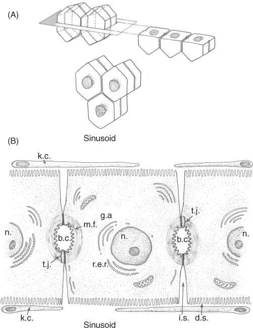
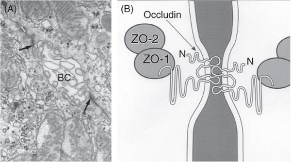
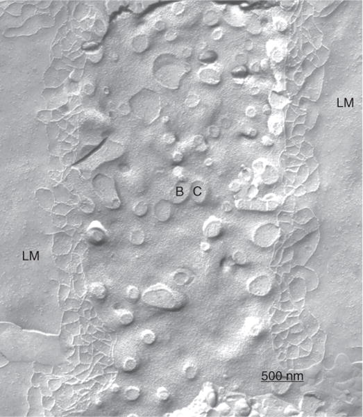
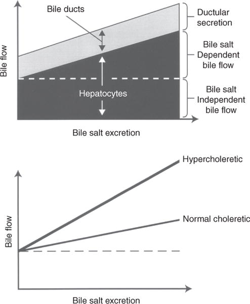
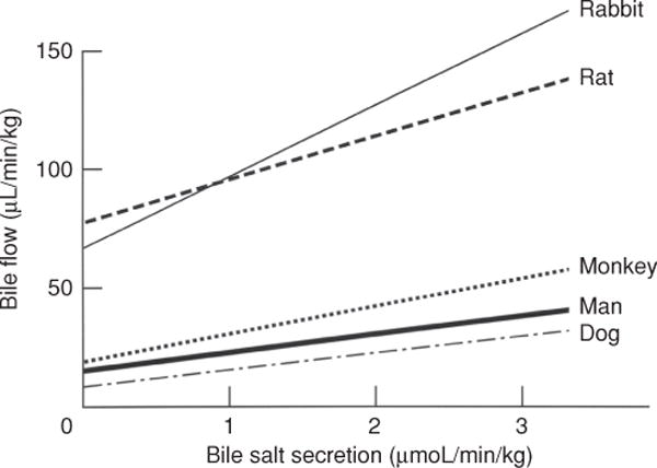
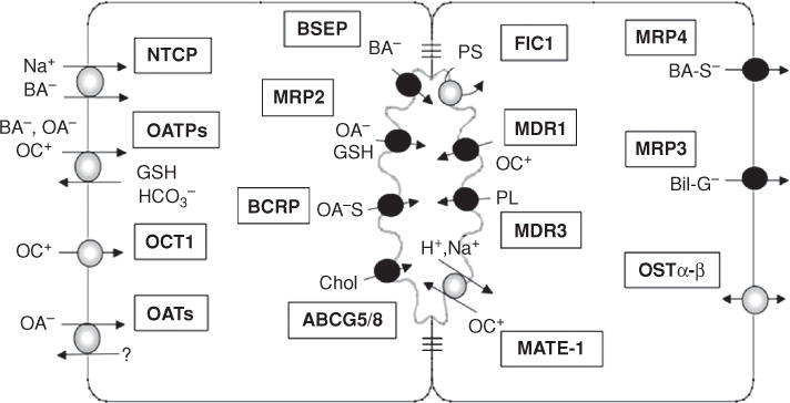
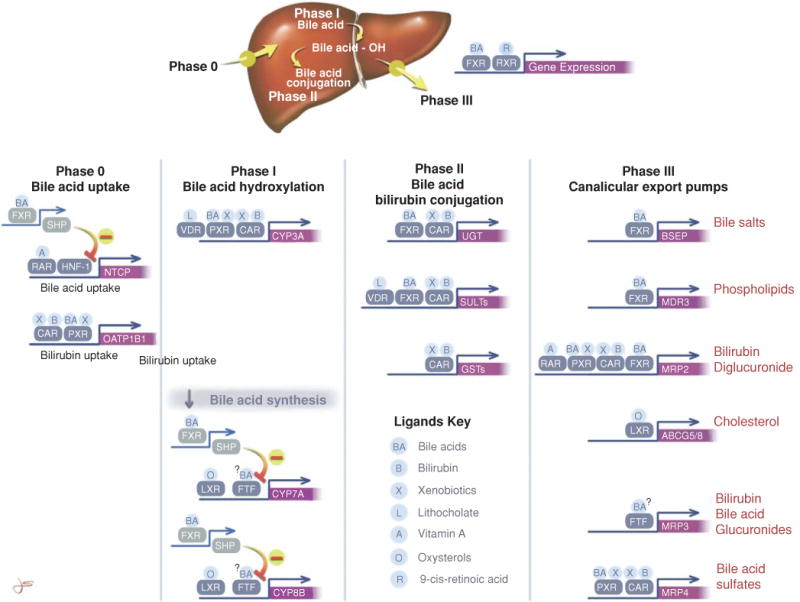
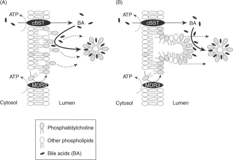
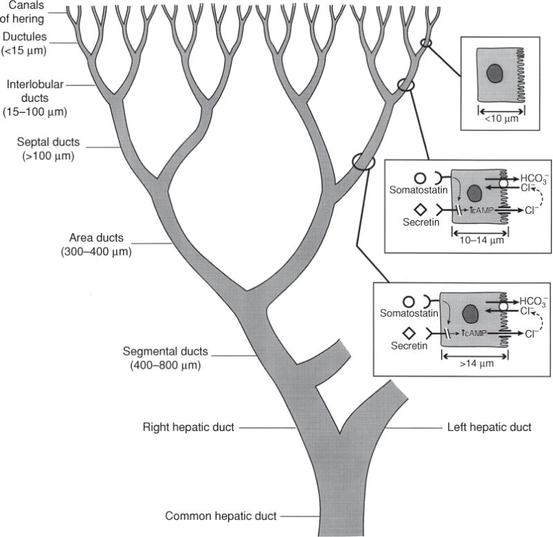
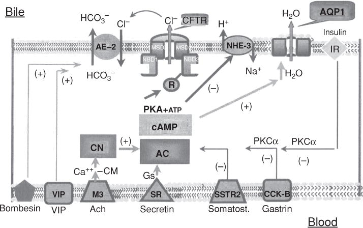
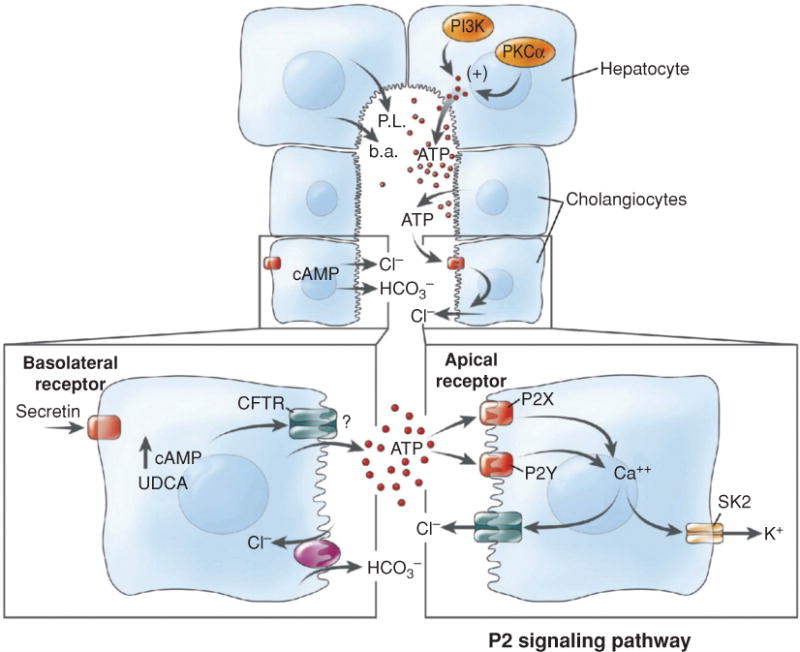
Bile is a unique and vital aqueous secretion of the liver that is formed by the hepatocyte and modified down stream by absorptive and secretory properties of the bile duct epithelium. Approximately 5% of bile consists of organic and inorganic solutes of considerable complexity. The bile-secretory unit consists of a canalicular network which is formed by the apical membrane of adjacent hepatocytes and sealed by tight junctions. The bile canaliculi (∼1 μm in diameter) conduct the flow of bile countercurrent to the direction of portal blood flow and connect with the canal of Hering and bile ducts which progressively increase in diameter and complexity prior to the entry of bile into the gallbladder, common bile duct, and intestine. Canalicular bile secretion is determined by both bile salt-dependent and independent transport systems which are localized at the apical membrane of the hepatocyte and largely consist of a series of adenosine triphosphate-binding cassette transport proteins that function as export pumps for bile salts and other organic solutes. These transporters create osmotic gradients within the bile canalicular lumen that provide the driving force for movement of fluid into the lumen via aquaporins. Species vary with respect to the relative amounts of bile salt-dependent and independent canalicular flow and cholangiocyte secretion which is highly regulated by hormones, second messengers, and signal transduction pathways. Most determinants of bile secretion are now characterized at the molecular level in animal models and in man. Genetic mutations serve to illuminate many of their functions.


Figures

Figure 1
Figure 1
(A) Three-dimensional projection of arrangement of hexagonal hepatocytes in liver plates illustrating the position of bile canaliculi which form a “chicken wire” mesh of interconnecting conduits of the primary secretion of bile. (B) Adjoining hepatocytes illustrating the location of the bile canaliculus (b.c.), intercellular space (i.c.), Disse’s space (d.s.), and fenestrated endothelial lining cells and Kupffer cells (k.c.). Tight junctions seal the lumen of the bile canaliculus (t.j.) whose luminal membrane is surrounded by microfilaments (m.f.) and other cytoskeletal elements that provide a contractile mechanism for canalicular peristalsis. Golgi apparatus (g.a.) and rough endoplasmic reticulum (r.e.r) are also illustrated. Reprinted, with permission, from Ref. (71).
Figure 2
Figure 2
The hepatocyte tight junction complex. (A) Electron micrograph of the bile canaliculus formed between two adjacent hepatocytes and whose lumen is filled with microvilli and sealed by the tight junctions (arrows). (B) Schematic of the tight junction complex showing that occludins and claudins are transmembrane proteins forming the junction seal, whereas zonula occluden proteins 1 and 2 (Z01 and Z02) are cytoplasmic proteins that may serve as anchors for occludins. The latter protein forms the interconnecting strands illustrated in the freeze fracture in Figure 3. See Ref. (384) for more details of tight junction anatomy. Reprinted, with permission, from Ref. (76).
Figure 3
Figure 3
Freeze fracture replica of the bile canaliculus (BC). The tight junction elements represent the only anatomical barrier between bile and the intercellular space lined by the lateral membrane (LM) of the hepatocyte. Magnifcation × 39,186. Reprinted, with permission, from Ref. (72).
Figure 4
Figure 4
Graphic representation of determinants of bile flow. Top: canalicular bile flow consists of a bile salt-dependent (BSDF) and a bile salt-independent (BSIF) fraction. The BSDF increases linearly as a function of bile salt excretion. Both canalicular bile flow and secretion from the bile ducts contribute to total bile flow. Bottom: choleretic potential varies among bile salts. Bile flow is linearly related to bile salt excretion, but hypercholeretic bile salts increase bile flow more rapidly than do bile salts with normal choleretic potential. Reprinted, with permission, from Ref. (76).
Figure 5
Figure 5
Relationship between bile salt concentration and bile flow in various animal species. Note that extrapolation to zero bile salt excretion yields a positive intercept for BSIF that is greater in rats and rabbits than in the three other species. Reprinted, with permission, from Ref. (73)
Figure 6
Figure 6
Membrane transporters that determine the uptake and excretion of bile salts and other organic solutes in hepatocytes. (Na+, sodium; BA, bile salts; OA, organic anions; OC+, organic cations; GSH, glutathione; HC03, bicarbonate; OA-S, sulfated organic anions; Chol, cholesterol; H+, proton; PL, phospholipid; PS, phosphatidyl serine; BA-S, sulfated bile salts; Bil-G, bilirubin glucuronide). Also see Table 4 for full terminology and function for these and other transporter determinants of bile secretion. Reprinted, with permission, from Ref. (79).
Figure 7
Figure 7
The hepatic clearance of bile salts and other organic solutes is determined by four steps or phases; Phase 0, hepatic uptake; Phase I, hydroxylation by cytochrome 3A and other CYP450s; Phase II, conjugation reactions with glucuronides, glutathione, sulfates, or acetates; and Phase III, export from the liver by adenosine triphosphate-dependent ATP-binding cassette (ABC) transporters. The figure also shows the coordinated ligand-activated regulation of gene expression that determines the hepatic clearance of bile salts, bilirubin, and xenobiotics. Some of the major nuclear receptors that regulate the expression of these key genes are shown. Unless otherwise indicated by ↓ or – symbols, these ligands stimulate gene expression. Normally, many of these nuclear receptors form heterodimeric complexes with the retinoid X receptor (RXR). This complex then binds to specific response elements in the gene promoter. Other nuclear receptors such as short heterodimeric protein-1, fetal transcription factor (FTF), and hepatocyte nuclear factor 1 (HNF-1) do not form heterodimers with RXR and do not have specific ligands. Reprinted, with permission, from Ref. (78).
Figure 8
Figure 8
Two alternative mechanisms for the mechanism of phosphatidylcholine (PC) excretion into bile. (A) Bile salts are transported into the canalicular lumen by the canalicular bile salt transporter (cBST) (now termed BSEP), and PC accumulates on the luminal side of the canalicular membrane by the action of MDR3. Luminal bile salts then extract the phospholipid from the membrane into micelles. (B) MDR3 flops PC to the external domains of the canalicular membrane bilayer which extrude into the bile lumen and are destabilized by bile salts which pinch off the membrane. Reprinted, with permission, from Ref. (76) as modified from Oude Elferink RPJ, Tytgat GNJ, Groen AK. Faseb J, 11: 19, 1997(427).
Figure 9
Figure 9
The figure illustrates the heterogeneity of the structure and function of the biliary tree and bile duct epithelial cells. Canalicular bile secreted by hepatocytes enters the biliary tree by joining upstream with the canals of Hering. As branches of the biliary tree join, the luminal diameter increases (values in parentheses) and the bile duct epithelial cells become larger. The range of receptors and transporters on medium and large bile duct cells is similar although secretin receptor expression and Cl/HCO3 exchange activity is greater in the median and large size bile duct cells. Reprinted, with permission, from Ref. (76).
Figure 10
Figure 10
Hormonal regulation of cholangiocyte HCO3 excretion based on studies in rodents. Secretin induces ductular bicarbonate-rich choleresis by activation of apical Cl/HCO3 exchanger via a cyclic adenosine monophosphate (cAMP) and PKA-dependent pathway; acetylcholine, by activation of calcineurin, induces a “sensitization” of adenylcyclase to secretin leading to a maximal stimulation of the Cl/HCO3 exchanger. Vasoactive intestinal peptide (VIP) and bombesin stimulate cholangiocyte bicarbonate secretion via a cAMP and cyclic guanosine monophosphate (cGMP)-independent pathway. Somatostatin, gastrin, and insulin inhibit both basal and hormonal induced bicarbonate cholangiocyte secretion via a PKC-α-dependent pathway. Ach, acetylcholine; M3, muscarinic receptor 3; SR, secretin receptor; CM, calmodulin; AC, adenyl cyclase; PKA, protein kinase A; PKCα, protein kinase c alpha; AE-2, Cl/HCO3 exchanger; CFTR, cystic fibrosis transmembrane conductance regulator; NHE-3, sodium hydrogen exchanger isoform 3; AQP1, aquaporin 1; IR, insulin receptor. Reprinted, with permission, from Ref. (183).
Figure 11
Figure 11
The role of extracellular adenosine triphosphate (ATP) in bile formation. Proposed model of P2 signaling. Bile formation begins via transport of bile salts, phospholipids, and ATP from the hepatocyte canalicular membrane. Hepatocyte ATP release is positively regulated by phosphatidylinositol 3-kinase (PI3K) and protein kinase C. Secretin stimulates increases in cholangiocyte cyclic adenosine monophosphate (cAMP) levels via stimulation of basolateral receptors resulting in Cl efflux through CFTR and an increase in Cl/HCO3 exchange. Increases in cAMP, as well as exposure to the bile salt ursodeoxycholate (UDCA), may also increase ATP release through a CFTR-dependent mechanism. BA, bile acids; PL, phospholipids; SK2, Ca2+-activated K+ channel; CFTR, cystic fibrosis transmembrane conductance regulator. Reprinted, with permission, from Ref. (159).

Similar articles

Cited by

References

    1. Abu-Hayyeh S, Martinez-Becerra P, Sheikh Abdul Kadir SH, Selden C, Romero MR, Rees M, Marschall HU, Marin JJ, Williamson C. Inhibition of Na+-taurocholate Co-transporting polypeptide-mediated bile acid transport by cholestatic sulfated progesterone metabolites. J Biol Chem. 2010;285:16504–16512. - PMC - PubMed
    1. Accatino L, Pizarro M, Solís N, Koenig CS, Vollrath V, Chianale J. Modulation of hepatic content and biliary excretion of P-glycoproteins in hepatocellular and obstructive cholestasis in the rat. J Hepatol. 1996;25:349–361. - PubMed
    1. Adachi Y, Kobuyashi H, Kurumi Y. ATP-dependent taurocholate transport by rat liver canalicular membrane vesicles. Hepatology. 1991;14:655–659. - PubMed
    1. Agre P, Bonhivers M, Borgnia MJ. The aquaporins, blueprints for cellular plumbing systems. J Biol Chem. 1998;273:14659–14662. - PubMed
    1. Akita H, Suzuki H, Hirohashi T, Takikawa H, Sugiyama Y. Transport activity of human MRP3 expressed in Sf9 cells: Comparative studies with rat MRP3. Pharm Res. 2002;19:34–41. - PubMed

Substances