Review
. 2013 Jul;3(3):1191-212.
doi: 10.1002/cphy.c120023.

Bile acid metabolism and signaling

Affiliations
Review

Bile acid metabolism and signaling

John Y L Chiang. Compr Physiol. 2013 Jul.

Abstract

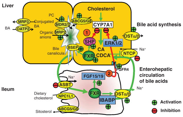
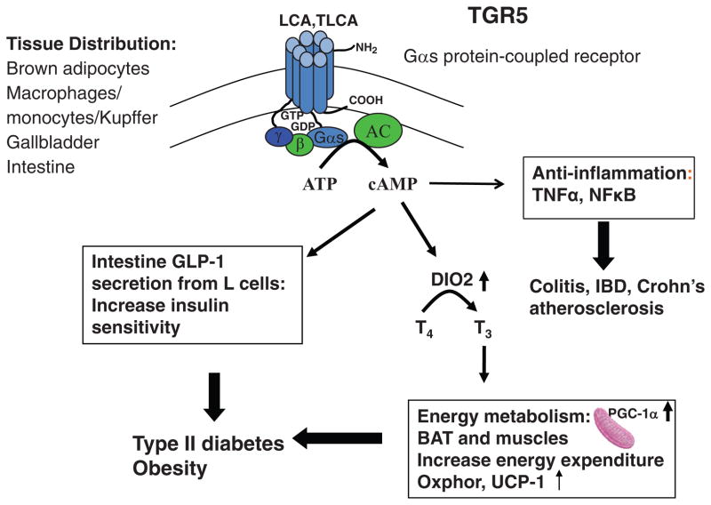
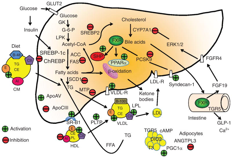
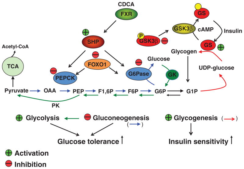
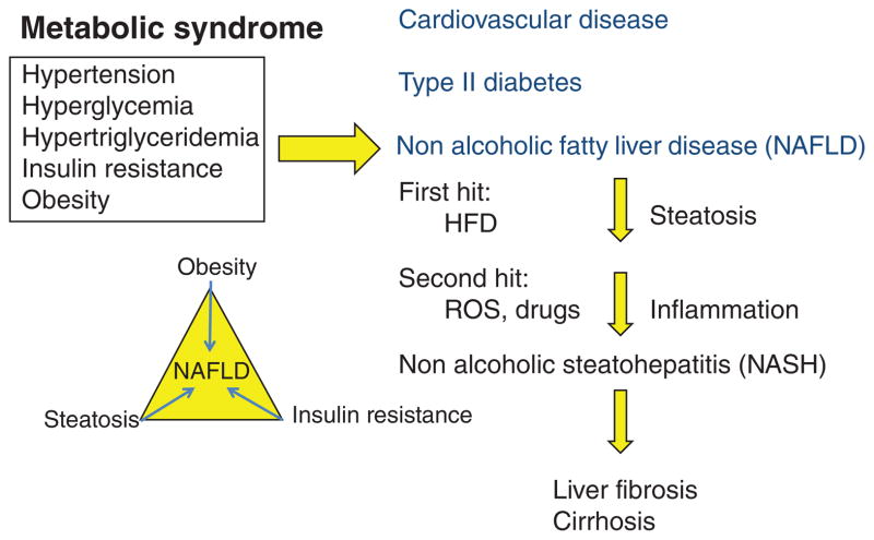
Bile acids are important physiological agents for intestinal nutrient absorption and biliary secretion of lipids, toxic metabolites, and xenobiotics. Bile acids also are signaling molecules and metabolic regulators that activate nuclear receptors and G protein-coupled receptor (GPCR) signaling to regulate hepatic lipid, glucose, and energy homeostasis and maintain metabolic homeostasis. Conversion of cholesterol to bile acids is critical for maintaining cholesterol homeostasis and preventing accumulation of cholesterol, triglycerides, and toxic metabolites, and injury in the liver and other organs. Enterohepatic circulation of bile acids from the liver to intestine and back to the liver plays a central role in nutrient absorption and distribution, and metabolic regulation and homeostasis. This physiological process is regulated by a complex membrane transport system in the liver and intestine regulated by nuclear receptors. Toxic bile acids may cause inflammation, apoptosis, and cell death. On the other hand, bile acid-activated nuclear and GPCR signaling protects against inflammation in liver, intestine, and macrophages. Disorders in bile acid metabolism cause cholestatic liver diseases, dyslipidemia, fatty liver diseases, cardiovascular diseases, and diabetes. Bile acids, bile acid derivatives, and bile acid sequestrants are therapeutic agents for treating chronic liver diseases, obesity, and diabetes in humans.


Figures

Figure 1
Figure 1
Bile acid biosynthetic pathways. Two major bile acid biosynthetic pathways are shown. The neutral (or classic) pathway is initiated by cholesterol 7α-hydroxylase (CYP7A1) located in the endoplasmic reticulum of the liver, whereas the acidic (or alternative) pathway is initiated by mitochondrial sterol 27-hydroxylase (CYP27A1). There are three sterol hydroxylases that convert cholesterol to oxysterols: CYP27A1 in macrophages and other tissues, microsomal sterol 25-hydroxylase in liver microsomes, and sterol 24-hydroxylase (CYP46A1) in the brain. Oxysterol 7α-hydroxylase (CYP7B1) is nonspecific and catalyzes hydroxylation of 27- and 25-hydroxycholesterol to 3β, 7α-dihydroxy-5-cholestenoic acid and 5-cholesten-3β, 7α, 25-triol, respectively. A brain-specific oxysterol 7α-hydroxylase (CYP39A1) catalyzes hydroxylation of 24-hydroxycholesterol to 5-cholesten-3β, 7α, 24(S)-triol. These oxysterols could be converted to CDCA if transported to the liver. In the liver, 3β-hydoxysteroid dehydrogenase (3βHSD, HSD3B7) convert 7α-hydroxycholesterol to 7α-hydroxy-4-cholesten-3-one (C4), which is converted to 7α, 12α-dihydroxy-4-cholesten-3-one by a sterol 12α-hydroxylase (CYP8B1), leading to synthesis of cholic acid (CA). Without 12α-hydroxylation, the pathway produces CDCA. Aldos-keto reductase 1D1 (AKR1D1) and AKR1C1 catalyze isomerization and saturation of the steroid ring. Then CYP27A1 catalyzes steroid side-chain oxidation to form cholestanoic acids, THCA, and DHCA. Bile acid-Co-A synthase (BACS) or very long-chain Co-A synthase (VLCS) in the endoplasmic reticulum (ER) ligates Co-A to the carboxyl groups. Bile acid thioesters are transported into peroxisomes, where an α-methylacyl-CoA racemase (AMACR) converts the methyl group from 25(R) to 25(S) conformation, and three peroxisomal β-oxidation enzymes, branched-chain acyl-CoA oxidase, D-bifunctional enzyme, and thiolase (or sterol carrier protein x) catalyze oxidative cleavage of a propionyl group from the steroid side-chain to form cholyl-CoA and chenodeoxycholyl-CoA. Cytosolic or peroxisomal bile acid: amino-acid transferase (BAAT) catalyzes conjugation of amino acids, glycine or taurine to the carboxyl group of cholyl-CoA and chenodeoxycholyl-CoA to form tauro- or glycol-conjugated CA or CDCA. ER: endoplasmic reticulum.
Figure 2
Figure 2
Enterohepatic circulation of bile acids. An average man produces ~0.5g bile acid per day by synthesis in the liver, and secretes ~0.5g/day. This daily turnover of bile acids accounts for about 5% of total bile acid pool. The remaining 95% of bile acids in the pool are recycled 4 to 12 times a day. Most bile acids are reabsorbed in the ileum by active transport, while a small amount is reabsorbed by passive diffusion in the upper intestine to portal blood for circulation to the liver. Small amounts of bile acids spilled over into the systemic circulation are recovered in kidney.
Figure 3
Figure 3
Bile acid synthesis regulates cholesterol homeostasis in hepatocytes. Cholesterol homeostasis is maintained by dietary uptake of cholesterol, de novo cholesterol synthesis from acetyl-CoA, and conversion of cholesterol to bile acids. Oxysterols are derived from cholesterol and bile acids. When intracellular cholesterol/oxysterol levels are high, steroid response element binding protein 2 (SREBP-2) precursor (125 kDa) interacts with insulin induced gene 1/2 (Insig1/2) and is retained in endoplasmic reticulum (ER) membrane. When intracellular oxysterol levels are low, SREBP cleavage and activating protein (SCAP) escorts SREBP-2 precursor to the Golgi apparatus, where sterol sensitive proteases S1P and S2P are activated to cleave a N-terminal fragment (65 kDa), which is translocated to the nucleus to bind to the steroid response elements in the gene promoters of all cholesterogenic genes and stimulates de novo cholesterol synthesis. Oxysterols activate LXRα, which induces CYP7A1 gene transcription to stimulate bile acid synthesis in mice, but not humans. Bile acids (CDCA) activate farnesoid X receptor (FXR) to inhibit CYP7A1 gene transcription and bile acid synthesis. This may lead to increased cholesterol levels and inhibited de novo cholesterol synthesis and absorption of dietary cholesterol.
Figure 4
Figure 4
Nuclear receptors. The general structure of nuclear receptors is shown on the top. The NR1 family of genes involved in metabolic regulation, and their respective endogenous ligands are shown. The putative nuclear receptor response element binding sequence, arranged in direct repeat (DR), everted repeat (ER), and inverted repeat (IR), is shown. Ligand-activated receptors recruit coactivators to replace corepressors and results in transactivation of target gene expression. AF-1-2, activation function-1 and -2; CAR, constitutive androstane receptor; FXR, farnesoid X receptor; LXR, liver orphan receptor; PPAR, peroxisome proliferator-activated receptor; PXR, pregnane X receptor; NLS, nuclear localization sequence.
Figure 5
Figure 5
Farnesoid X receptor (FXR) regulates enterohepatic circulation of bile acids. Major bile acid transporters in human hepatocytes and enterocytes are shown. Enzymes and transporters regulated by FXR are indicated. In hepatocytes, bile acids activate FXR to inhibit CYP7A1 gene transcription by two pathways: (i) FXR induces small heterodimer partner (SHP), which inhibits CYP7A1 by inhibiting nuclear receptors liver related homologue-1 (LRH-1) or hepatocyte nuclear factor (HNF4), which bind to the CYP7A1 promoter. (ii) In enterocytes, FXR induces intestinal hormone fibroblast growth factor 19 (FGF19), which is circulated to hepatocytes to activate FGF receptor 4 (FGFR4) signaling to inhibit CYP7A1 via activation of the extracellular stress-activated receptor kinase 1/2 (ERK1/2) pathway. FXR induces bile salt expert pump (BSEP) to efflux bile acids into bile; multidrug resistance protein 2/3 (MDR2/3) to efflux phosphatidylcholine (PC) to bile; and MDR related protein 2 (MRP2) to efflux organic anions including glucuronidated- and sulfated-bile acids, organic anions, and drugs into bile. Bile acids also facilitate efflux of cholesterol to bile by ATP binding casette G5/G8(ABCG5/G8). In the bile, bile acids, PC, and cholesterol form mixed micelles, which are stored in the gallbladder. In the brush border membrane of the ileum, bile acids are reabsorbed by the apical sodium bile salt transporter (ASBT). In enterocytes, bile acids activate FXR, which induces ileum bile acid binding protein (IBABP) to bind bile salts and may facilitate intracellular transport of bile acids to organic solute transporter α/β (OSTα/β) located in the basolateral membrane for efflux of bile acids into portal circulation. Bile acids in portal blood are reabsorbed into hepatocytes by Na+-dependent taurocholate cotransport peptide (NTCP). FXR inhibits NTCP transcription as a feedback inhibition of bile acid uptake to prevent liver injury. In the sinusoidal membrane of enterocytes and hepatocytes, FXR also induces MRP3/4 to efflux bile acids as an adaptive response to cholestasis. In hepatocytes, FXR also induces OSTα/β to efflux bile acids into sinusoidal blood to prevent bile acid accumulation in hepatocytes. MRP3 may be induced by FXR as an adaptive response to cholestasis. Bile acids returned to hepatic sinusoid are also taken up by Na+-independent organic anion transport proteins (OATP2). Many of these membrane transporters (ASBT, OSTα/β, and MRP2/3) also are present in cholangiocytes for reabsorption of bile acids, and in renal proximal tubule cells for reabsorption of bile acids from blood circulation and excretion of hydrophilic bile acids.
Figure 6
Figure 6
Bile acid-activated TGR5 signaling. TGR5 is expressed in brown adipocytes, macrophages/monocytes and hepatic Kupffer cells, gallbladder epithelium, and intestine, with especially high levels in the colon. TGR5 is the first G protein-coupled receptor (GPCR) identified as a bile acid-activated membrane receptor. TGR5 is a Gαs GPCR activated by secondary bile acids, lithocholic acid (LCA) and TLCA to induce cAMP signaling through activation of adenylyl cyclase (AC). TGR5 signaling may increase insulin sensitivity through two mechanisms. (i) In brown adipose tissue, cAMP induces type 2 deiodinase (DIO2), which converts and activates thyroid hormone T4 to T3 to stimulate energy metabolism in mitochondria by activating oxidative phosphorylation (OXphor) and uncoupling protein-1 (UCP-1). (ii) In the intestine, cAMP stimulates glucagon like peptide-1 (GLP-1) in L cells, which stimulates insulin secretion in the pancreas. TGR5 also has anti-inflammatory functions by antagonizing of TNFα and NF-κB-dependent induction of proinflammatory cytokines in intestine and macrophages, thus, protecting against colitis, inflammatory bowel disease and Crohn’s disease, and also atherosclerosis.
Figure 7
Figure 7
Bile acid-activated S1P2 signaling. Tauro-conjugated bile acids stimulates sphingosine-1-phosphate receptor 2 (S1P2), a Gαi protein-coupled receptor, which activates the extracellular signal-regulated kinase 1/2 (ERK1/2) and AKT pathways. S1P2 signaling activates the insulin receptor/AKT pathway through activation of Src and epidermal growth factor receptors (EGFRs) leading to activation of the insulin receptor (IR), which phosphorylates and activates insulin receptor substrate-1 (IRS-1). IRS-1 phosphorylates phosphatidylinositol 3-kinase (PI3K), which phosphorylates phosphatidylinositol 4′, 5′ bis-phosphate to phosphatidylinositol 3′, 4′, 5′-trisphosphate (PIP3). PIP3 then ohosphorylates pyruvate dehydrogenase kinase-1 (PDK-1) to phosphorylate and activate AKT (also know as PKB). Phosphorylated AKT phosphorylates and inactivates glycogen synthase kinase 3β (GSK3β) and resulting in dephosphorylation and activation of glycogen synthase (GS), a key enzyme in glycogenesis. AKT also phosphorylates and inactivates a transcription factor, FoxO1 and resulted in inhibiting phosphoenolpyruvate carboxykinase (PEPCK) and glucose-6-phosphatase (G6Pase) in gluconeogenesis. S1P2 signaling also may activate FXR/SHP pathway to inhibit peroxisome proliferator-activated receptor αγ (PPARα/γ)/PGC-1α-mediated fatty acid oxidation and steroid response element binding protein 1c (SREBP-1c)-mediated fatty acid synthesis. Therefore, SiP2 signaling may reduce serum glucose and triglycerides, and improve insulin sensitivity by stimulating glycogensis and inhibiting gluconeogenesis and lipogenesis.
Figure 8
Figure 8
Farnesoid X receptor (FXR) regulation of hepatic glucose and lipid metabolism in liver, adipocytes, and intestine. Glucose and insulin stimulate glycolysis to form acetyl-CoA, which is a precursor of cholesterol and fatty acids. The FXR/small heterodimer partner (SHP) pathway may inhibit steroid response element binding protein 1c (SREBP-1c), which induces all genes involved in lipogenesis, acetyl CoA carboxylase (ACC), fatty acid synthase (FAS), and stearoyl CoA desaturase (SCD). The FXR/SHP pathway also inhibits SREBP-2, which induces all genes in de novo cholesterol synthesis. FXR activates mitochondria fatty acid β-oxidation by inducing peroxisome proliferator-activated receptor α (PPARα). FXR inhibits PCSK9, which is an inhibitor of LDL receptor. Thus FXR induces LDL-R and also syndecan-1 involved in cholesterol uptake. FXR inhibits mitochondria triglyceride transport protein (MTP), which is required for assembly of VLDL particles with ApoB100. FXR also induces ApoE, which is a high affinity ligand of LDL receptor and scavenger receptor B1 (SR-B1), and a component of VLDL and chylomicron remnants, which are taken up by ApoE receptors. FXR also induces phospholipid transport protein (PLTP) involved in reverse cholesterol transport of cholesterol from peripheral tissues to liver by HDL/SR-B1 receptor-mediated mechanisms. On the other hand, FXR inhibits ApoA1, a component of HDL, and ANGPTL3, which is involved in hydrolysis of triglycerides in liver and adipocytes. FXR induces FGF19 synthesis in the intestine, which activates FGFR4 receptor in hepatocytes to activate ERK1/2 signaling to inhibit CYP7A1 and bile acid synthesis. In colon, bile acids activate TGR5 signaling to stimulate GLP-1 release. GLP-1 increases insulin sensitivity. TGR5 in brown adipocytes stimulates cAMP production, which induce deiodinase 2 (DIO2) to convert T4 to T3, which stimulates mitochondrial energy metabolism via activation of PGC-1α.
Figure 9
Figure 9
FXR regulation of hepatic glucose metabolism. FXR signaling phosphorylates and inhibits glycogen synthase kinase 3β (GSK3β), which is an inhibitor of glycogen synthase activity. This results in stimulating glycogenesis. The FXR/SHP pathway inhibits PEPCK and glucose 6-phosphatase (G6Pase) to inhibit gluconeogenesis. This results in increasing glucose tolerance and insulin sensitivity.
Figure 10
Figure 10
Mechanism of nonalcoholic fatty liver disease (NAFLD). Metabolic syndrome is a constellation of five clinical symptoms, hypertension, hyperglycemia, hypertriglyceridemia, insulin resistance, and obesity. Metabolic syndrome is linked to cardiovascular disease, type II diabetes, and NAFLD. NAFLD is progressed to nonalcoholic steatohepatitis (NASH) by many factors. The first hit is high fat diet (HFD)-induced hepatic steatosis, followed by inflammation involving reactive oxidizing species, drugs, and endoplasmid reticulum stress to NASH. NASH patients have prevalence for liver fibrosis and cirrhosis, while NAFLD is linked to obesity, steatosis, and insulin resistance. HFD, high fat diet; ROS, reactive oxidizing species.

Similar articles

Cited by

References

    1. Adorini L, Pruzanski M, Shapiro D. Farnesoid X receptor targeting to treat nonalcoholic steatohepatitis. Drug Discov Today. 2012;17:988–997. - PubMed
    1. Agellon LB, Cheema SK. The 3′-untranslated region of the mouse cholesterol 7α-hydroxylase mRNA contains elements responsive to post-transcriptional regulation by bile acids. Biochem J. 1997;328:393–399. - PMC - PubMed
    1. Aldridge MA, Ito MK. Colesevelam hydrochloride: A novel bile acid-binding resin. Ann Pharmacother. 2001;35:898–907. - PubMed
    1. Alvarez L, Jara P, Sanchez-Sabate E, Hierro L, Larrauri J, Diaz MC, Camarena C, De La Vega A, Frauca E, Lopez-Collazo E, Lapunzina P. Reduced hepatic expression of Farnesoid X Receptor in hereditary cholestasis associated to mutation in ATP8B1. Hum Mol Genet. 2004;13:2451–2460. - PubMed
    1. Amaral JD, Viana RJ, Ramalho RM, Steer CJ, Rodrigues CM. Bile acids: Regulation of apoptosis by ursodeoxycholic ccid. J Lipid Res. 2009;50:1721–1734. - PMC - PubMed

Publication types

MeSH terms