. 2013 Nov;31(11):2492-505.
doi: 10.1002/stem.1484.

Lgr4 is a key regulator of prostate development and prostate stem cell differentiation

Affiliations

Lgr4 is a key regulator of prostate development and prostate stem cell differentiation

Weijia Luo et al. Stem Cells. 2013 Nov.

Abstract

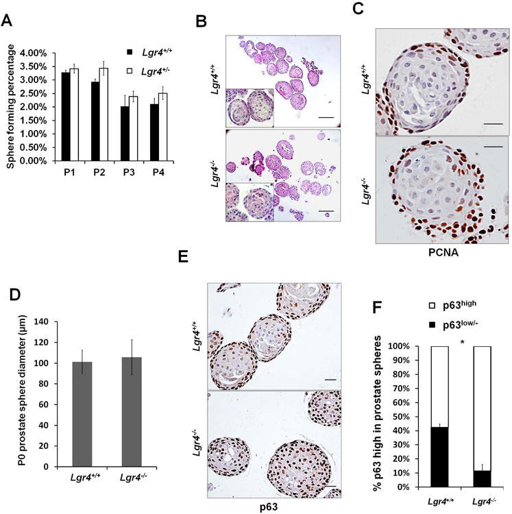
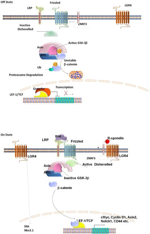
Mechanisms modulating prostate cell fate determination remain unexplored. The leucine-rich repeat containing G-protein-coupled receptors (Lgr) have been identified as important stem cell markers in various tissues. Here, we investigated the roles of Lgr4/Gpr48 in prostate stem cells (PSCs) and development. Lgr4 was ubiquitously expressed during early prostate development prior to lineage specification, with adult expression restricted to a few basal cells (principally Lin(-)Sca1(+)CD49f(+)). Lgr4(-/-) mice had compromised branching morphogenesis and delayed epithelial differentiation, leading to decreased prostate size and impaired luminal cell function. In vitro prostate sphere culture revealed that Lgr4(-/-) Lin(-)/Sca1(+)/CD49f(+) cells failed to generate p63(low) cells, indicating a differentiation deficiency. Furthermore, Lgr4 ablation arrested PSC differentiation of in vivo kidney capsule prostate grafts, suggesting that Lgr4 modulates PSC properties independent of hormonal and mesenchymal effects. Analysis of neonatal prostates and prostate spheres revealed a decrease in Wnt, Sonic Hedgehog, and Notch1 expression in Lgr4(-/-) cells. Lgr4 loss blocked differentiation of prostate sphere p63(hi) cells to p63(low). Treatment with exogenous Sonic Hedgehog partially restored the differentiation of p63(hi) cells in Lgr4(-/-) spheres. Taken together, our data revealed the roles of Lgr4 in early prostate development and in stem cell differentiation through regulation of the Wnt, Notch, and Sonic Hedgehog signaling pathways.

Keywords: Gpr48; Lgr4; Prostate development; Prostate stem cells.


Conflict of interest statement

The authors declare no conflicts of interest.

Figures

Figure 1
Figure 1. Lgr4 ablation compromised prostate development
A) Whole mount prostates stained for LacZ were sectioned (7μm thickness) and counterstained with Eosin to show Lgr4 expression in 1-4 week-old prostates. Scale bar 25μm. B) In 8 week-old prostate, Lgr4 expression (shown by LacZ staining) is partially co-localized with p63 (shown by IHC). Representative photograph of section from anterior lobe; pattern is characteristic of all lobes. Arrows indicate co-localization of Lgr4 and p63. Scale bar 25μm. C) Lgr4 is expressed in basal epithelial cells in the human prostate. Lgr4 expression in adult human prostate was determined by immunohistochemistry. Representative photograph of N=5. Scale bar = 100 μm. D) Lgr4 mRNA levels in different populations of primary prostate cells were determined by qPCR. E). Reduced prostate sizeof Lgr4−/− mice when compared to their wild type littermates at indicated ages. Scale bar 5mm. F) Prostatic lobes were dissected from 6 week-old Lgr4−/− mice and their wild type littermates, and their ductal networks were microdisected. Scale bar 5mm. G) The branching points of each prostatic lobe from 6 week mice were quantified. Bar graph shows the mean ± S. E. from 3 pairs of mice, p≤0.02. AP, anterior prostate; VP, ventral prostate; DLP, dorsal-lateral prostate. H) Hematoxylin & Eosin staining of prostate sections at indicated ages. Scale bar 100μm.
Figure 2
Figure 2. Impaired proliferation, epithelial differentiation and function in Lgr4−/− prostate
A) Ki67IHC shows proliferating cells in 1 to 6 week-old anterior prostate. Scale bar 50μm. B) Quantitation of Ki67 positive epithelial cells from 3 pairs of mice, 5 sections per prostate. Data expressed as mean ± S. E., * p≤0.001. C) CK8 IHC in anterior prostates at indicated ages. Photographs representative of CK8 pattern in all lobes. Scale bar 25μm.D) CK5 IHC in anterior prostates at indicated ages. Scale bar 25μm.E) Basal versus luminal epithelial cell ratios of 6 week-old anterior prostates were quantified. Bar graph expresses mean ± S. E. from 3 pairs of mice, 5 sections per prostate, p≤0.001. F) Probasin expression (green) in 4 week- and 6 week-old anterior prostate is shown by IF. Sections were counter stained with TO-PRO-3 (blue). Scale bar 100μm.G) Androgen receptor level in 2 week-old anterior prostates shown by IHC. Scale bar 50μm.
Figure 3
Figure 3. Lgr4 ablation diminished prostate progenitor cell epithelial differentiation in prostate sphere culture without affecting their self-renewal abilities
A) Number of prostate spheres from 3 pairs of mice in triplicate were counted for each generation. Prostate spheres were cultured for 4 generations. Graph shows the mean percentage of sphere forming cells ± S. E.. B) P0 prostate sphere morphology shown by Hematoxylin& Eosin staining. Scale bar 100μm. C) IHC of PCNA in P0 prostate spheres. Scale bar 20μm. D) Prostate sphere diameter was measured in at least 6 prostate spheres from 3 pairs of sphere samples. Bar graph shows the mean value ± S. D.E) p63 IHC of prostate spheres. Scale bar 20μm.F) p63high and p63low/− cells were counted from 8 prostate spheres. Bar graph shows the mean percentage ± S. E., p≤0.001.
Figure 4
Figure 4. Lgr4 inactivation in prostate stem cells disrupted epithelial differentiation and impaired prostate graft regenerationin vivo
A) Prostatic grafts after 2 months of regeneration in vivo. Scale bar 5mm. B) Prostatic graft wet weight. Data shows mean ± S. E. n=5, p≤0.002. C) Hematoxylin & Eosin staining of regenerated prostatic graft sections. Scale bar 200μm. D) Co-IF of CK5 (green) and CK8 (red) in regenerated prostatic grafts. Nuclei were counter stained with TO-PRO-3 (blue). Scale bar 20μm. E) CK8+ luminal epithelial cells and CK5+ basal epithelial cells were quantified from 5 pairs of regenerated prostatic grafts, 3 sections per graft. Bar graph shows mean ± S. E. basal versus luminal epithelial cell ratio. p≤0.001. F) CK5 (green) and CK8 (red) co-IF in adult mouse prostates after 8 days of androgen castration (left panel) and followed by 14 days of androgen replenishment (right panel). Nuclei were counter stained with TO-PRO-3 (blue). Scale bar 20μm.
Figure 5
Figure 5. Lgr4 regulates PSC proliferation and differentiation through Wnt/β-catenin signaling
Wnt/β-catein Signaling pathways in early prostate development were compared by qPCR between Lgr4+/+ and Lgr4−/− in P1 prostate spheres A) and in PD1 prostates B). Three pairs of prostate spheres or prostate samples were tested in these experiments. * p≤0.05.C) IF of β-catenin (green) and p63 (red) in P1 prostate spheres. Nuclei were counter stained with TO-PRO-3 (blue). Scale bar 20μm.D) Wnt/β-catenin activity indicated by GFP expression in wild-type and Lgr4−/− P1 prostate spheres treated with RSPO3 or control media (Mock). Scale bar 20μm. E) Prostate spheres treated 10 days with or without Wnt3a and/or RSPO3. Scale bar 20μm. F) Prostate sphere diameter was measured in at least 6 prostate spheres from 3 pairs of sphere samples. Bar graph shows the mean value ± S. E., *p≤0.05, ** p≤0.01. G) p63 (red) IF in prostate spheres treated with Wnt3a and/or RSPO3. Nuclei were counter stained with TOPRO-3 (blue). Scale bar 10μm.H) Quantification of p63 high cells in prostate spheres treated with or without Wnt3a and/or RSPO3. p63high and p63low/− cells were quantified from 6 prostate spheres. Bar graph shows the mean value ± S. E., *p≤0.05, ** p≤0.01.
Figure 6
Figure 6. Notch and Shh pathways are key targets regulated by Lgr4 in PSCs and early prostate development
A) Notch1, Hes1, and Notch ligand expression in P1 prostate spheres. B) Notch1, Hes1, and Notch ligand expression in postnatal day 1 prostates. C) Notch1 (green) and p63 (red) co-IFin P1 prostate spheres. Nuclei were counter stained with TOPRO-3 (blue). Scale bar 20μm. D) Chromatin immuno-precipitation on Notch1 promoter in P1 prostate spheres. E) Notch1 mRNA level was assessed by qPCR in 4 pairs of prostate sphere samples treated with or without RSPO3. Bar graph shows the mean value ± S. E., *p≤0.05F) Shh expression in P1 prostate spheres. G) Shh signaling in postnatal day 1 prostates. H) p63 (red) IF in prostate spheres cultured with or without Shh treatment. Nuclei were counter stained with TOPRO-3 (blue). Scale bar 20μm. I) p63high and p63low/− cells were quantified from 6 prostate spheres treated with or without Shh. Bar graph shows the mean percentage ± S. E. of either population in prostate spheres, p≤0.02.
Figure 7
Figure 7. Lgr4 working model in PSCs
In the absence of R-spondin and Wnt (top), ZNRF3 ubiquitinates the Wnt receptor Frizzled and the co-receptor Lrp6 (Hao et al. 2012), leading to their proteasomal degradation; Dishevelled remains inactive, and active GSK-3β phosphorylates β-catenin leading to ubiquitination and degradation of β-catenin. Upon R-spondin binding to Lgr4 (bottom), ZNRF3 is inhibited, leading to increased membrane levels of Frizzled and LRP6. Addition of Wnt leads to activation of Frizzled, with consequent activation of Dishevelled, inhibition of GSK-3, and β-catenin accumulation, nuclear translocation, and association with LEF-1/TCF transcription factors. This results in the transcription of target genes including Notch 1. In addition, Lgr4 positively regulates Sonic Hedgehog (Shh) and Nkx3.1 gene expression through unknown mechanisms.

References

    1. Kasper S. Exploring the origins of the normal prostate and prostate cancer stem cell. Stem cell reviews. 2008;4:193–201. - PMC - PubMed
    1. Matusik RJ, Jin RJ, Sun Q, et al. Prostate epithelial cell fate. Differentiation; research in biological diversity. 2008;76:682–698. - PubMed
    1. Marker PC. Does prostate cancer co-opt the developmental program? Differentiation; research in biological diversity. 2008;76:736–744. - PubMed
    1. Ousset M, Van Keymeulen A, Bouvencourt G, et al. Multipotent and unipotent progenitors contribute to prostate postnatal development. Nature cell biology. 2012;14:1131–1138. - PubMed
    1. Choi N, Zhang B, Zhang L, et al. Adult murine prostate basal and luminal cells are self-sustained lineages that can both serve as targets for prostate cancer initiation. Cancer cell. 2012;21:253–265. - PMC - PubMed

Publication types

Substances