. 2013 Nov;31(11):2478-91.
doi: 10.1002/stem.1487.

Cyclin D3 critically regulates the balance between self-renewal and differentiation in skeletal muscle stem cells

Affiliations
Free PMC article

Cyclin D3 critically regulates the balance between self-renewal and differentiation in skeletal muscle stem cells

Giulia De Luca et al. Stem Cells. 2013 Nov.
Free PMC article

Abstract

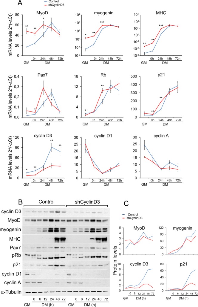
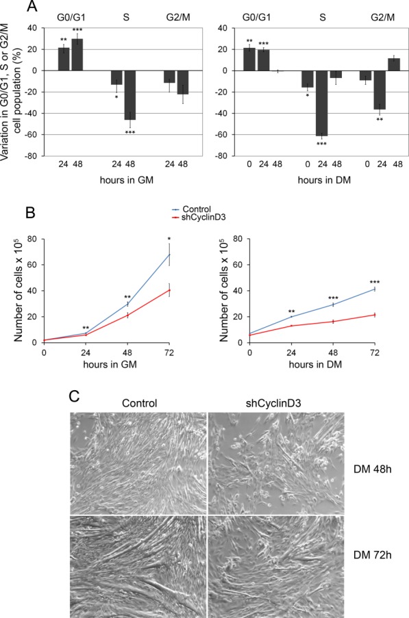
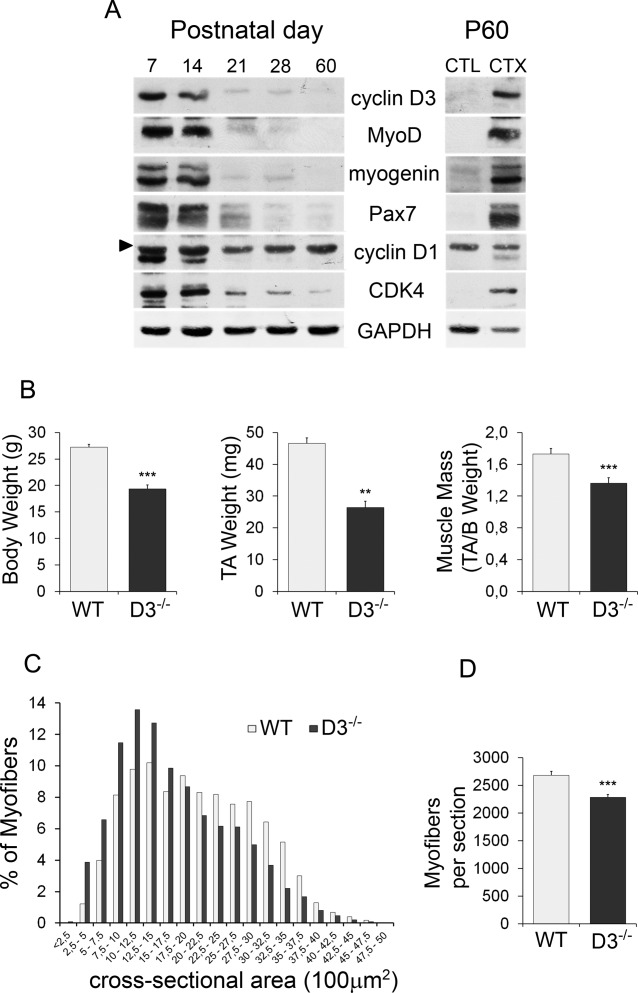
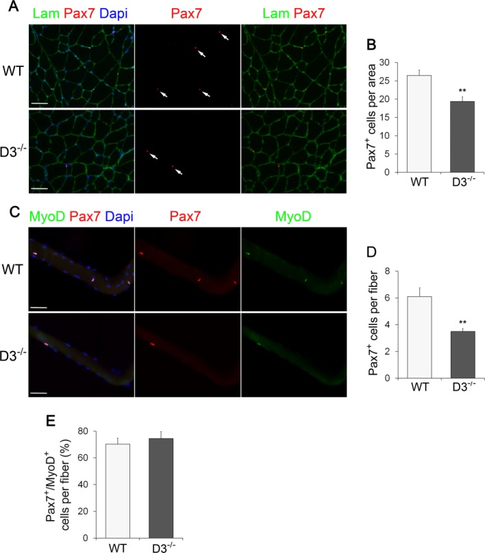
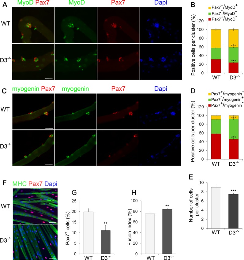
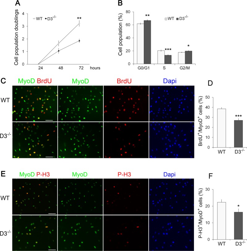
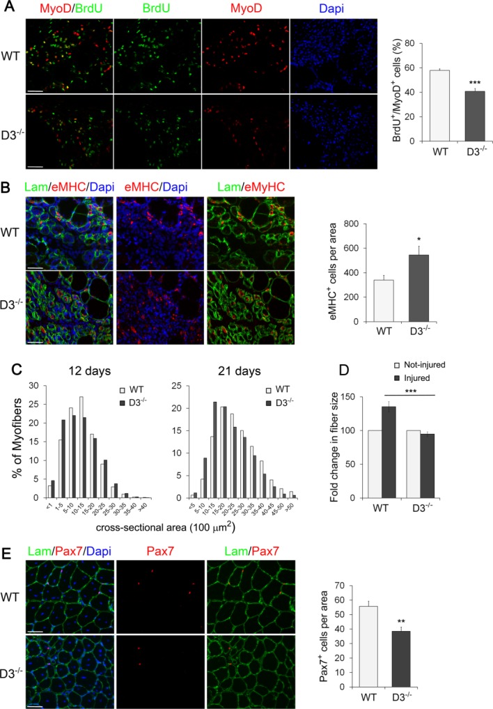
Satellite cells are mitotically quiescent myogenic stem cells resident beneath the basal lamina surrounding adult muscle myofibers. In response to injury, multiple extrinsic signals drive the entry of satellite cells into the cell cycle and then to proliferation, differentiation, and self-renewal of their downstream progeny. Because satellite cells must endure for a lifetime, their cell cycle activity must be carefully controlled to coordinate proliferative expansion and self-renewal with the onset of the differentiation program. In this study, we find that cyclin D3, a member of the family of mitogen-activated D-type cyclins, is critically required for proper developmental progression of myogenic progenitors. Using a cyclin D3-knockout mouse we determined that cyclin D3 deficiency leads to reduced myofiber size and impaired establishment of the satellite cell population within the adult muscle. Cyclin D3-null myogenic progenitors, studied ex vivo on isolated myofibers and in vitro, displayed impaired cell cycle progression, increased differentiation potential, and reduced self-renewal capability. Similarly, silencing of cyclin D3 in C2 myoblasts caused anticipated exit from the cell cycle and precocious onset of terminal differentiation. After induced muscle damage, cyclin D3-null myogenic progenitors exhibited proliferation deficits, a precocious ability to form newly generated myofibers and a reduced capability to repopulate the satellite cell niche at later stages of the regeneration process. These results indicate that cyclin D3 plays a cell-autonomous and nonredundant function in regulating the dynamic balance between proliferation, differentiation, and self-renewal that normally establishes an appropriate pool size of adult satellite cells.

Keywords: Cell cycle; Muscle stem cell; Myogenesis; Regeneration; Satellite cell; Self-renewal.


Figures

Figure 1
Figure 1
Cyclin D3 knockdown in myoblasts leads to precocious onset of differentiation. C2.7 myoblasts transduced either with the retrovirus expressing cyclin D3-specific shRNA (shCyclin D3) or with the empty retrovirus (control) were seeded at 2 × 10⁵ cells into 90-mm dishes, and transferred to DM after 48 hours. Total RNA and whole-cell extracts were prepared from cells collected in GM 24 hours after seeding or at indicated times after exposure to DM. (A): RT-qPCR analysis was performed on the genes indicated. Graphs represent average 2−ΔCT values ± SEM from four independent experiments. (B): Western blot analysis was performed using antibodies specific for the indicated proteins. (C): Quantification of protein expression by densitometry analysis of Western blots shown in (B); values are presented as fold change relative to control myoblasts cultured in GM (set to 1), after normalization to the corresponding values of α-tubulin levels. Asterisks denote significance (*, p < .05; **, p < .01; ***, p < .001). Abbreviations: DM, differentiation medium; GM, growth medium; MHC, myosin heavy chain; Rb, retinoblastoma gene; pRb, retinoblastoma protein.
Figure 2
Figure 2
Cyclin D3 knockdown in myoblasts results in reduced proliferation, accelerated exit from the cell cycle, and impaired myotube formation. C2.7 myoblasts transduced either with the retrovirus expressing shCyclinD3 or with the control retrovirus were seeded at 2 × 10⁵ cells into 90-mm dishes and either cultured in GM for 72 hours or transferred to DM 24 hours after seeding. (A): Cells cultured in GM (left panel) or in DM (right panel) were harvested at the indicated time points, stained with propidium iodide, and analyzed by flow cytometry. Data are presented as changes in the percentage of shCyclinD3 cells, in particular phases of cell cycle at each time point, relative to control cells. Error bars represent SEM (±SEM) for five independent experiments. (B): Cells cultured in GM (left panel) or in DM (right panel) were counted at the indicated time points. Graphs represent average cell numbers ± SEM (n = 5). (C): shCyclin D3 or control myoblasts were seeded at 2 × 10⁵ cells into 90-mm dishes, and transferred to DM after 48 hours. Phase-contrast microphotographs (whole ×20 field) show cells induced to differentiate for 2–3 days. Asterisks denote significance (*, p < .05; **, p < .01; ***, p < .001). Abbreviations: DM, differentiation medium; GM, growth medium.
Figure 3
Figure 3
Cyclin D3 deficiency in vivo results in postnatal muscle defects. (A): Cyclin D3 is highly expressed in muscle during postnatal and regenerative myogenesis. Western blot analysis of extracts prepared from hind limb muscles of 7–60-day-old mice and from uninjured (CTL) or injured tibialis anterior muscles of 60-day-old (P60) mice. Injury was induced by injection of CTX, and muscle was collected after 3 days. Immunoblots were probed with the indicated antibodies. The arrow head indicates a non-specific band (B): Average body weight, TA muscle weight, and TA muscle mass of 70–85-day-old wild-type (WT) and cyclin D3/ (D3/) male mice. Error bars represent ±SEM for n = 6. (C): Frequency histogram showing the myofiber cross-sectional area measured on transversal sections of TA muscles from WT and D3/ male mice at P60 (n = 3; WT: 4,033 myofibers, D3/: 3,701 myofibers). (D): Average number ±SEM of myofibers per TA cryosection in P60 WT and D3/ male mice (n = 3). Asterisks denote significance (**, p < .01; ***, p < .001). Abbreviations: CTX, cardiotoxin; TA, tibialis anterior; WT, wild type.
Figure 4
Figure 4
Ablation of cyclin D3 leads to a decline in the adult satellite cell pool size. (A): Transverse sections of tibialis anterior muscles from WT and D3−/− male mice at P60 were stained with Laminin to identify myofibers (green) and Pax7 to identify sublaminar satellite cells (red) indicated by arrows. Nuclei were counterstained with Dapi (blue). (B): Average number of Pax7-positive cells per cross-sectional tissue area (mm2) in WT and D3−/− mice. Error bars represent ±SEM for n = 3. (C): Single myofibers freshly isolated from extensor digitorum longus muscles of 8–12-week-old WT and D3−/− mice were stained 6 hours ex vivo for Pax7 (red) and MyoD (green), and counterstained with Dapi (blue). (D): Average number of Pax7+ satellite cells per myofiber in WT and D3−/− mice. Error bars represent ±SEM for n = 5 WT and n = 7 D3−/−. (E): Quantification of Pax7+/MyoD+ satellite cells per myofiber in WT and D3−/− mice. Values are expressed as the mean percentage of Pax7+ cells that were MyoD-positive (means ± SEM for n = 4). Asterisks denote significance (**, p < .01). Scale bar = 50 µm. Abbreviations: Lam, Laminin; Dapi, 4′,6-diamidino-2-phenylindole; WT, wild type.
Figure 5
Figure 5
Cyclin D3 loss affects SC differentiation and self-renewal. (A): Batches of single extensor digitorum longus (EDL) myofibers from WT and D3−/− mice were coimmunostained for Pax7 (red) and MyoD (green) after 72 hours in suspension culture. Counterstaining with Dapi (blue) was used to identify all nuclei present on the myofiber. (B): The graph represents the quantification of the Pax7+/MyoD, Pax7+/MyoD+, or Pax7/MyoD+ cells contained in clusters of four or more cells. Data are expressed as the percentage in each category of the total positive cells per cluster (means ± SEM). Clusters (270) from WT (n = 4) and D3−/− (n = 3) mice were analyzed, for a total of 2,000 cells counted for each genotype. (C): Myogenin (green) and Pax7 (red) coimmunostaining of WT and D3−/− EDL myofibers 72 hours after isolation, and counterstaining with Dapi (blue). (D): The graph represents the quantification of the Pax7+/Myogenin, Pax7+/Myogenin+, or Pax7/Myogenin+ cells. Data are expressed as in (B). Clusters (180) from WT (n = 3) and D3−/− (n = 5) mice were analyzed, for a total of 1,300 cells counted for each genotype. (E): Quantification of cluster size. The total number of cells present in the WT and D3−/− clusters analyzed in (B) was calculated and expressed as mean number of cells per cluster ± SEM. (F): Satellite cells derived from WT and D3−/− EDL myofibers were seeded in eight-well permanox chamber slides (1.2 × 10⁴/well) and transferred to DM after 24 hours. Cells were cultured in differentiation medium for 72 hours before fixing and immunostaining for MHC (green) to visualize myotubes and Pax7 to identify mononuclear undifferentiated reserve cells (red). Counterstaining with Dapi was used to visualize all nuclei (blue). (G): Relative number of Pax7-expressing cells. Values are expressed as the mean percentage ± SEM of total nuclei (WT: n = 6; D3−/−: n = 5). (H): Quantification of fusion index. Values are expressed as the mean ratio ± SEM of nuclei present in myotubes to the total number of nuclei (n = 5). Asterisks denote significance (**, p < .01; ***, p < .001). Scale bar = 50 µm. Abbreviations: Dapi, 4′,6-diamidino-2-phenylindole; MHC, myosin heavy chain; WT, wild type.
Figure 6
Figure 6
Cyclin D3 null satellite cells show proliferative deficits in vitro. (A): Primary myoblasts from dissociated limb muscles of WT and D3−/− mice were seeded in eight-well permanox chamber slides (104/well) and counted in 10 independent microscopic fields after 24, 48, and 72 hours in culture (>95% myogenic cells, as assessed by MyoD staining). The graph represents the mean number ± SEM (n = 4) of cell population doublings at 48 and 72 hours, relative to 24 hours (log 2 [number of cells at 48 or 72 hours]/number of cells at 24 hours). (B): WT and D3−/− primary myoblasts cultured for 48 hours were detached and stained with propidium iodide. Flow cytometry analysis of the cell cycle reveals a lower percentage of D3−/− cells in S-phase and a higher percentage in G0/G1 and G2/M compared with WT cells (means ± SEM, n = 6). (C, E): WT and D3−/− primary myoblasts cultured for 48 hours were labeled with 10 µM BrdU for 4 hours before fixation and immunostaining to detect BrdU (red), MyoD (green), and PH3 (red) as a marker of mitotic cells. Nuclei were counterstained with Dapi (blue). (D, F): The percentages of BrdU or P-H3 positive cells/total MyoD-positive cells are shown. Error bars represent ±SEM (n = 4). Asterisks denote significance (*, p < .05; **, p < .01; ***, p < .001). Scale bar = 50 µm. Abbreviations: Dapi, 4′,6-diamidino-2-phenylindole; P-H3, Phospho-Histone H3; WT, wild type.
Figure 7
Figure 7
Loss of cyclin D3 affects muscle regeneration. (A): Acute muscle injury was induced in WT and D3−/− male mice at P60 by intramuscular injection of cardiotoxin. Mice were injected BrdU intraperitoneally 6 hours before sacrifice. Left, cross-sections of tibialis anterior (TA) muscles after 3 days of regeneration stained for MyoD (red) and BrdU (green) to label proliferating myogenic progenitors. Nuclei were counterstained with Dapi (blue). The graph on the right represents the mean percentage ± SEM of MyoD-positive cells that were BrdU+ (WT: n = 5; D3−/−: n = 4). (B) Left, muscle cross-sections as in (A) were stained for eMHC (red) to identify newly generated myofibers and Laminin (green) to identify damaged fibers. Nuclei were counterstained with Dapi (blue). Right, quantitative analyses of nascent fiber number normalized to cross-sectional area of regenerating tissue (mean ± SEM for n = 4). (C) Frequency histograms showing the cross-sectional area of WT and cyclin D3-null myofibers 12 days and 21 days after cardiotoxin injection (12 days: n = 3; WT: 7,866 myofibers; D3/: 7,633 myofibers—21 days: n = 4; WT: 4,826 myofibers; D3/: 5,006 myofibers). (D): Fold change in regenerated fiber size from WT and D3−/− TA muscles 21 days after injury relative to uninjured fibers in adjacent areas of the tissue (means ± SEM, n = 4). (E): Left, cross-sections from regenerating TA muscles 21 days after injury stained for Pax7 (red) and Laminin (green). Nuclei were counterstained with Dapi (blue). Right, quantification of Pax7-positive cells associated with regenerated myofibers, normalized to 1 mm2 of tissue cross-sectional area (means ± SEM; WT, n = 5; D3/, n = 3). Asterisks denote significance (*, p < .05; **, p < .01; ***, p <.001). Scale bar = 50 µm. Abbreviations: Dapi, 4′,6-diamidino-2-phenylindole; eMHC, embryonic myosin heavy chain; Lam, Laminin; WT, wild type.

Similar articles

Cited by

References

    1. Collins CA, Olsen I, Zammit PS, et al. Stem cell function, self-renewal, and behavioral heterogeneity of cells from the adult muscle satellite cell niche. Cell. 2005;122:289–301. - PubMed
    1. Zammit PS. All muscle satellite cells are equal, but are some more equal than others? J Cell Sci. 2008;121(Pt 18):2975–2982. - PubMed
    1. Kuang S, Gillespie MA, Rudnicki MA. Niche regulation of muscle satellite cell self-renewal and differentiation. Cell Stem Cell. 2008;2:22–31. - PubMed
    1. Brack AS, Rando TA. Tissue-specific stem cells: Lessons from the skeletal muscle satellite cell. Cell Stem Cell. 2012;10:504–514. - PMC - PubMed
    1. Wang YX, Rudnicki MA. Satellite cells, the engines of muscle repair. Nat Rev Mol Cell Biol. 2012;13:127–133. - PubMed

Publication types