. 2013 Dec;31(12):2813-26.
doi: 10.1002/stem.1488.

Dysfunction of endothelial progenitor cells from smokers and chronic obstructive pulmonary disease patients due to increased DNA damage and senescence

Affiliations

Dysfunction of endothelial progenitor cells from smokers and chronic obstructive pulmonary disease patients due to increased DNA damage and senescence

Koralia E Paschalaki et al. Stem Cells. 2013 Dec.

Abstract

Cardiovascular disease (CVD) is a major cause of death in smokers, particularly in those with chronic obstructive pulmonary disease (COPD). Circulating endothelial progenitor cells (EPC) are required for endothelial homeostasis, and their dysfunction contributes to CVD. To investigate EPC dysfunction in smokers, we isolated and expanded blood outgrowth endothelial cells (BOEC) from peripheral blood samples from healthy nonsmokers, healthy smokers, and COPD patients. BOEC from smokers and COPD patients showed increased DNA double-strand breaks and senescence compared to nonsmokers. Senescence negatively correlated with the expression and activity of sirtuin-1 (SIRT1), a protein deacetylase that protects against DNA damage and cellular senescence. Inhibition of DNA damage response by silencing of ataxia telangiectasia mutated (ATM) kinase resulted in upregulation of SIRT1 expression and decreased senescence. Treatment of BOEC from COPD patients with the SIRT1 activator resveratrol or an ATM inhibitor (KU-55933) also rescued the senescent phenotype. Using an in vivo mouse model of angiogenesis, we demonstrated that senescent BOEC from COPD patients are dysfunctional, displaying impaired angiogenic ability and increased apoptosis compared to cells from healthy nonsmokers. Therefore, this study identifies epigenetic regulation of DNA damage and senescence as pathogenetic mechanisms linked to endothelial progenitors' dysfunction in smokers and COPD patients. These defects may contribute to vascular disease and cardiovascular events in smokers and could therefore constitute therapeutic targets for intervention.

Keywords: Ataxia telangiectasia-mutated kinase; Cellular senescence; DNA damage response; Endothelial progenitor cells; Sirtuin; Smoking.


Figures

Figure 1
Figure 1
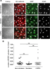
BOEC isolation and characterization. (A): Representative images of BOEC colonies in culture derived from healthy nonsmokers, healthy smokers, and COPD patients. Immunofluorescence (IF) staining for VE-cadherin (red), VWF (green), and CD45 (red). TOPRO-3 (white) was used as nuclear stain. BOEC from all groups were positive for the endothelial markers VE-cadherin and VWF and negative for the leukocytic CD45 marker (bright-field images scale bars = 100 μm; IF images scale bars = 20 μm). (B): Number of BOEC colonies in healthy nonsmokers, healthy smokers, and patients with COPD. The number of discrete BOEC colonies that appeared in the peripheral blood mononuclear cell culture was counted relative to the number of mL of blood obtained from healthy nonsmokers (n = 18), healthy smokers (n = 11), and COPD patients (n = 20) (open triangle: ex-smokers, black triangle: current smokers). Abbreviations: BOEC, blood outgrowth endothelial cells; COPD, chronic obstructive pulmonary disease; VWF, von Willebrand factor; VE, vascular endothelial.
Figure 2
Figure 2
Increased senescence and DNA damage in smokers and COPD patients. SA-β-gal activity, p21, and p16 were assessed as markers of cellular senescence. (A): Quantification of SA-β-gal activity was assessed in blood outgrowth endothelial cells (BOEC) from healthy nonsmokers (n = 10), healthy smokers (n = 6), and COPD patients (n = 12), by counting the number of blue (senescent) cells relative to the total cell number and expressed as a percentage (%). BOEC from healthy smokers and COPD patients exhibited significantly increased senescence compared to healthy nonsmokers. Representative images from a healthy nonsmoker (72 years old), a healthy smoker (64 years old), and a COPD patient (65 years old) are shown (scale bars = 100 μm). (B): p21 protein levels were measured by Western blotting in whole cell lysates from BOEC isolated from healthy nonsmokers (n = 3), healthy smokers (n = 3), and COPD patients (n = 3). α-tubulin was used as loading control. p21 protein levels were significantly increased in smokers and COPD patients compared to nonsmokers. Representative images of Western blots are shown. (C, D): Immunofluorescence staining of BOEC for p16 (red, left panels) and p21 (red, right panels). TOPRO-3 (blue) was used as nuclear staining. Representative images from the three groups are shown. (E, F): DNA damage was assessed by measuring two markers of DSB formation, the γ-H2AX (left panels) and 53BP1 (right panels) by immunofluorescence staining (n = 3–5). Representative images from the three groups are shown. The number of cells with distinct nuclear immunofluorescent foci (see arrows) relative to the total cell number was counted in two to four optic fields, using a ×20 and ×40 objective lens (scale bars = 20 μm). An increased number of cells with focal nuclear staining of γ-H2AX was observed in healthy smokers and COPD patients compared to healthy nonsmokers (E, bottom). An increased number of cells with focal nuclear staining of 53BP1 was observed in healthy smokers and COPD patients compared to healthy nonsmokers (F, bottom). *, p < .05; **, p < .01 (open triangle: ex-smokers, black triangle: current smokers). Abbreviations: COPD, chronic obstructive pulmonary disease; SA-β-gal, senescence-associated-β-galactosidase; 53BP1, 53 binding protein 1.
Figure 3
Figure 3
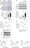
SIRT1 regulates senescence in blood outgrowth endothelial cells (BOEC); Decreased SIRT1 levels and activity in BOEC from healthy smokers and COPD patients compared to healthy nonsmokers. (A, B): BOEC from healthy nonsmokers (n = 4) were transfected with small interfering RNA (siRNA) against SIRT1 or control siRNA for 48 hours. Cells were exposed to 50 μmol/L H2O2 for 1 hour and cultured for 48 additional hours to induce oxidative-stress premature senescence. Cellular senescence was assessed by SA-β-gal activity. Protein levels of SIRT1 and acetylation of p53 at Lys-382 were measured by Western blotting. GAPDH was used as loading control. Inhibition of SIRT1 in BOEC induced increased SA-β-gal activity and acetylation of p53 at Lys-382, both under baseline and oxidant conditions. (C): SIRT1 protein levels were measured in BOEC from healthy nonsmokers (n = 9), healthy smokers (n = 6), and patients with COPD (n = 8) by Western blotting (representative image shown). GAPDH was used as loading control. SIRT1 protein levels were significantly reduced in smokers and COPD patients compared to nonsmokers. (D): SIRT1 activity was measured in nuclear extracts from BOEC isolated from healthy nonsmokers (n = 6), healthy smokers (n = 6), and COPD patients (n = 7) using a SIRT1 fluorescent activity assay kit. SIRT1 activity was significantly reduced in healthy smokers and COPD patients compared to healthy nonsmokers. (E–G): SIRT1 protein levels correlated with SIRT1 activity in samples from all groups. SIRT1 protein levels and activity negatively correlated with SA-β-gal activity in samples from all groups (scale bars = 100 μm) *, p < .05; **, p < .01; ***, p < .001. Abbreviations: COPD, chronic obstructive pulmonary disease; SA-β-gal senescence-associated-β-galactosidase; SIRT1, sirtuin-1.
Figure 4
Figure 4
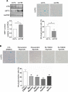
Inhibition of ATM signaling upregulates SIRT1 levels and suppresses endothelial senescence induced by oxidative stress. (A): HUVEC were transfected with small interfering RNA (siRNA) against ATM or control siRNA and grown in low serum medium for 72 hours. ATM and SIRT1 protein levels were measured by Western blotting. GAPDH was used as loading control. Cellular senescence was assessed by SA-β-gal activity. SIRT1 protein levels were significantly increased (bottom left) and senescence was significantly reduced (bottom right) in ATM-deficient cells compared to controls (n = 4–5). Representative images of Western blots and SA-β-gal activity are shown. (B): BOEC from healthy nonsmokers were treated and assessed as described in panel (A). (C): After 48 hours of siRNA treatment for ATM and control, HUVEC were exposed to 50 μmol/L H2O2 for 1 hour and cultured for 48 additional hours to induce oxidative-stress premature senescence. Inhibition of ATM in HUVEC by siRNA induced increased SIRT1 protein levels (n = 3–6). ATM silencing also caused a reduction in SA-β-gal activity in cells exposed to conditions of oxidative stress. Representative images of Western blots and SA-β-gal staining are shown. (D): BOEC from healthy nonsmokers were treated and assessed as described in panel (C) to induce oxidative-stress premature senescence (scale bars = 100 μm) *, p < .05; **, p < .01. Abbreviations: ATM, ataxia telangiectasia mutated; BOEC, blood outgrowth endothelial cells; HUVEC, human umbilical vein endothelial cells; SA-β-gal senescence-associated-β-galactosidase; SIRT1, sirtuin-1.
Figure 5
Figure 5
Inhibition of ATM suppresses senescence in blood outgrowth endothelial cells (BOEC) from COPD patients; treatment with SIRT1 activators and ATM inhibitors. (A): BOEC from COPD patients were transfected with siRNA and grown in low serum medium for 72 hours. SIRT1 and ATM were measured by Western blotting and cellular senescence by SA-β-gal activity. SIRT1 protein levels were significantly increased (bottom left), and senescence was significantly reduced (bottom right) in ATM-deficient BOEC (n = 3). Representative images of Western blots and SA-β-gal staining are shown. (B): BOEC from COPD patients were seeded in basal medium (plus 10% fetal bovine serum) for 24 hours. BOEC were treated with different concentrations of the SIRT1 activator RSV (10 and 50 μmol/L) or the selective ATM inhibitor KU (5 and 10 μmol/L) for 48 hours. Treatment with both concentrations of RSV or KU significantly reduced the grade of senescence compared to control treated BOEC from COPD patients (n = 4) (scale bars = 100 μm) *, p < .05; **, p < .01; ***, p < .001. Abbreviations: ATM, ataxia telangiectasia mutated; COPD, chronic obstructive pulmonary disease; DMSO, Dimethyl sulfoxide; KU, KU-55933; RSV, resveratrol; SA-β-gal senescence-associated-β-galactosidase; SIRT1, sirtuin-1.
Figure 6
Figure 6
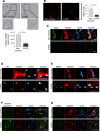
Senescent blood outgrowth endothelial cells (BOEC) from COPD patients show reduced angiogenic ability and increased apoptosis. (A–G): BOEC from nonsmokers (n = 4) or COPD patients (n = 3) were labeled with Dil-Vybrant cell solution and were subcutaneously injected in severe combined immunodeficiency mice. After 7 days, plugs were harvested, frozen, cryosectioned, and stained with hematoxylin and eosin (H&E) (A) or analyzed by immunofluorescence microscopy (B, C). Experiments were performed for each sample in duplicate. Cellular infiltrate was quantified in H&E sections (A, bottom) and Vybrant positive cells were quantified from Z-stack images taken with ×20 objective lens (B, right). (C): BOEC from nonsmokers formed capillary-like structures in the Matrigel plugs. Immunofluorescent analysis for CD31 staining (green) revealed lining of the Vybrant positive cells and formation of capillary-like structures in the Matrigel plugs with BOEC from nonsmokers but not in COPD samples. (D–G): Immunofluorescent analysis for p16 staining (green, top left), 53BP1 (green, top right), cleaved caspase-3 (green, bottom left), and TUNEL staining (green, bottom right) showed increased expression of all markers in BOEC from COPD patients compared to nonsmokers (scale bars = 50 μm). Abbreviations: COPD, chronic obstructive pulmonary disease; 53BP1, 53 binding protein 1.

References

    1. Asahara T, Murohara T, Sullivan A, et al. Isolation of putative progenitor endothelial cells for angiogenesis. Science. 1997;275:964–967. - PubMed
    1. Critser PJ, Voytik-Harbin SL, Yoder MC. Isolating and defining cells to engineer human blood vessels. Cell Prolif. 2011;44(suppl 1):15–21. - PMC - PubMed
    1. Yoder MC, Mead LE, Prater D, et al. Redefining endothelial progenitor cells via clonal analysis and hematopoietic stem/progenitor cell principals. Blood. 2007;109:1801–1809. - PMC - PubMed
    1. Fuchs S, Ghanaati S, Orth C, et al. Contribution of outgrowth endothelial cells from human peripheral blood on in vivo vascularization of bone tissue engineered constructs based on starch polycaprolactone scaffolds. Biomaterials. 2009;30:526–534. - PubMed
    1. Hendrickx B, Verdonck K, Van den Berge S, et al. Integration of blood outgrowth endothelial cells in dermal fibroblast sheets promotes full thickness wound healing. Stem Cells. 2010;28:1165–1177. - PubMed

Publication types

MeSH terms