

Vascular masking for improved unfolding in 2D SENSE-accelerated 3D contrast-enhanced MR angiography
- PMID: 23897776
- PMCID: PMC3818335
- DOI: 10.1002/jmri.24266
Vascular masking for improved unfolding in 2D SENSE-accelerated 3D contrast-enhanced MR angiography
Abstract
Purpose: To describe and evaluate the method we refer to as "vascular masking" for improving signal-to-noise ratio (SNR) retention in sensitivity encoding (SENSE)-accelerated contrast-enhanced magnetic resonance angiography (CE-MRA).
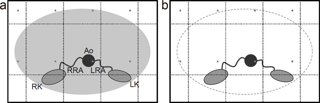
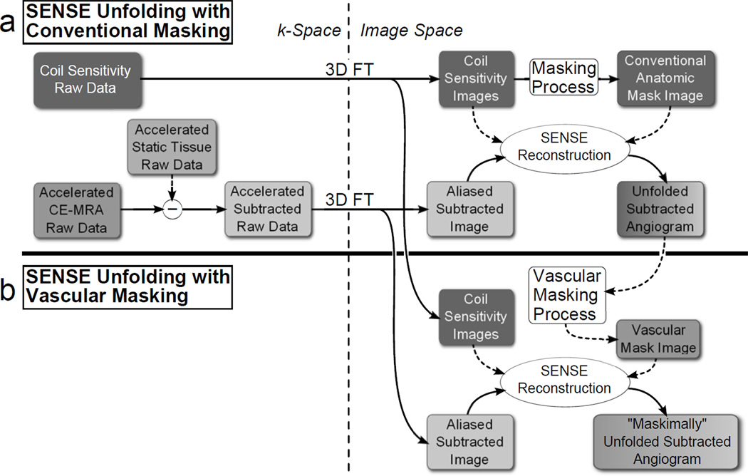
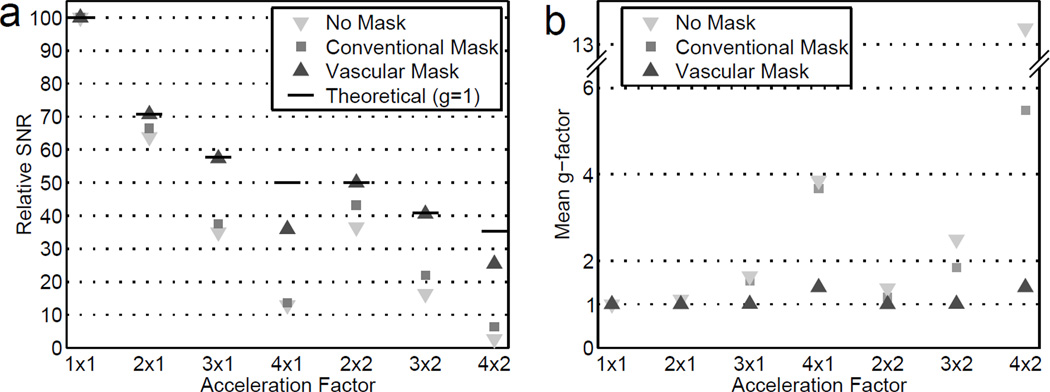
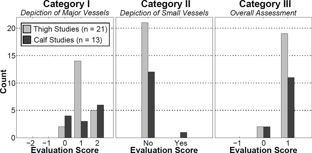
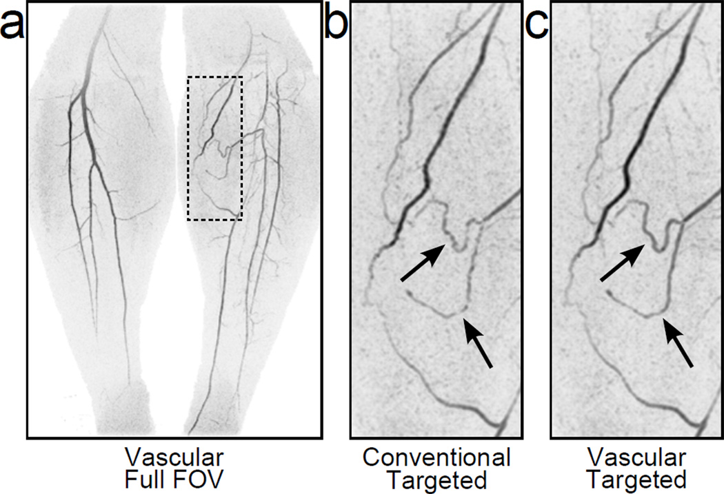
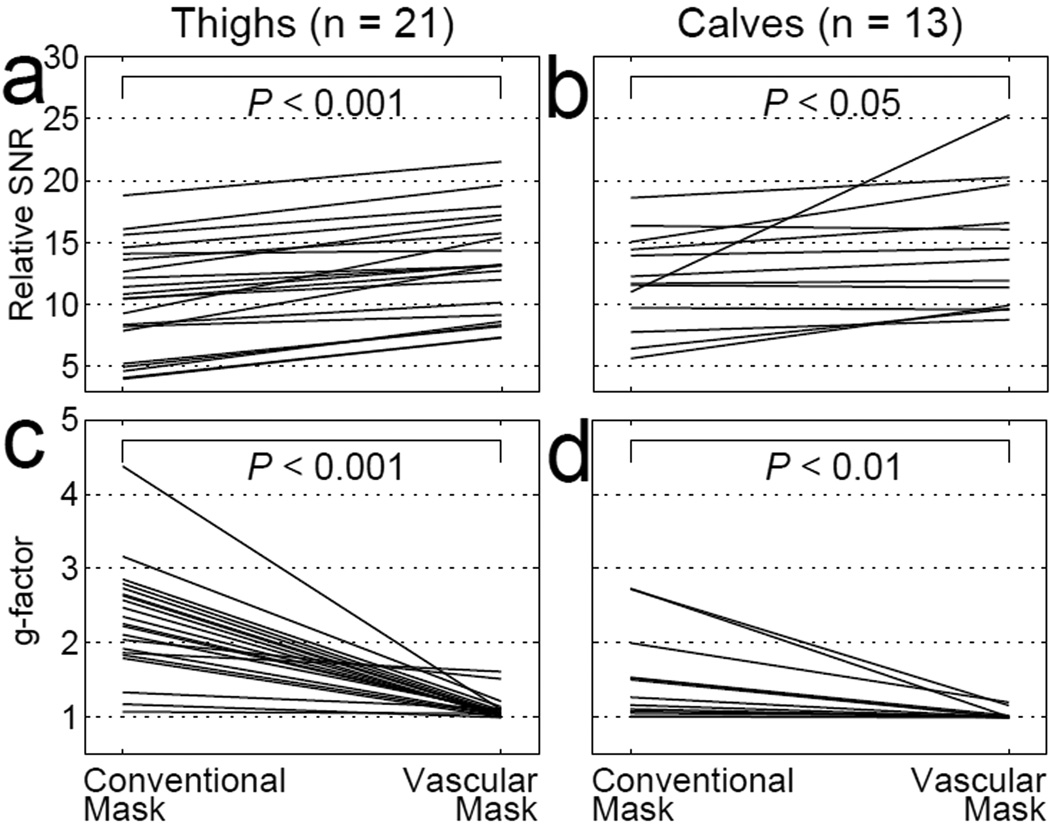
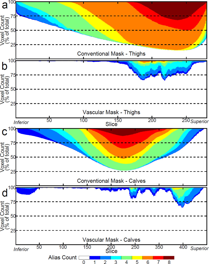
Materials and methods: Vascular masking is a technique that restricts the SENSE unfolding of an accelerated subtraction angiogram to the voxels within the field of view known to have enhancing signal. This is a more restricted voxel set than that identified with conventional masking, which excludes only voxels in the air around the object. Thus, improved retention of SNR is expected. Evaluation was done in phantom and in vivo studies by comparing SNR and the g-factor in results reconstructed using vascular versus conventional masking. A radiological evaluation was also performed comparing conventional and vascular masking in R = 8 accelerated CE-MRA studies of the thighs (n = 21) and calves (n = 13).
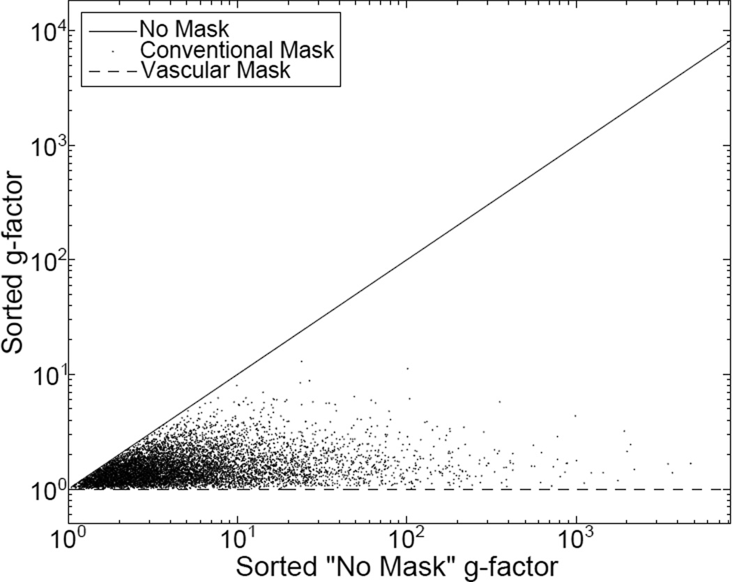
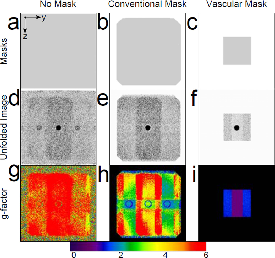
Results: Images reconstructed with vascular masking showed a significant reduction in g-factor and improved retention of SNR versus those reconstructed with conventional masking. In the radiological evaluation, vascular masking consistently provided reduced background noise, improved luminal signal smoothness, and better small vessel conspicuity.
Conclusion: Vascular masking provides improved SNR retention and improved depiction of the vasculature in accelerated, subtraction 3D CE-MRA of the thighs and calves.
Keywords: SENSE; contrast-enhanced MR angiography (CE-MRA); masking.
Copyright © 2013 Wiley Periodicals, Inc.
Figures

Similar articles
-
Direct comparison of sensitivity encoding (SENSE) accelerated and conventional 3D contrast enhanced magnetic resonance angiography (CE-MRA) of renal arteries: effect of increasing spatial resolution.J Magn Reson Imaging. 2010 Jan;31(1):149-59. doi: 10.1002/jmri.22002. J Magn Reson Imaging. 2010. PMID: 20027583
-
Highly compressed SENSE accelerated relaxation-enhanced angiography without contrast and triggering (REACT) for fast non-contrast enhanced magnetic resonance angiography of the neck: Clinical evaluation in patients with acute ischemic stroke at 3 tesla.Magn Reson Imaging. 2024 Oct;112:27-37. doi: 10.1016/j.mri.2024.04.009. Epub 2024 Apr 9. Magn Reson Imaging. 2024. PMID: 38599503
-
High spatial and temporal resolution dynamic contrast-enhanced magnetic resonance angiography using compressed sensing with magnitude image subtraction.Magn Reson Med. 2014 May;71(5):1771-83. doi: 10.1002/mrm.24842. Epub 2013 Jun 25. Magn Reson Med. 2014. PMID: 23801456 Free PMC article.
-
[Diagnosis of renal artery stenosis with magnetic resonance angiography and stenosis quantification].J Mal Vasc. 2000 Dec;25(5):312-320. J Mal Vasc. 2000. PMID: 11148391 Review. French.
-
MR angiography with three-dimensional MR digital subtraction angiography.Top Magn Reson Imaging. 1996 Dec;8(6):366-88. Top Magn Reson Imaging. 1996. PMID: 9402678 Review.
Cited by
-
Three-station three-dimensional bolus-chase MR angiography with real-time fluoroscopic tracking.Radiology. 2014 Jul;272(1):241-51. doi: 10.1148/radiol.14131603. Epub 2014 Mar 14. Radiology. 2014. PMID: 24635676 Free PMC article.
-
Technical Aspects of Contrast-enhanced MR Angiography: Current Status and New Applications.Magn Reson Med Sci. 2018 Jan 10;17(1):3-12. doi: 10.2463/mrms.rev.2017-0053. Epub 2017 Aug 31. Magn Reson Med Sci. 2018. PMID: 28855470 Free PMC article.
References
-
- Pruessmann KP, Weiger M, Scheidegger MB, Boesiger P. SENSE: sensitivity encoding for fast MRI. Magn Reson Med. 1999;42:952–962. - PubMed
-
- Griswold MA, Jakob PM, Heidemann RM, et al. Generalized autocalibrating partially parallel acquisitions (GRAPPA) Magn Reson Med. 2002;47:1202–1210. - PubMed
-
- Sodickson DK, McKenzie CA, Li W, Wolff S, Manning WJ, Edelman RR. Contrast-enhanced 3D MR angiography with simultaneous acquisition of spatial harmonics: A pilot study. Radiology. 2000;217:284–289. - PubMed
-
- Weiger M, Pruessmann KP, Kassner A, et al. Contrast-enhanced 3D MRA using SENSE. J Magn Reson Imaging. 2000;12:671–677. - PubMed
-
- Quick HH, Vogt FM, Maderwald S, et al. High spatial resolution whole-body MR angiography featuring parallel imaging: initial experience. Rofo. 2004;176:163–169. - PubMed
Publication types
MeSH terms
Substances
Grants and funding
LinkOut - more resources
Full Text Sources
Other Literature Sources
