. 2013 Sep 27;288(39):28021-33.
doi: 10.1074/jbc.M113.498758. Epub 2013 Jul 29.

Histone deacetylase (HDAC) 10 suppresses cervical cancer metastasis through inhibition of matrix metalloproteinase (MMP) 2 and 9 expression

Affiliations

Histone deacetylase (HDAC) 10 suppresses cervical cancer metastasis through inhibition of matrix metalloproteinase (MMP) 2 and 9 expression

Chenlin Song et al. J Biol Chem. .

Abstract

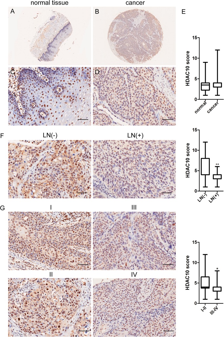
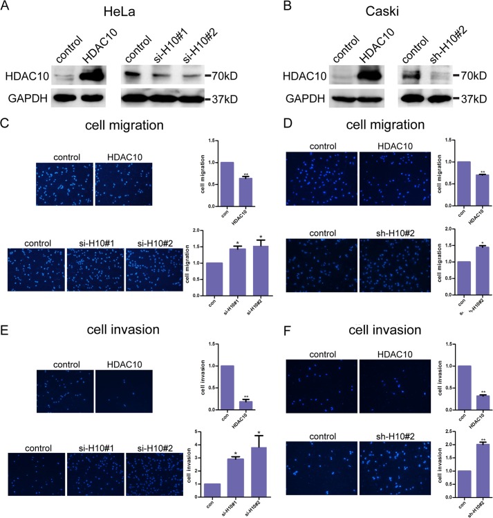
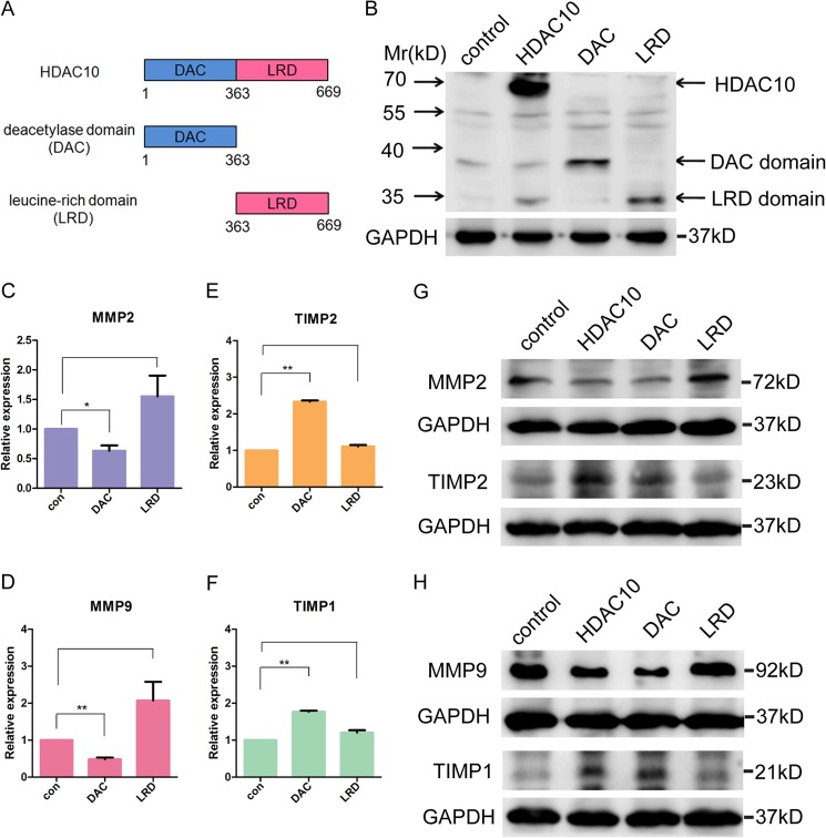
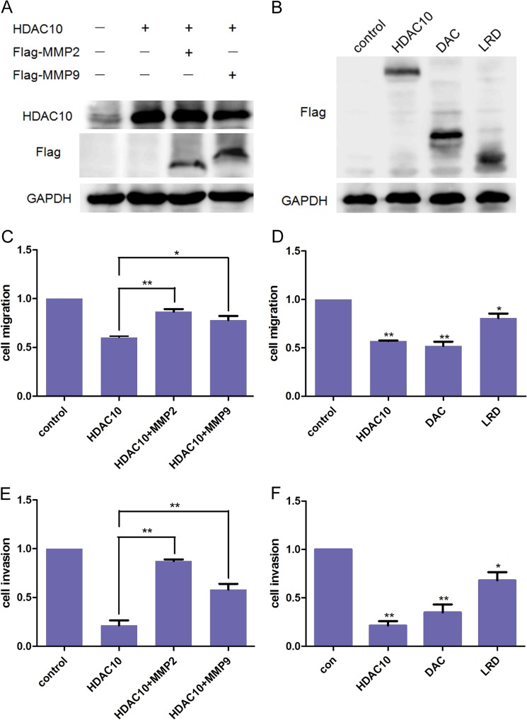
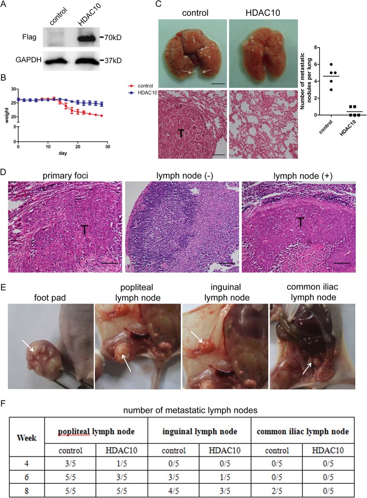
Aberrant expression of histone deacetylases (HDACs) is associated with carcinogenesis. Some HDAC inhibitors are widely considered as promising anticancer therapeutics. A major obstacle for development of HDAC inhibitors as highly safe and effective anticancer therapeutics is that our current knowledge on the contributions of different HDACs in various cancer types remains scant. Here we report that the expression level of HDAC10 was significantly lower in patients exhibiting lymph node metastasis compared with that in patients lacking lymph node metastasis in human cervical squamous cell carcinoma. Forced expression of HDAC10 in cervical cancer cells significantly inhibited cell motility and invasiveness in vitro and metastasis in vivo. Mechanistically, HDAC10 suppresses expression of matrix metalloproteinase (MMP) 2 and 9 genes, which are known to be critical for cancer cell invasion and metastasis. At the molecular level, HDAC10 binds to MMP2 and -9 promoter regions, reduces the histone acetylation level, and inhibits the binding of RNA polymerase II to these regions. Furthermore, an HDAC10 mutant lacking histone deacetylase activity failed to mimic the functions of full-length protein. These results identify a critical role of HDAC10 in suppression of cervical cancer metastasis, underscoring the importance of developing isoform-specific HDAC inhibitors for treatment of certain cancer types such as cervical squamous cell carcinoma.

Keywords: Cell Invasion; Cell Migration; Histone Deacetylase; Matrix Metalloproteinase (MMP); Metastasis.


Figures

FIGURE 1.
FIGURE 1.
HDAC10 expression inversely correlates with lymph node metastasis in cervical cancer. A–D, HDAC10 levels in representative normal and carcinoma tissues. Immunohistochemistry using rabbit anti-HDAC10 antibody was performed on normal cervical tissues (A and C) and cervical squamous cell carcinoma tissues (B and D). E, plot representation of scores according to immunohistochemical expression of HDAC10 in normal tissues and the counterpart tumor tissues. A total of 60 specimens were analyzed. The scores were calculated by intensity × percentage of stained cells. F, correlation between HDAC10 expression levels and lymph node (LN) metastasis status in carcinoma tissues. G, correlation between HDAC10 expression levels and TNM stage in carcinoma tissues. All data were analyzed by Student's t test. *, p < 0.05; **, p < 0.01. Scale bars, 100 μm. Error bars represent S.D.
FIGURE 2.
FIGURE 2.
HDAC10 inhibits cervical cancer cell migration and invasion. A and B, HDAC10 overexpression and knockdown. A, HeLa cells were transfected with empty vectors or vectors encoding HDAC10 (left panel) and control siRNA or two siRNAs targeting HDAC10 (right panel). The cell lysates were analyzed by Western blotting with anti-HDAC10 antibodies. B, Caski cells were infected with lentivirus encoding HDAC10 (left panel) or short hairpin RNAs targeting HDAC10 (right panel). The expression of HDAC10 was analyzed by Western blotting with anti-HDAC10 antibodies. C and D, cell migration. HeLa cells (C) or Caski cells (D) of the control (con), HDAC10 overexpression, or HDAC10 knockdown group were collected and added to Transwell inserts. 16 h later, cells on the undersurface were fixed, stained, photographed, and counted. E and F, cell invasion. The upper surface of the inserts was precoated with Matrigel for 4 h. Control, HDAC10-overexpressing, and HDAC10 knockdown HeLa cells (E) and Caski cells (F) were added to the precoated Transwell chambers. Several hours later, cells were detected. Photos are shown on the left, and the dots are nuclei of the cells. Student's t test was performed, and the statistical analysis is shown on the right. Every experiment was repeated three times. *, p < 0.05; **, p < 0.01. Error bars represent S.D.
FIGURE 3.
FIGURE 3.
HDAC10 inhibits MMP2 and -9 expression. A–D, HDAC10 down-regulates MMP2 and -9 expression and up-regulates TIMP1 and -2 expression. HeLa cells were transiently transfected with empty vector, HDAC10-encoding vector, and HDAC10 siRNA, respectively. The mRNA levels of these genes were detected by quantitative RT-PCR analysis in control (con) cells, HDAC10-overexpressing, and HDAC10 knockdown cells. *, p < 0.05; **, p < 0.01 versus the control group. E and F, Western blotting was used to analyze the expression of MMP2, -9, -11, TIMP1, and -2 in the control, HDAC10 overexpression, and HDAC10 knockdown groups in HeLa cells (E) and Caski cells (F). GAPDH serves as a loading control. G and H, the expression levels of MMP2 (G) and MMP9 (H) in carcinoma tissues were detected in HDAC10 low and HDAC10 high groups by tissue microarray (HDAC10 low, score <6; HDAC10 high, score ≥6). Data were analyzed by Student's t test. *, p < 0.05; **, p < 0.01. Scale bars, 100 μm. Error bars represent S.D.
FIGURE 4.
FIGURE 4.
Histone deacetylase activity is required for down-regulation of MMP2 and -9 expression. A, domain structure of HDAC10. The N-terminal region contains a DAC domain, and the C-terminal region contains an LRD. B, plasmids encoding FLAG-tagged full-length HDAC10, DAC domain, and LRD were constructed. The expression of the gene and its fragments was confirmed by Western blotting with an anti-FLAG antibody. C–F, quantitative real time PCR was used to detect the mRNA level of MMP2 and -9 and TIMP1 and -2 in control HeLa cells and cells transfected with plasmids encoding DAC domain or LRD. *, p < 0.05; **, p < 0.01 versus the control group (con). G and H, the change in MMP2, -9, TIMP1, and -2 expression levels after overexpressing full-length HDAC10, DAC domain, and LRD. The protein levels were analyzed by Western blotting. GAPDH was used as a loading control. Error bars represent S.D.
FIGURE 5.
FIGURE 5.
HDAC10 binds to the promoter regions of MMP2 and -9 and deacetylates histones H3 and H4 in these regions. A–J, results of ChIP assay. A and B, HDAC10 binds to MMP2 and -9 promoter regions. Top, diagrams of MMP2 and -9 promoter regions. The transcriptional factor binding sites and the primers designed are shown. Bottom, the amount of DNA precipitated by either control IgG or anti-HDAC10 antibody was expressed as a percentage of the total input genomic DNA. C and D, HDAC10 decreases acetylation of histones H3 and H4 in MMP2 and -9 promoter regions. Acetylated histone H3 and H4 antibodies were used to precipitate DNA in ChIP assay. Primers associated with the transcription factor binding sites in MMP2 and -9 promoter regions were used for quantitative real time PCR. The data are from three independent experiments. E and F, reduced acetylation of H3 and H4 blocks the association of RNA polymerase II to the transcription factors binding sites. DNA of HeLa cells in the control group and HDAC10 overexpression group was precipitated by anti-RNA polymerase II (pol II) antibody in a ChIP assay. G and H, HeLa cells overexpressing full-length HDAC10, DAC domain, and LRD were collected and analyzed using a ChIP assay. DNA was precipitated by acetylated histone H3 and H4 antibodies. Primers recognizing MMP2 (−1298 to −1073) (G) and MMP9 (−670 to −486) (H) were used to analyze the amount of DNA precipitated. Columns, results of triplicates; error bars, S.D. *, p < 0.05; **, p < 0.01 versus the control group. I and J, co-immunoprecipitation (IP) assay. Cell lysates of the control and HDAC10 overexpression groups were incubated with anti-FLAG-agarose beads. The immunoprecipitates were analyzed by Western blotting with antibodies recognizing FLAG, AP1, or p65. CREB, cAMP-response element-binding protein. IB, immunoblot.
FIGURE 6.
FIGURE 6.
HDAC10 inhibits HeLa cell migration and invasion by down-regulating MMP2 and -9 expression. A and B, Western blotting. A, HeLa cells were transfected with HDAC10 expression vector alone or co-transfected with HDAC10 and MMP2 or MMP9 expression vectors. B, plasmids encoding full-length HDAC10, DAC domain, or LRD were transfected into HeLa cells. The effect of overexpression was detected 48 h after transfection. C and D, cell migration. Cells of the groups described above were collected and added to the Transwell chambers at a density of 5 × 10⁴ cells/well. 16 h later, cells on the undersurface of the membrane were detected. E and F, cell invasion. Transwell chambers were precoated, and 1 × 10⁵ HeLa cells were added to each well. The results were analyzed 20 h later. The results are from three independent experiments. The data were analyzed by Student's t test. *, p < 0.05; **, p < 0.01 versus the control group (con). Error bars represent S.D.
FIGURE 7.
FIGURE 7.
HDAC10 inhibits cell metastasis in vivo. A, HDAC10 overexpression. HeLa cells were infected with lentivirus encoding FLAG-tagged HDAC10 or control virus. 72 h after infection, cells were collected, and cell lysate was analyzed by Western blotting with anti-FLAG antibody. GAPDH was used as a loading control. B, weights of the mice. Weights of the mice in each group were measured and recorded every other day after tail vein injection until the mice were killed. Data were calculated, and the average and S.D. of weights in the control group and HDAC10 overexpression group are shown in the diagram. C, 4 weeks after tail vein injection, lungs were isolated and examined. Left, upper panel, photos of lungs. Scale bar, 5 mm. Lower panel, H&E staining of lung metastatic tumors. Scale bar, 100 μm. Right, the number of metastatic nodules on each lung in the control and HDAC10 overexpression groups were counted, and the results are shown (n = 5). D, H&E staining results of primary foci on the foot pad and negative and positive lymph nodes. Scale bar, 100 μm. E, images of primary tumor and metastatic popliteal, inguinal, and common iliac lymph nodes were taken 8 weeks after foot pad injection. F, 4, 6, and 8 weeks after injection, the numbers of positive lymph nodes were determined and recorded (n = 5). Error bars represent S.D.

References

    1. The Merck Manual Home Health Handbook (2013) Cervical Cancer, www.merckmanuals.com/home/womens_health_issues/cancers_of_the_female_rep...
    1. Armenian Medical Network (2013) Cervical Cancer, www.health.am/cr/cervical-cancer/
    1. Boothello R. (2011) Matrix Metalloproteinases: Its Implications in Cardiovascular Disorders, pharmaxchange.info/press/2011/11/matrix-metalloproteinases-its-implicati...
    1. Wang X. X., Cheng Q., Zhang S. N., Qian H. Y., Wu J. X., Tian H., Pei D. S., Zheng J. N. (May 22, 2013) PAK5-Egr1-MMP2 signaling controls the migration and invasion in breast cancer cell. Tumour Biol. 10.1007/s13277-013-0824-x - DOI - PubMed
    1. Yu G., Li H., Wang X., Wu T., Zhu J., Huang S., Wan Y., Tang J. (2013) MicroRNA-19a targets tissue factor to inhibit colon cancer cells migration and invasion. Mol. Cell. Biochem. 380, 239–247 - PubMed

Publication types

MeSH terms