. 2013 Aug 5;202(3):441-51.
doi: 10.1083/jcb.201305041. Epub 2013 Jul 29.

Tubulin glycylases and glutamylases have distinct functions in stabilization and motility of ependymal cilia

Affiliations

Tubulin glycylases and glutamylases have distinct functions in stabilization and motility of ependymal cilia

Montserrat Bosch Grau et al. J Cell Biol. .

Abstract

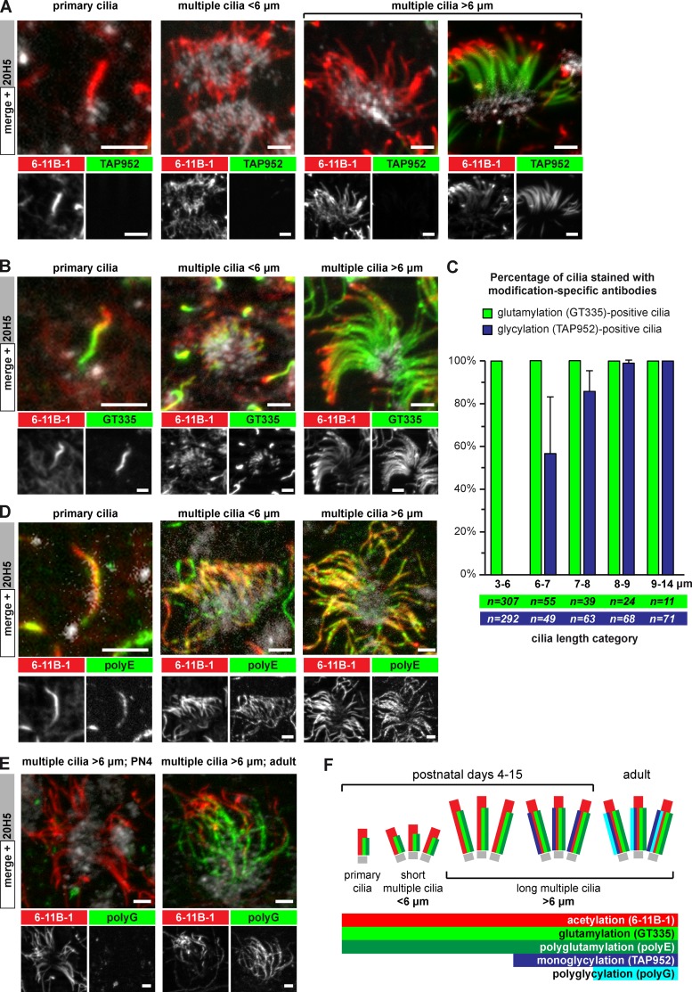
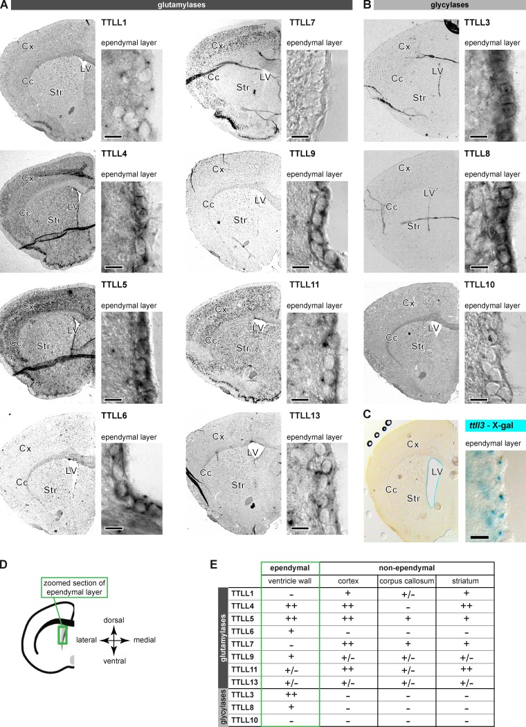
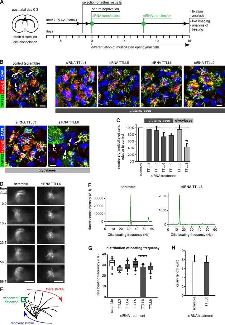
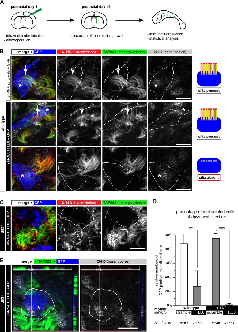
Microtubules are subject to a variety of posttranslational modifications that potentially regulate cytoskeletal functions. Two modifications, glutamylation and glycylation, are highly enriched in the axonemes of most eukaryotes, and might therefore play particularly important roles in cilia and flagella. Here we systematically analyze the dynamics of glutamylation and glycylation in developing mouse ependymal cilia and the expression of the corresponding enzymes in the brain. By systematically screening enzymes of the TTLL family for specific functions in ependymal cilia, we demonstrate that the glycylating enzymes TTLL3 and TTLL8 were required for stability and maintenance of ependymal cilia, whereas the polyglutamylase TTLL6 was necessary for coordinated beating behavior. Our work provides evidence for a functional separation of glutamylating and glycylating enzymes in mammalian ependymal cilia. It further advances the elucidation of the functions of tubulin posttranslational modifications in motile cilia of the mammalian brain and their potential importance in brain development and disease.


Figures

Figure 1.
Figure 1.
Glycylation and glutamylation are dynamically regulated in developing mouse ependymal cilia. (A) Maximum projection of image stacks taken from whole-mount ventricles at postnatal day (PN) 4. Basal bodies are labeled with 20H5, acetylated tubulin and thus cilia with 6-11B-1, and monoglycylation with TAP952. (B) Ependymal cells as in A; green channel shows glutamylation (GT335). (C) Quantitative analysis of the presence of monoglycylation (A; TAP952) and glutamylation (B; GT335) as a function of total ciliary length (6-11B-1). Average values with standard deviation are represented for four animals. Total sample numbers are indicated. (D) Ependymal cells as in A; green channel shows polyglutamylation (polyE). (E) Ependymal cells with long (>6 µm) motile cilia at PN4 and in adult mice. Polyglycylation (polyG) was restricted to cilia in adult mice. (F) Schematic representation of the distributions of PTMs in developing ependymal cilia. Bars, 2.5 µm.
Figure 2.
Figure 2.
A subset of TTLL enzymes is expressed in adult mouse ependymal cells. In situ hybridization revealed the expression patterns of TTLL genes encoding glutamylases (A) or glycylases (B) in coronal brain sections (Cc, corpus callosum; Cx, cortex; LV, lateral ventricle; Str, striatum). Controls in Fig. S1, B and C. (C) TTLL3 expression visualized by X-Gal staining in the ttll3−/− mouse. (D) Schematic representation of the right hemisphere with the green box indicating the localization of zoom images showing the ependymal layer in A–C. (E) Summary of expression analysis of the TTLL genes. Only weak (+) or strong (++) expression levels were considered specific. Bars (A–C): 10 µm.
Figure 3.
Figure 3.
Depletion of ependymal-specific glutamylases and glycylases induces different ciliary phenotypes in ependymal cells. (A) Flow scheme of the experimental paradigm of all siRNA experiments. (B) Analysis of multiciliated ependymal cells after siRNA. Cilia were co-labeled in fixed cells for polyglutamylation (polyE) and monoglycylation (TAP952). Bars, 10 µm. (C) Quantification of the relative numbers of multiciliated ependymal cells in areas with high cell density after siRNA treatment. The total number of cells (nuclei, DAPI) was related to the number of multiciliated cells polyE/TAP952 (B). Three independent experiments with more than 1,000 cells were analyzed, and controls (scramble siRNA) were set as 100%. Error bars represent SEM. After one-way ANOVA with Tukey’s post-hoc analysis, differences with P < 0.05 (*) were considered significant. (D) Image sequence of beating cilia after treatment with siRNA (A; 15 d), and labeling with Tubulin Tracker green. Ciliary beating was recorded at 120 frames per second (frame series of 75 ms; Videos 1 and 2). Bar, 10 µm. (E) Schematic representation of ciliary beating with the region of interest (green box) used for measurements. (F) Beating frequency distribution obtained by Fourier transformation of the beating frequency recording (E) of the cilia shown in D. (G) Box plot of the distribution of ciliary beating frequencies after siRNA (A). For each siRNA, three independent experiments, each with more than 25 cells, were recorded. Error bars show SEM; P < 10−6 (***) in Fisher variance test was considered significant. (H) The length of motile cilia after siRNA treatment measured on fixed cells (B) showed no difference between scramble and TTLL6 siRNA (Welch’s t test).
Figure 4.
Figure 4.
Long-term ablation of glycylases leads to nonciliated ependymal cells. (A) Flow scheme of the experimental paradigm used for shRNA-mediated depletion of TTLL8 in vivo. (B) Ependymal layer of wild-type mice. GFP-positive cells (blue or contours) express shRNA. Motile cilia were labeled for acetylation (6-11B-1), monoglycylation (TAP952), and basal bodies (20H5). Expression of TTLL8-shRNA partially led to loss of motile cilia; cells still contain multiple basal bodies. Quantification in D. (C) Expression of TTLL8-shRNA in the ependymal epithelium of ttll3−/− mice. Transfected cells (blue or contours) have no cilia. (D) Quantification of multiple motile cilia on shRNA-expressing cells 14 d after electroporation (B, C, and E). Three independent experiments per condition were performed (total number of counted cells are given below). Mean values with SEM and statistics (Welch t test) are represented (**, P < 0.01; ***, P < 0.001). (E) 3D images of TTLL8-depleted cells in the ependymal layer show fully developed and correctly arranged multiple basal bodies in cells without motile cilia. Panels on the top and the right represent the Z-stack of the image. (B, C, and E) Arrows, cilia; asterisks, basal bodies in GFP-positive cells. Bars, 10 µm.
Figure 5.
Figure 5.
Normal ciliogenesis precedes loss of cilia in ependymal cells depleted of glycylases. (A) Scheme of the experiment. (B) Lateral ventricle walls with ependymal cells 3 d after electroporation (A), expressing either scramble or TTLL8-shRNA (blue or contours). Cilia were visualized with acetylated tubulin (6-11B-1) and basal bodies (20H5). Arrows, cilia; asterisks, basal bodies of GFP-positive (blue) cells. Bars, 10 µm. (C) Quantification of GFP-positive cells with motile cilia 3 d after electroporation (B). Analysis was done as in Fig. 4 D. No significant differences between control and TTLL8 shRNA were detected (Welch’s t test).

Similar articles

Cited by

References

    1. Backer C.B., Gutzman J.H., Pearson C.G., Cheeseman I.M. 2012. CSAP localizes to polyglutamylated microtubules and promotes proper cilia function and zebrafish development. Mol. Biol. Cell. 23:2122–2130 10.1091/mbc.E11-11-0931 - DOI - PMC - PubMed
    1. Bell P.B., Jr, Safiejko-Mroczka B. 1995. Improved methods for preserving macromolecular structures and visualizing them by fluorescence and scanning electron microscopy. Scanning Microsc. 9:843–857, discussion :858–860 - PubMed
    1. Boutin C., Diestel S., Desoeuvre A., Tiveron M.-C., Cremer H. 2008. Efficient in vivo electroporation of the postnatal rodent forebrain. PLoS ONE. 3:e1883 10.1371/journal.pone.0001883 - DOI - PMC - PubMed
    1. Bré M.H., Redeker V., Quibell M., Darmanaden-Delorme J., Bressac C., Cosson J., Huitorel P., Schmitter J.M., Rossler J., Johnson T., et al. 1996. Axonemal tubulin polyglycylation probed with two monoclonal antibodies: widespread evolutionary distribution, appearance during spermatozoan maturation and possible function in motility. J. Cell Sci. 109:727–738 - PubMed
    1. Carlén M., Meletis K., Göritz C., Darsalia V., Evergren E., Tanigaki K., Amendola M., Barnabé-Heider F., Yeung M.S.Y., Naldini L., et al. 2009. Forebrain ependymal cells are Notch-dependent and generate neuroblasts and astrocytes after stroke. Nat. Neurosci. 12:259–267 10.1038/nn.2268 - DOI - PubMed

Publication types