. 2013 Aug 5;202(3):527-43.
doi: 10.1083/jcb.201211045. Epub 2013 Jul 29.

An ESCRT-spastin interaction promotes fission of recycling tubules from the endosome

Affiliations

An ESCRT-spastin interaction promotes fission of recycling tubules from the endosome

Rachel Allison et al. J Cell Biol. .

Abstract

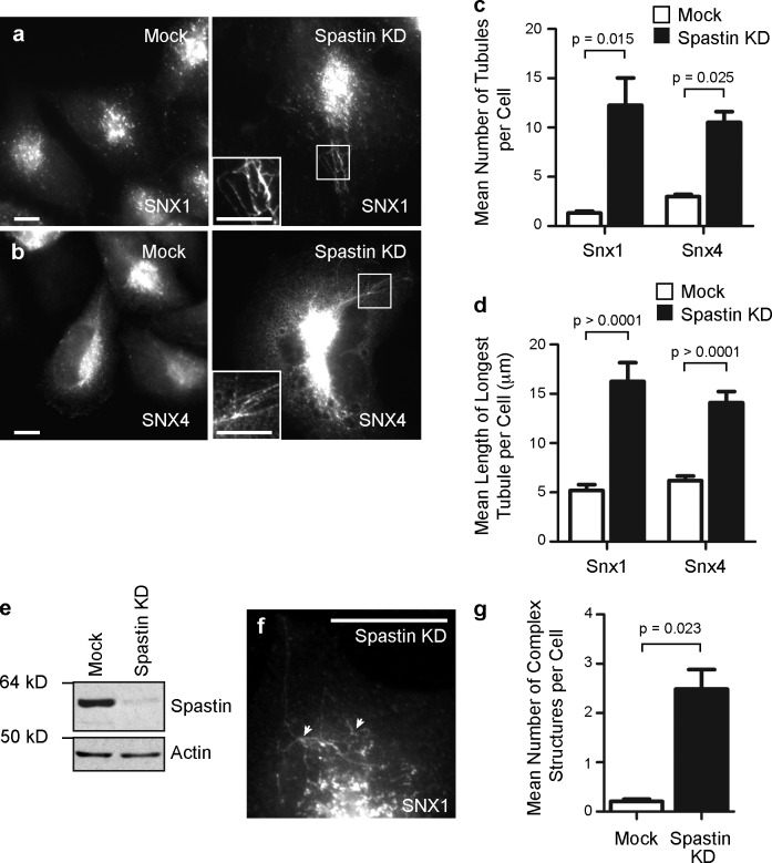
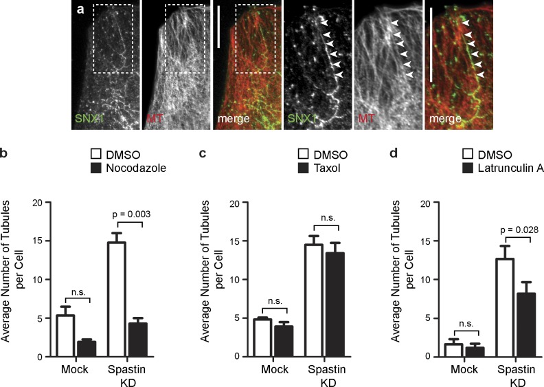
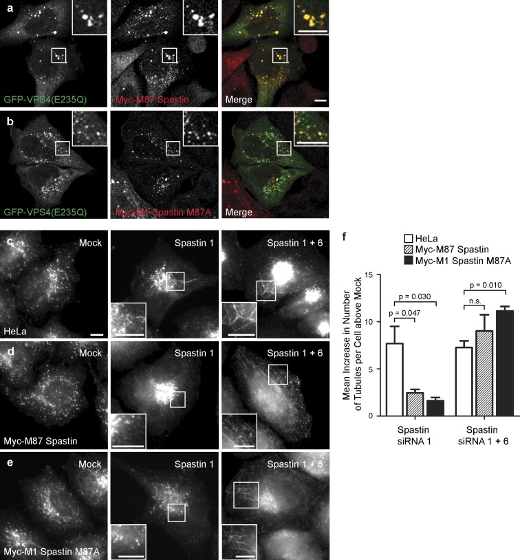
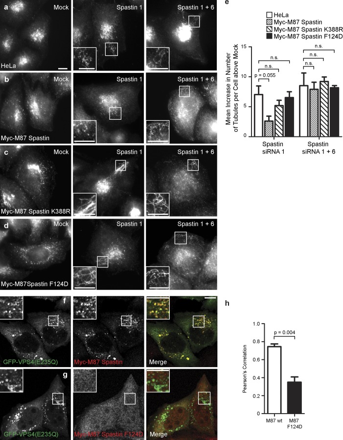
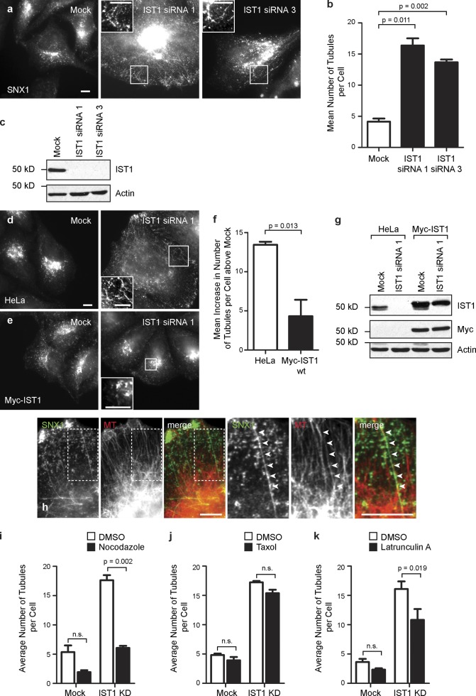
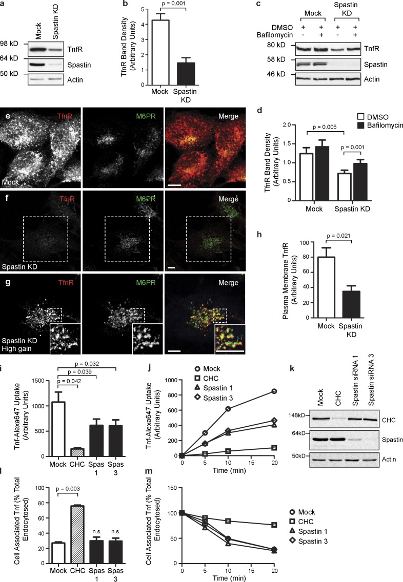
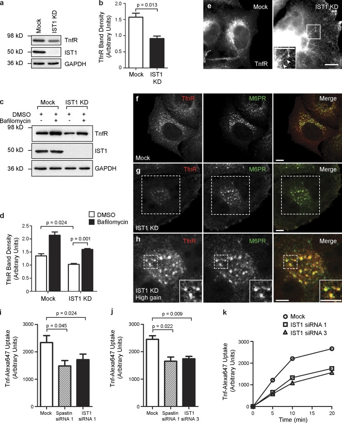
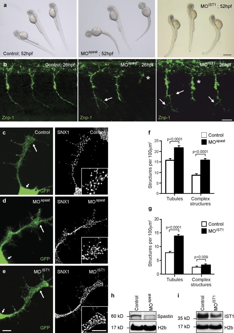
Mechanisms coordinating endosomal degradation and recycling are poorly understood, as are the cellular roles of microtubule (MT) severing. We show that cells lacking the MT-severing protein spastin had increased tubulation of and defective receptor sorting through endosomal tubular recycling compartments. Spastin required the ability to sever MTs and to interact with ESCRT-III (a complex controlling cargo degradation) proteins to regulate endosomal tubulation. Cells lacking IST1 (increased sodium tolerance 1), an endosomal sorting complex required for transport (ESCRT) component to which spastin binds, also had increased endosomal tubulation. Our results suggest that inclusion of IST1 into the ESCRT complex allows recruitment of spastin to promote fission of recycling tubules from the endosome. Thus, we reveal a novel cellular role for MT severing and identify a mechanism by which endosomal recycling can be coordinated with the degradative machinery. Spastin is mutated in the axonopathy hereditary spastic paraplegia. Zebrafish spinal motor axons depleted of spastin or IST1 also had abnormal endosomal tubulation, so we propose this phenotype is important for axonal degeneration.


Figures

Figure 1.
Figure 1.
Spastin regulates endosomal tubulation. (a and b) HeLa cells were subject to mock transfection or transfection with a spastin siRNA pool (spastin knockdown [KD]) and labeled versus endogenous SNX1 (a) or transfected SNX4-mCherry (b). The insets in a and b are magnified images of the boxed regions indicated. (c) The mean number of SNX1 and SNX4 tubules per cell was quantified (n = 5 independent experiments for SNX1, and n = 3 for SNX4, 30 cells counted per experimental condition in each experiment). (d) The length of the longest tubule per cell was measured in 30 cells, and the mean values were plotted. (e) Depletion of spastin was confirmed by immunoblotting. Note that M1 spastin is not routinely seen in immunoblots such as this, in which the exposure is optimized to show the much stronger M87 band. Actin labeling is shown to verify equal sample loading. (f) Complex structures, defined as having at least three tubules emanating from a central punctum (arrowheads), were commonly seen in spastin-depleted cells. (g) The mean number of complex structures per cell was quantified (n = 3, 30 cells counted per condition in each experiment). Bars, 10 µm. Error bars show SEMs.
Figure 2.
Figure 2.
Endosomal tubulation in spastin-depleted cells requires intact MTs. (a) HeLa cells depleted of spastin by transfection with an siRNA pool were labeled with antibodies to endogenous SNX1 and α-tubulin (MT). In this and subsequent color images, the color of the lettering in the black and white images indicates the color of that image in the corresponding merged image. Arrowheads in the magnified images of the boxed areas indicate an aligned SNX1 tubule and MT. (b–d) Mock-transfected cells or cells depleted of spastin by transfection with an siRNA pool were treated with vehicle (DMSO) or the MT-depolymerizing agent nocodazole (b), the MT stabilizing agent taxol (c), or the actin-depolymerizing agent latrunculin (d), and then, the number of SNX1 tubules per cell was counted (30 cells per condition), and the mean values of three (or four for latrunculin treatment) independent experiments were plotted. Representative immunofluorescence images from these experiments are shown in Fig. S2. KD, knockdown. Bars, 10 µm. Error bars show SEMs.
Figure 3.
Figure 3.
M1 and M87 spastin are recruited to endosomes and regulate endosomal tubulation. (a and b) GFP-VPS4-E235Q was transiently transfected into cell lines stably expressing myc-tagged M87 spastin (a) or myc-tagged M1 spastin M87A (b), and then, the cells were labeled with an anti-myc antibody. (c–f) HeLa cells (c), HeLa cells stably expressing myc-tagged M87 spastin (d), or HeLa cells expressing myc-tagged M1 spastinM87A (e) were subjected to mock transfection, transfected with an siRNA oligonucleotide directed against endogenous spastin (spastin 1, to which the myc-tagged transcripts were resistant), or with a combination of two siRNA oligonucleotides that together targeted endogenous and transfected spastin (spastin 1 and 6). The cells were labeled with an antibody to endogenous SNX1, and the number of SNX1 tubules per cell was counted (30 cells per condition). To control for any variation in the baseline number of tubules per cell in different cell lines, for each cell line, the mean tubule count per cell for siRNA-treated cells was normalized by subtracting the mean tubule count per cell in the corresponding mock-transfected cells. The resulting values for the mean increase in tubule number per cell in siRNA-treated cells were then plotted in f; n = 3 independent experiments. Cellular depletion of exogenous and/or endogenous spastin in these experiments was verified by immunofluorescence and immunoblotting (Fig. S3). Insets are magnifications of boxed regions. Bars, 10 µm. Error bars show SEMs.
Figure 4.
Figure 4.
ATPase activity and interaction with ESCRT-III are required for spastin to regulate endosomal tubulation. (a–d) Wild-type HeLa cells (a), HeLa cell lines stably expressing myc-tagged M87 spastin (b), myc-tagged M87 spastinK388R (c), or myc-tagged M87 spastinF124D (d) were subjected to mock transfection or were transfected with an siRNA oligonucleotide directed against endogenous spastin (spastin 1, to which the myc-tagged transcripts were resistant) or with siRNA oligonucleotides directed against endogenous and exogenous spastin (spastin 1 and 6). Cellular depletion of exogenous and/or endogenous spastin in these experiments was verified by immunofluorescence and immunoblotting (Fig. S3). The number of SNX1 tubules per cell was counted (30 cells per condition). To control for any variation in the baseline number of tubules per cell in different cell lines, for each cell line, the mean tubule count per cell for siRNA-treated cells was normalized by subtracting the mean tubule count per cell in the corresponding mock-transfected cells. The resulting values for the mean increase in tubule number per cell in siRNA-treated cells were then plotted in e; n = 3 independent experiments. (f–h) GFP-tagged VPS4-E235Q was transiently transfected into HeLa cells stably expressing myc-tagged wild-type (wt) M87 spastin (f) or myc-tagged M87 spastinF124D (g), which has dramatically reduced binding to CHMP1B and IST1. The cells were labeled with anti-myc antibodies. The extent of colocalization between GFP-VPS4-E235Q and the spastin proteins was estimated by calculating the Pearson’s correlation coefficient for red and green pixels in each cell, using Volocity software (h; n = 3 experiments, 20 cells per condition quantified in each experiment). Insets are magnifications of boxed regions. Bars, 10 µm. Error bars show SEMs.
Figure 5.
Figure 5.
IST1 regulates endosomal tubulation. (a and b) Wild-type HeLa cells were subjected to mock transfection or transfected with either of the two IST1 siRNA oligonucleotides indicated and then labeled with SNX1. The number of SNX1 tubules per cell was counted (30 cells per condition), and the mean values of three independent experiments were plotted in b. (c) Depletion of IST1 was verified by immunoblotting. (d–g) Wild-type HeLa cells (d) or HeLa cells stably expressing myc-tagged siRNA-resistant IST1 (e) were subjected to mock transfection or were transfected with an siRNA targeting endogenous IST1. The number of SNX1 tubules per cell was counted as in a. To control for any variation in the baseline number of tubules per cell in the two cell lines, for each cell line, the mean tubule count per cell for siRNA-treated cells was normalized by subtracting the mean tubule count per cell in the corresponding mock-transfected cells. The resulting values for the mean increase in tubule number per cell in siRNA-treated cells were then plotted in f; n = 3 independent experiments. wt, wild type. (g) Depletion of endogenous IST1 was verified by immunoblotting. (h) HeLa cells depleted of IST1 by transfection with IST1 siRNA1 were labeled with antibodies to endogenous SNX1 and α-tubulin (MT). Arrowheads in the magnified images of the boxed areas indicate an aligned SNX1 tubule and MT. (i–k) Mock-transfected cells or cells depleted of IST1 by transfection with siRNA1 were treated with vehicle (DMSO), nocodazole (i), taxol (j), or latrunculin A (k), and then, the number of SNX1 tubules per cell was counted (30 cells per condition), and the mean values of three independent experiments were plotted. KD, knockdown. Insets are magnifications of boxed regions. Bars, 10 µm. Error bars show SEMs.
Figure 6.
Figure 6.
Spastin is required for sorting TfnR away from degradation. (a and b) Mock-transfected HeLa cells or HeLa cells transfected with a pool of spastin siRNA (spastin knockdown [KD]) were immunoblotted for the antibodies indicated. TfnR band density was quantified, and the mean density in three independent experiments was plotted in b. (c and d) Mock-transfected HeLa cells or HeLa cells transfected with a pool of spastin siRNA were treated with vehicle (DMSO) or DMSO + bafilomycin and then immunoblotted versus the antibodies indicated. TfnR band density was quantified, and the mean density in eight independent experiments was plotted in d. (e–g) Confocal micrographs of HeLa cells subjected to mock transfection (e) or transfected with a spastin siRNA pool (f and g) and then labeled with the markers shown. Confocal micrograph gain settings were identical in e and f, but in g, which shows a higher magnification image of the dashed area indicated in f, gain settings were increased to reveal that the remaining TfnR signal strongly colocalized with M6PR. Note that tubular TfnR structures are not readily seen under confocal microscopy, as the tubules tend to leave the plane of section. Insets are magnified regions of the boxed areas. (h) Mock-transfected HeLa cells or HeLa cells transfected with a pool of spastin siRNA were fixed and labeled (without permeabilization) with a FITC-conjugated antibody against TfnR, and then, the cell-associated fluorescent signal was quantified by FACS analysis. The mean fluorescence values for three independent experiments were plotted. (i–k) Uptake of Alexa Fluor 647–conjugated Tfn was measured by FACS over a 20-min time course in mock-transfected HeLa cells, cells transfected with siRNA targeting clathrin heavy chain (CHC), or cells transfected with one of two siRNAs directed against spastin (spas1, spastin 1; spas3, spastin 3). Mean Tfn uptake at the 20-min time point (n = 3 experiments) is shown in i, and a representative time course experiment is shown in j. Depletion of the relevant proteins targeted by siRNA was confirmed by immunoblotting, and a representative example is shown in k. (l and m) Recycling of internalized fluorescently labeled Tfn was measured over a 20-min time course, and the mean cell-associated Tfn at the 20-min time point (n = 3 experiments) is shown in l, with a representative time course experiment shown in m. Bars, 10 µm. Error bars show SEMs.
Figure 7.
Figure 7.
Ist1 is required for sorting TfnR away from degradation. (a and b) Mock-transfected HeLa cells or HeLa cells transfected with IST1 siRNA1 (IST1 knockdown [KD]) were immunoblotted for the antibodies indicated. TfnR band density was quantified, and the mean density in three independent experiments was plotted in b. GAPDH immunoblotting serves as a loading control. (c and d) Mock-transfected HeLa cells or HeLa cells transfected with IST1 siRNA1 were treated with vehicle (DMSO) or DMSO + bafilomycin and then immunoblotted versus the antibodies indicated. TfnR band density was quantified, and the mean density in three independent experiments was plotted in d. (e) Mock-transfected HeLa cells or HeLa cells transfected with IST1 siRNA1 were labeled for TfnR. Increased TfnR tubulation is seen in the IST1-depleted cells (see arrowheads in inset magnified region of the boxed area). The exposure settings in the IST1 knockdown image have been increased to compensate for the reduced TfnR levels in these cells. (f–h) Confocal micrographs of HeLa cells subjected to mock transfection (f) or transfected with IST1 siRNA1 (g and h) and then labeled with the markers shown. Confocal micrograph gain settings were identical in f and g, but in h, which shows higher magnification images of the dashed areas indicated in g, gain settings were increased to reveal that the remaining TfnR signal strongly colocalized with M6PR. Insets are magnified areas of the boxed regions. (i–k) Uptake of Alexa Fluor Tfn 647 was measured by FACS over a 20-min time course in mock-transfected HeLa cells, cells transfected with spastin siRNA1 (as a positive control), and IST1 siRNA1 (i) or IST1 siRNA3 (j). Mean Tfn uptake at the 20-min time point is shown (n = 3 experiments plotted for each histogram). A representative time course experiment is shown in k. Bars, 10 µm. Error bars show SEMs.
Figure 8.
Figure 8.
Spastin regulates endosomal tubulation in spinal motor axonal growth cones. (a) Gross phenotype of 52-hpf control, spastin morphant (MOspast), and IST1 morphant (MOIST1) zebrafish larvae. Bar, 500 µm. (b) Whole-mount immunohistochemistry of 26-hpf zebrafish control, MOspast, and MOIST1 embryos labeled with the motor neuron marker synaptotagmin 2 (znp-1). Images are lateral views of the trunk; anterior is to the left. Axonal stumps (asterisks) and abnormal branching (arrows) of spinal motor neuron axons were observed in all spastin and IST1 morphants. Bar, 25 µm. (c–i) Spinal neurons were cultured from control (c), spastin morphant (d), and IST1 morphant (e) 24-hpf Tg(Mnx1:mGFP) embryos, in which GFP is selectively expressed in motor neurons. Cultured neurons were labeled with SNX1 and GFP antibodies, and the mean number of SNX1-positive tubules and complex tubular structures per 100-µm2 region of each motor axon growth cone (large arrows in GFP-labeled images and inset boxes in SNX1-labeled images; small arrows indicate the cell body) was calculated by counting ∼30 cells per condition. Bars, 5 µm. (f and g) The mean values obtained in three (f) or five (g) independent experiments were plotted. (h and i) In these experiments, spastin and IST1 depletion was verified by immunoblotting. Anti–histone H2b immunoblotting is shown to verify equal protein loading. Error bars show SEMs.

References

    1. Agromayor M., Carlton J.G., Phelan J.P., Matthews D.R., Carlin L.M., Ameer-Beg S., Bowers K., Martin-Serrano J. 2009. Essential role of hIST1 in cytokinesis. Mol. Biol. Cell. 20:1374–1387 10.1091/mbc.E08-05-0474 - DOI - PMC - PubMed
    1. Babst M., Katzmann D.J., Estepa-Sabal E.J., Meerloo T., Emr S.D. 2002a. Escrt-III: an endosome-associated heterooligomeric protein complex required for mvb sorting. Dev. Cell. 3:271–282 10.1016/S1534-5807(02)00220-4 - DOI - PubMed
    1. Babst M., Katzmann D.J., Snyder W.B., Wendland B., Emr S.D. 2002b. Endosome-associated complex, ESCRT-II, recruits transport machinery for protein sorting at the multivesicular body. Dev. Cell. 3:283–289 10.1016/S1534-5807(02)00219-8 - DOI - PubMed
    1. Bajorek M., Morita E., Skalicky J.J., Morham S.G., Babst M., Sundquist W.I. 2009a. Biochemical analyses of human IST1 and its function in cytokinesis. Mol. Biol. Cell. 20:1360–1373 10.1091/mbc.E08-05-0475 - DOI - PMC - PubMed
    1. Bajorek M., Schubert H.L., McCullough J., Langelier C., Eckert D.M., Stubblefield W.M., Uter N.T., Myszka D.G., Hill C.P., Sundquist W.I. 2009b. Structural basis for ESCRT-III protein autoinhibition. Nat. Struct. Mol. Biol. 16:754–762 10.1038/nsmb.1621 - DOI - PMC - PubMed

Publication types

MeSH terms