. 2013 Aug 5;202(3):495-508.
doi: 10.1083/jcb.201302078. Epub 2013 Jul 29.

JIP1 regulates the directionality of APP axonal transport by coordinating kinesin and dynein motors

Affiliations

JIP1 regulates the directionality of APP axonal transport by coordinating kinesin and dynein motors

Meng-meng Fu et al. J Cell Biol. .

Abstract

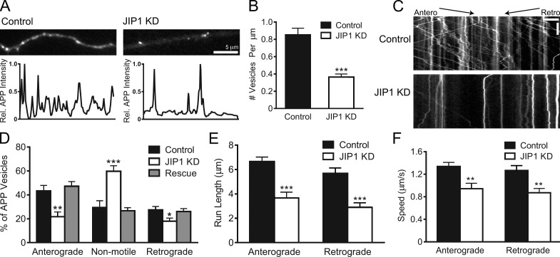
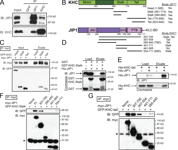
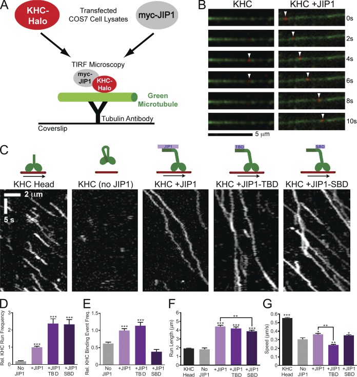
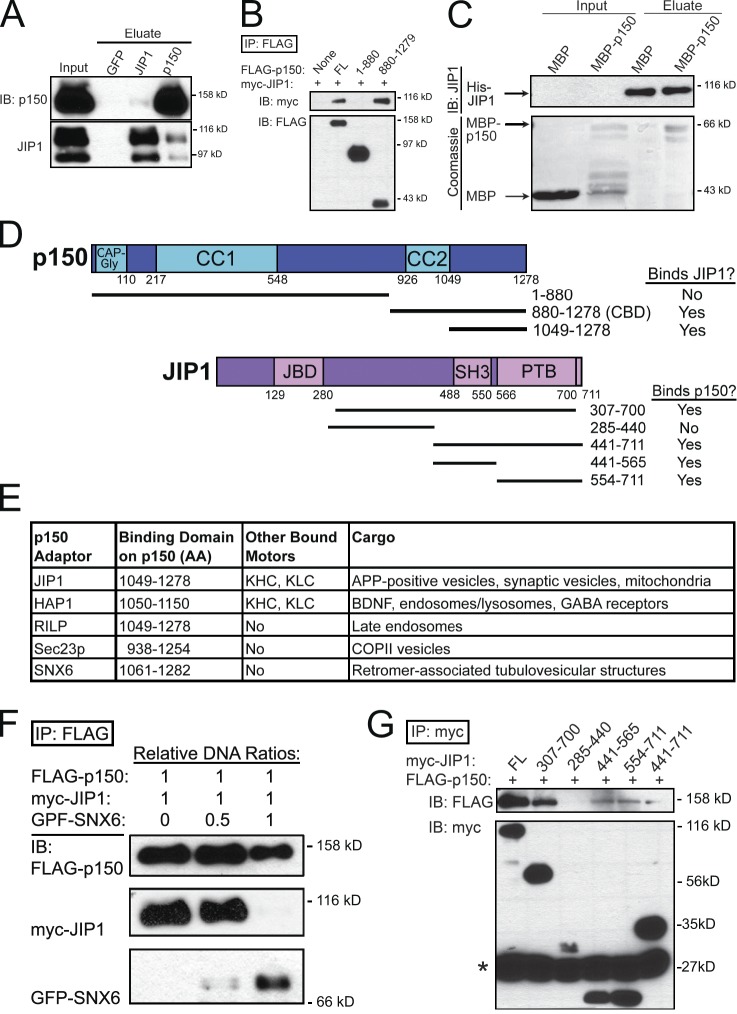
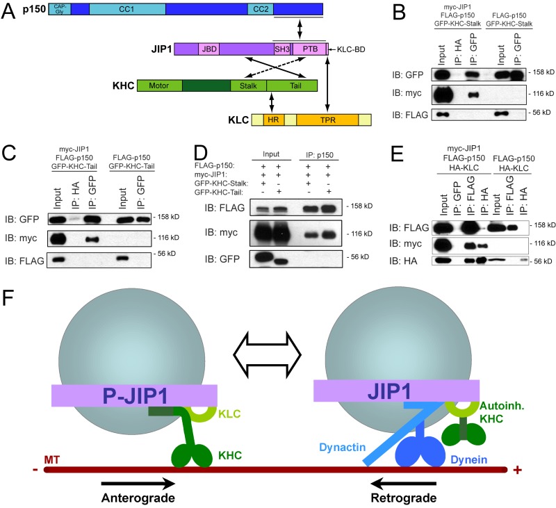
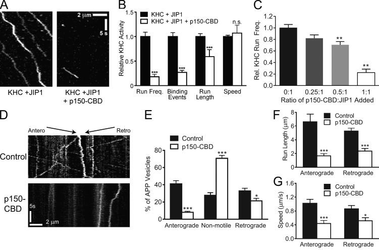
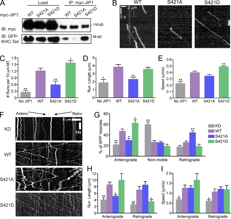
Regulation of the opposing kinesin and dynein motors that drive axonal transport is essential to maintain neuronal homeostasis. Here, we examine coordination of motor activity by the scaffolding protein JNK-interacting protein 1 (JIP1), which we find is required for long-range anterograde and retrograde amyloid precursor protein (APP) motility in axons. We identify novel interactions between JIP1 and kinesin heavy chain (KHC) that relieve KHC autoinhibition, activating motor function in single molecule assays. The direct binding of the dynactin subunit p150(Glued) to JIP1 competitively inhibits KHC activation in vitro and disrupts the transport of APP in neurons. Together, these experiments support a model whereby JIP1 coordinates APP transport by switching between anterograde and retrograde motile complexes. We find that mutations in the JNK-dependent phosphorylation site S421 in JIP1 alter both KHC activation in vitro and the directionality of APP transport in neurons. Thus phosphorylation of S421 of JIP1 serves as a molecular switch to regulate the direction of APP transport in neurons.


Figures

Figure 1.
Figure 1.
JIP1 knockdown disrupts both anterograde and retrograde transport of APP-positive vesicles. (A) Representative images and line scans of APP-YFP intensity show that JIP1-knockdown DRGs contain fewer APP-positive vesicles in the axon than control DRGs. (B) JIP1 knockdown in DRGs significantly decreased the number of APP-positive vesicles in the axon. Control, 0.85 ± 0.08/µm; JIP1 siRNA, 0.36 ± 0.04/µm. (B–F) Data represent three independent experiments (n = 15–23 neurons). (C) Kymographs of APP-YFP motility in DRG transfected with siRNA against JIP1. Kymographs represent cumulative organelle movement (displacement on the x-axis) over time (y-axis). Arrested vesicles appear as vertical lines, whereas motile vesicles appear as diagonal lines toward either the right (anterograde) or left (retrograde). Bars: (horizontal) 5 µm; (vertical) 15 s. (D) JIP1 depletion significantly alters the directional distribution of APP-positive vesicles, causing decreases in the percentages of anterograde and retrograde vesicles and an increase in the percentage of arrested vesicles. Transport changes induced by JIP1 depletion are fully rescued by expression of a human JIP1 cDNA resistant to the siRNA. (E and F) JIP1 depletion significantly decreases mean run lengths and speeds of APP-positive vesicles in both anterograde and retrograde directions. Means represent only vesicles categorized as motile (i.e., anterograde or retrograde in D). Error bars show the mean ± SEM; *, P < 0.05; **, P < 0.01; ***, P < 0.001.
Figure 2.
Figure 2.
JIP1 binds directly to both stalk and tail domains of KHC independently of KLC. (A) JIP1 coimmunoprecipitates with KHC in mouse brain homogenate. A monoclonal JIP1 antibody immunoprecipitates the expected 110-kD band as well as a 90-kD band that likely represents a splice isoform. Both bands are also recognized by multiple JIP1 monoclonal antibodies (R&D Systems and BD). (B) Schematics of KHC (KIF5C) and JIP1 constructs and summary of mapping results. JIP1 contains JBD (JNK-binding domain), SH3 (src homology), and PTB domains. (C) JIP1 binds to both stalk and tail domains of KHC. Lysates from COS7 cells cotransfected with myc-JIP1 and GFP-KHC fragments were immunoprecipitated with an anti-myc antibody. (D) JIP1 binds directly to KHC stalk. Purified His-JIP1 incubated with glutathione beads bound to either GST or GST-KHC-stalk (aa 560–682) selectively bound to GST-KHC-stalk but not GST. (E) JIP1 binds directly to KHC tail. Purified His-KHC-tail (KIF5B aa 823–944) with or without His-JIP1 was coincubated with anti-JIP1 antibody, which specifically coimmunoprecipitated His-KHC-tail. (F) KHC stalk binds to the C terminus of JIP1 independently of KLC. Lysates from COS7 cells cotransfected with GFP-KHC-stalk and myc-JIP1 fragments were immunoprecipitated with an anti-myc antibody. KHC stalk coimmunoprecipitated with myc-JIP1[554–711] (myc-JIP1-SBD). Asterisk shows antibody light chain bands. (G) KHC tail binds to JIP1 independently of KLC. Lysates from COS7 cells transfected with GFP-KHC-tail and myc-JIP1 fragments were immunoprecipitated with an anti-myc antibody. KHC tail coimmunoprecipitated with myc-JIP1[285–440] (myc-JIP1-TBD). Asterisk shows antibody heavy chain bands.
Figure 3.
Figure 3.
JIP1 binding relieves KHC autoinhibition in in vitro TIRF motility assays. (A) Schematic of in vitro TIRF motility assay. Lysate from COS7 cells transfected with KHC-Halo and incubated with red fluorescent TMR ligand was combined with lysate from cells expressing myc-JIP1 constructs and applied to flow chambers containing green fluorescent microtubules, which were immobilized on glass coverslips with anti-tubulin antibody. KHC-Halo motility was imaged using a TIRF microscope. (B) Time-lapsed images acquired from a flow chamber containing KHC-Halo (red) lysate alone (left) show a brief non-motile binding event (arrowheads) to a microtubule (green). Images from a flow chamber containing KHC-Halo and myc-JIP1 lysates (right) show processive movement (arrowheads) along the microtubule. (C) Representative kymographs show activation of KHC-Halo by full-length JIP1, JIP1-TBD, or JIP1-SBD. 100 total frames (∼33 s) are shown. (D) Addition of full-length JIP1, JIP1-TBD, and JIP1-SBD increases the run frequency of full-length KHC-Halo. The absolute number of runs per 10 µm of microtubule was normalized to the KHC-Halo +JIP1 condition for each experiment. (D–G) Data represent three or more independent experiments per condition (n = 52–181 microtubules and n = 109–758 runs) and statistical comparisons were made relative to the KHC-Halo alone (no JIP1) condition unless otherwise indicated. (E) Addition of full-length JIP1 or JIP1-TBD increases the relative frequency of non-motile microtubule-binding events by full-length KHC-Halo. The number of non-motile binding events per 10 µm microtubule length was normalized relative to the KHC-Halo +JIP1 condition for each independent experiment. (F) Addition of full-length JIP1, JIP1-TBD or JIP1-SBD increases KHC-Halo run lengths. (G) Addition of full-length JIP1 or JIP1-SBD but not JIP1-TBD increases speed of KHC-Halo runs. Error bars show the mean ± SEM; *, P < 0.05; **, P < 0.01; ***, P < 0.001.
Figure 4.
Figure 4.
JIP1 binds directly to the p150Glued subunit of dynactin. (A) Endogenous JIP1 and p150Glued coimmunoprecipitate from mouse brain homogenate. (B) JIP1 binds to the C-terminal p150Glued-CBD. Lysates from COS7 cells cotransfected with myc-JIP1 and FLAG-p150Glued fragments were immunoprecipitated with an anti-FLAG antibody. JIP1 coimmunoprecipitated with C-terminal p150Glued[880–1278], but not with N-terminal p150Glued[1–880]. (C) JIP1 binds directly to C-terminal p150Glued. When applied to His-Bind resin bound to His-JIP1, MBP-p150Glued[1049–1278] selectively bound whereas MBP did not. (D) Summary of p150Glued and JIP1 mapping results. (E) A diverse set of cargo adaptors binds to p150Glued-CBD. (F) SNX6 and JIP1 bind competitively to p150Glued. Lysates from COS7 cells transfected with fixed amounts of FLAG-p150 and myc-JIP1 DNA and progressively increasing amounts of GFP-SNX6 DNA were immunoprecipitated with anti-FLAG antibody. FLAG-p150 coimmunoprecipitates predominantly with myc-JIP1 in the low GFP-SNX6 condition, but mostly with GFP-SNX6 in the high GFP-SNX6 condition. (G) C-terminal JIP1 binds to p150Glued. Lysates from COS7 cells cotransfected with FLAG-p150Glued and myc-JIP1 fragments were immunoprecipitated with an anti-myc antibody. p150Glued coimmunoprecipitated with both myc-JIP1[441–565] and myc-JIP1[554–711]; the presence of both these domains in JIP1 (myc-JIP1[441–711]) did not strengthen the interaction. Asterisk shows antibody light chain bands.
Figure 5.
Figure 5.
Anterograde and retrograde JIP1 motor complexes are mutually exclusive. (A) Summary schematic of direct binding interactions between JIP1, Kinesin-1, and dynactin. (B) JIP1 cannot bind simultaneously to both p150Glued and KHC stalk. Lysates from COS7 cells triple transfected with myc-JIP1, FLAG-p150Glued, and GFP-KHC-stalk were immunoprecipitated with an anti-GFP antibody. FLAG-p150Glued and GFP-KHC-stalk do not interact with each other either in the absence or presence of myc-JIP1. (C) JIP1 cannot bind simultaneously to both p150Glued and KHC tail. Lysates from COS7 cells triple transfected with myc-JIP1, FLAG-p150Glued, and GFP-KHC-tail were immunoprecipitated with an anti-GFP antibody. FLAG-p150Glued and GFP-KHC-tail do not interact with each other either in the absence or presence of myc-JIP1. (D) JIP1 cannot bind simultaneously to p150Glued and KHC. Lysates from COS7 cells triple transfected with myc-JIP1, FLAG-p150Glued, and either GFP-KHC-tail or GFP-KHC-stalk were immunoprecipitated with an anti-p150Glued antibody. Though robust levels of FLAG-p150Glued and associated myc-JIP1 are coimmunoprecipitated, no interacting GFP-KHC-stalk or GFP-KHC-tail can be detected. (E) JIP1 can bind simultaneously to both p150Glued and KLC. Lysates from COS7 cells triple transfected with myc-JIP1, FLAG-p150Glued, and HA-KLC were immunoprecipitated with either an anti-FLAG or anti-HA antibody. In the absence of myc-JIP1, FLAG-p150Glued and HA-KLC do not interact. The addition of myc-JIP1 facilitates the indirect interaction between p150Glued and KLC as both FLAG and HA antibodies immunoprecipitate triple complexes of FLAG-p150Glued, myc-JIP1, and HA-KLC. (F) Model of two mutually exclusive JIP1 motile complexes. The anterograde JIP1 complex activates KHC motility via direct binding to both stalk and tail domains (left) but cannot bind simultaneously to p150Glued; KLC may remain bound via the C-terminal tail of JIP1 (Verhey et al., 2001). The retrograde JIP1 complex binds directly to p150Glued to facilitate dynein-mediated transport and may retain autoinhibited KHC via simultaneous binding to KLC (right).
Figure 6.
Figure 6.
p150Glued-CBD disrupts JIP1-mediated KHC motility in vitro and anterograde APP-positive vesicle transport in DRGs. (A) Representative kymographs show that addition of p150Glued-CBD disrupts enhancement of KHC-Halo motility by JIP1. Lysate from COS7 cells transfected with myc-JIP1 were combined with FLAG-p150Glued-CBD lysate and immediately combined with KHC-Halo lysate. This lysate mixture was applied to flow chambers containing immobilized fluorescent microtubules and imaged. 100 total frames (∼33 s) are shown. (B) Addition of p150Glued-CBD decreases the number of motile KHC events mediated by JIP1. Motility measurements in the presence of FLAG-p150Glued-CBD were normalized to the condition containing only myc-JIP1 and KHC-Halo and represent three independent experiments (n = 60–100 microtubules and n = 23–214 runs). (C) p150Glued-CBD competitively inhibits JIP1-mediated KHC motility in vitro. At constant levels of myc-JIP1 lysate, addition of incrementally higher levels of p150Glued-CBD lysate leads to complementary decreases in relative KHC-Halo run frequency. Data represents three independent experiments (n = 6–52 microtubules). (D) Kymographs of APP-DsRed motility in DRGs transfected with a bicistronic construct coexpressing FLAG-p150Glued-CBD and GFP. Approximately 80 total frames (∼20 s) are shown. (E) Expression of p150Glued-CBD significantly decreases the percentage of anterograde APP-positive vesicles and correspondingly increases the percentage of arrested vesicles. (E–G) Data represent four independent experiments (n = 12–14 neurons). (F and G) Expression of p150Glued-CBD significantly decreases run length and speed of both anterograde and retrograde APP-positive vesicles. Means represent only vesicles categorized as motile (i.e., anterograde or retrograde in E). Error bars show the mean ± SEM; *, P < 0.05; **, P < 0.01; ***, P < 0.001.
Figure 7.
Figure 7.
Mutations of the JNK phosphorylation site S421 in JIP1 alter KHC activation in vitro and APP directionality in neurons. (A) Mutations at JIP1-S421 alter KHC-tail–binding ability. COS7 cells were cotransfected with GFP-KHC-tail and wild-type or mutant myc-JIP1 and immunoprecipitated with anti-myc antibody. (B) Representative kymographs of KHC-Halo motility show weak activation by myc-JIP1-S421A and enhanced activation by myc-JIP1-S421D in in vitro motility assays. 100 total frames (∼33 s) are shown. (C) KHC-Halo run frequencies in vitro decrease in the presence of JIP1-S421A and increase in the presence of JIP1-S421D. (C–E) Data from three independent experiments (n = 48–101 microtubules and n = 18–254 runs) are shown and statistical comparisons were made versus the wild-type JIP1 condition. (D) KHC-Halo run length is decreased with addition of JIP1-S421A. (E) KHC-Halo speed is increased with addition of JIP1-S421D. (F) Representative kymographs of APP-YFP motility in DRGs transfected with siRNA targeted to mouse JIP1 and rescued with a bicistronic construct coexpressing human wild-type or mutant JIP1 as well as the fluorescent transfection marker BFP. (G) DRGs expressing JIP1-S421D have increased percentages of anterograde APP vesicles, whereas DRGs expressing JIP1-S421A have increased percentages of retrograde APP vesicles. (G–I) Data from three independent experiments (n = 7–9 neurons and n = 78–224 runs) are shown with statistical comparisons made against the wild-type rescue condition. (H) APP-positive vesicles in DRGs expressing JIP1-S421A have decreased anterograde run length, whereas those expressing JIP1-S421D have decreased retrograde run length. (I) No significant differences are observed in APP speeds in DRGs expressing JIP1-S421 phosphomutants. Error bars show the mean ± SEM; *, P < 0.05; **, P < 0.01.

Comment in

References

    1. Abe N., Almenar-Queralt A., Lillo C., Shen Z., Lozach J., Briggs S.P., Williams D.S., Goldstein L.S., Cavalli V. 2009. Sunday driver interacts with two distinct classes of axonal organelles. J. Biol. Chem. 284:34628–34639 10.1074/jbc.M109.035022 - DOI - PMC - PubMed
    1. Arimoto M., Koushika S.P., Choudhary B.C., Li C., Matsumoto K., Hisamoto N. 2011. The Caenorhabditis elegans JIP3 protein UNC-16 functions as an adaptor to link kinesin-1 with cytoplasmic dynein. J. Neurosci. 31:2216–2224 10.1523/JNEUROSCI.2653-10.2011 - DOI - PMC - PubMed
    1. Blasius T.L., Cai D., Jih G.T., Toret C.P., Verhey K.J. 2007. Two binding partners cooperate to activate the molecular motor Kinesin-1. J. Cell Biol. 176:11–17 10.1083/jcb.200605099 - DOI - PMC - PubMed
    1. Bowman A.B., Kamal A., Ritchings B.W., Philp A.V., McGrail M., Gindhart J.G., Goldstein L.S. 2000. Kinesin-dependent axonal transport is mediated by the sunday driver (SYD) protein. Cell. 103:583–594 10.1016/S0092-8674(00)00162-8 - DOI - PubMed
    1. Cai D., Verhey K.J., Meyhöfer E. 2007. Tracking single Kinesin molecules in the cytoplasm of mammalian cells. Biophys. J. 92:4137–4144 10.1529/biophysj.106.100206 - DOI - PMC - PubMed

Publication types