. 2013 Aug 5;202(3):509-26.
doi: 10.1083/jcb.201211148. Epub 2013 Jul 29.

The synaptobrevin homologue Snc2p recruits the exocyst to secretory vesicles by binding to Sec6p

Affiliations

The synaptobrevin homologue Snc2p recruits the exocyst to secretory vesicles by binding to Sec6p

David Shen et al. J Cell Biol. .

Abstract

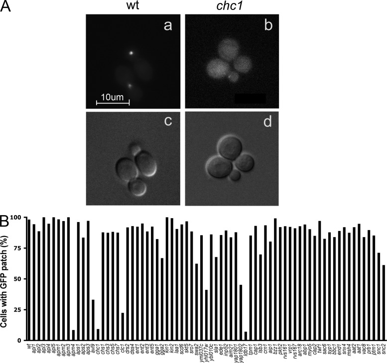
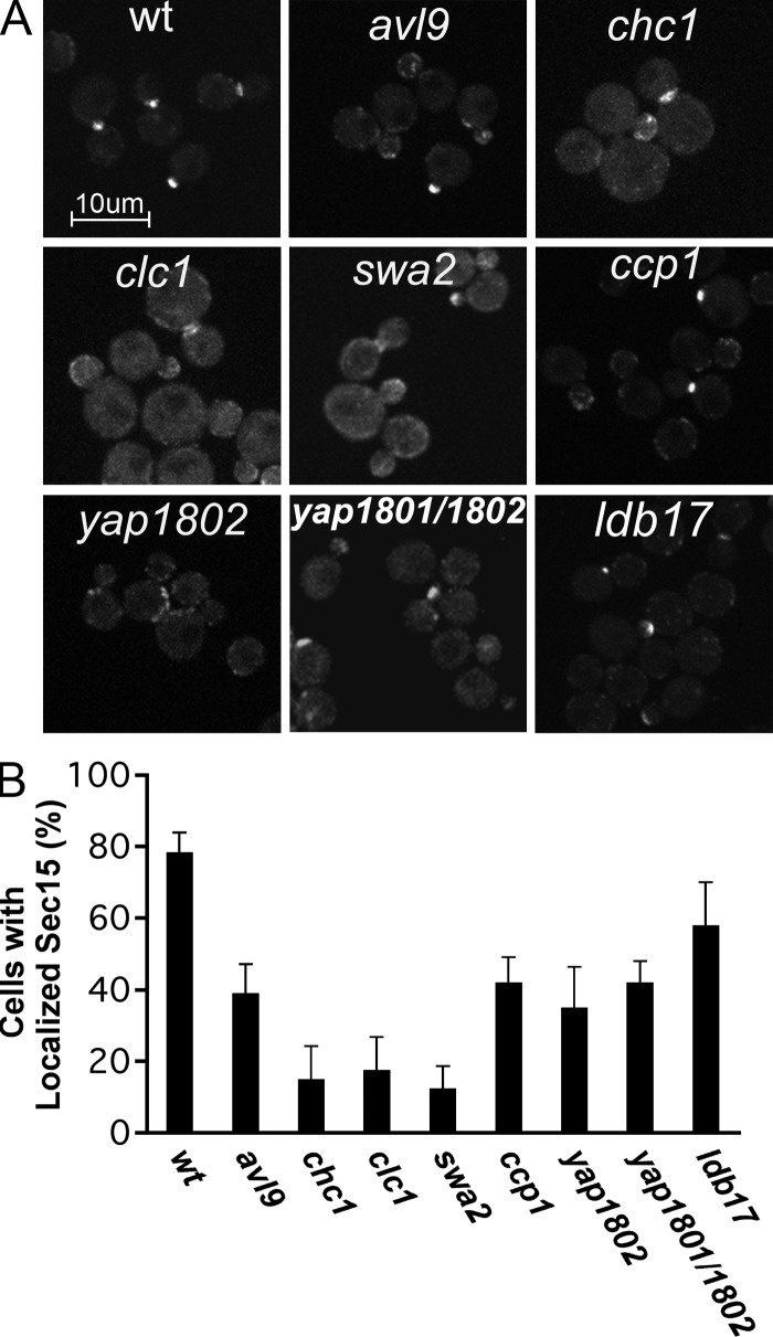
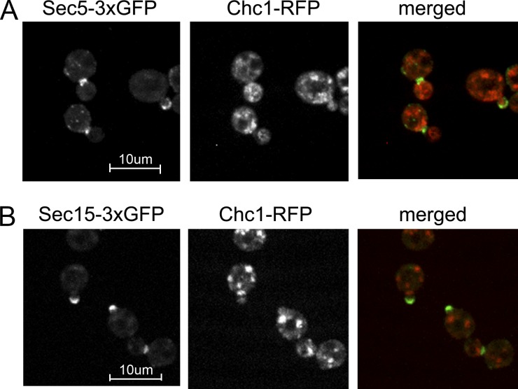
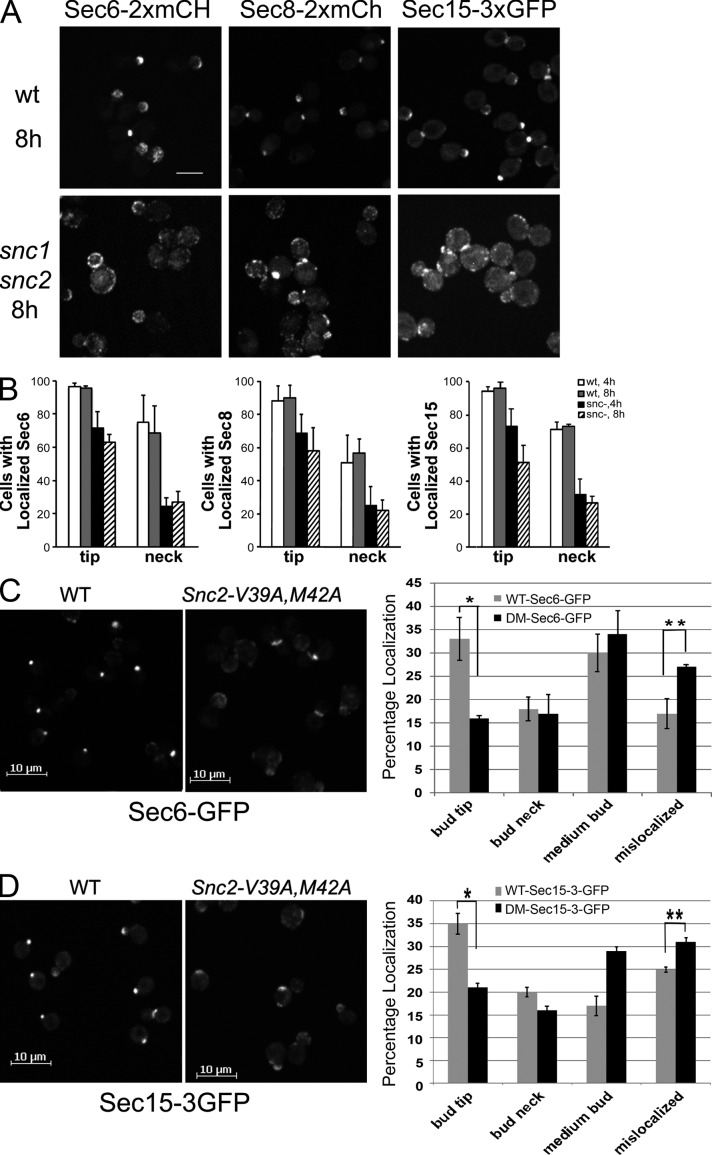
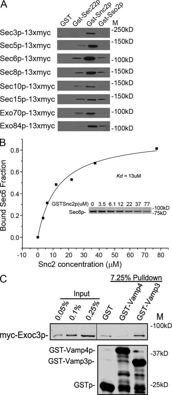
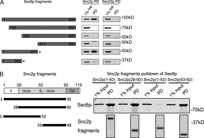
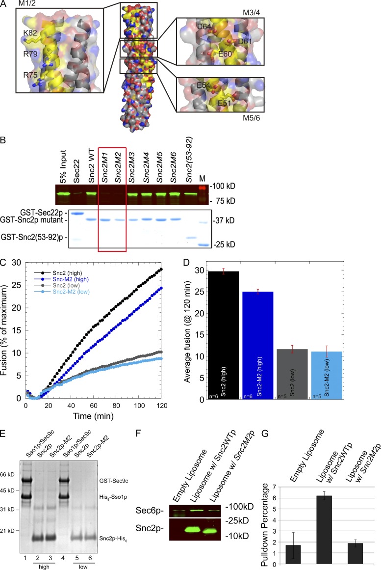
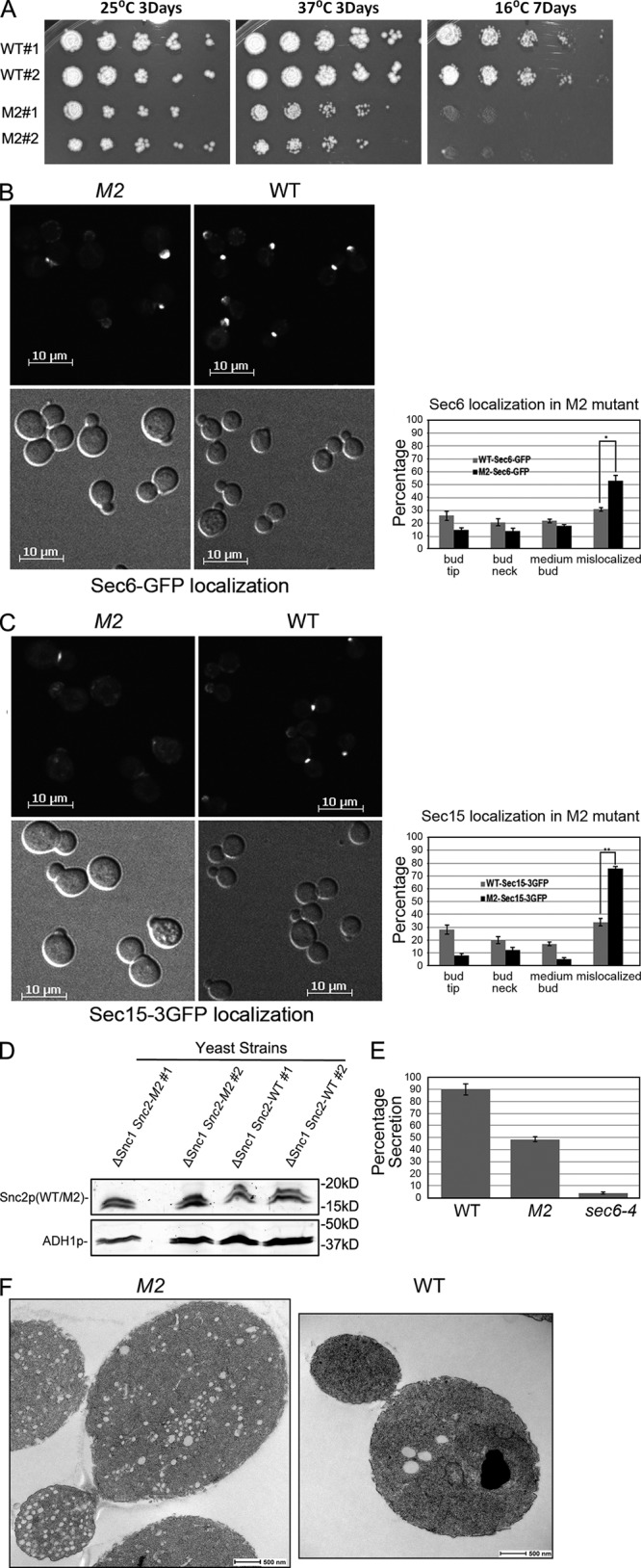
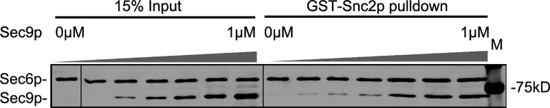
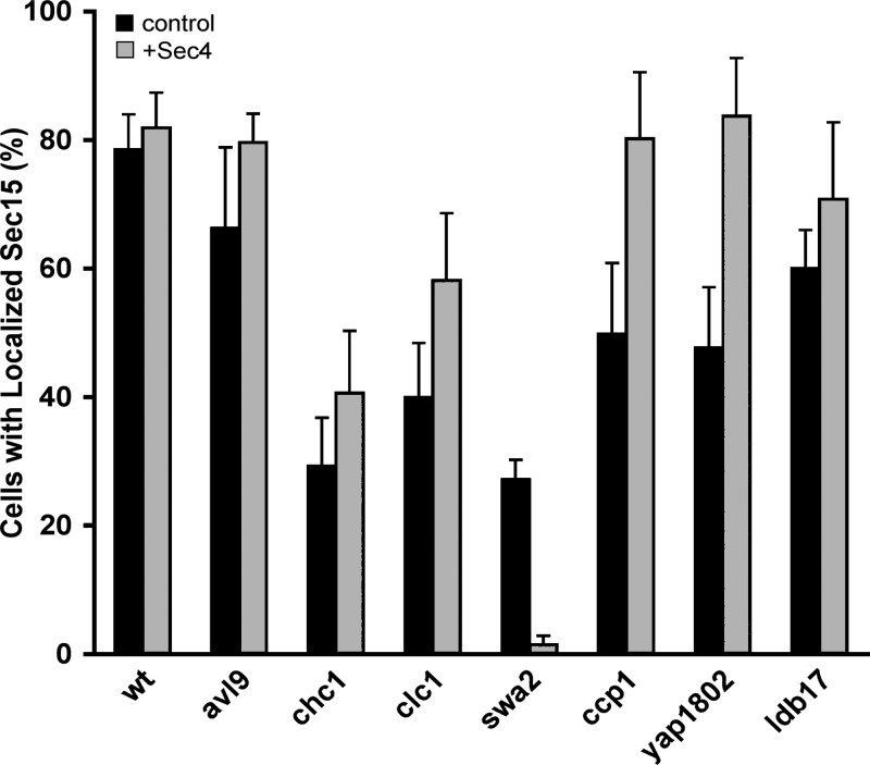
A screen for mutations that affect the recruitment of the exocyst to secretory vesicles identified genes encoding clathrin and proteins that associate or colocalize with clathrin at sites of endocytosis. However, no significant colocalization of the exocyst with clathrin was seen, arguing against a direct role in exocyst recruitment. Rather, these components are needed to recycle the exocytic vesicle SNAREs Snc1p and Snc2p from the plasma membrane into new secretory vesicles where they act to recruit the exocyst. We observe a direct interaction between the exocyst subunit Sec6p and the latter half of the SNARE motif of Snc2p. An snc2 mutation that specifically disrupts this interaction led to exocyst mislocalization and a block in exocytosis in vivo without affecting liposome fusion in vitro. Overexpression of Sec4p partially suppressed the exocyst localization defects of mutations in clathrin and clathrin-associated components. We propose that the exocyst is recruited to secretory vesicles by the combinatorial signals of Sec4-GTP and the Snc proteins. This could help to confer both specificity and directionality to vesicular traffic.


Figures

Figure 1.
Figure 1.
Visual screening of Sec15p-GFP localization in deletion strains. Strains were selected from the yeast deletion library and transformed with a GAL-SEC15-GFP construct. After 5 h of galactose induction, cells were collected and imaged by fluorescence microscopy. (A) Sec15p-GFP was concentrated at exocytic sites including bud tips and bud necks in WT cells (a), yet appeared diffuse in chc1Δ cells (b). The corresponding DIC images are shown in c and d. The appearance of Sec15p-GFP patches was quantified by scoring ∼200 cells for each strain (B). Values indicate the percentage of cells showing GFP-labeled patches at the bud tip or bud neck. Bar, 10 µm.
Figure 2.
Figure 2.
Localization of Sec15-3xGFP in selected strains. Sec15p-3xGFP was expressed at normal levels from the endogenous SEC15 promoter in the seven strains selected from the screen and a yap1801 yap1802 double mutant. The localization of Sec15p-3xGFP in wild type (wt) or cells carrying the indicated deletions is shown (A). Bar, 10 µm. The localization of Sec15p-3xGFP was quantified (B). Data represent mean ± SD from at least three independent experiments.
Figure 3.
Figure 3.
Localization of Chc1p and exocyst subunits. Cells coexpressing Chc1p-RFP and (A) Sec5p-3xGFP (NY2983) or (B) Sec15p-3xGFP (NY2984) were grown in selection medium to OD600 0.5 and imaged. Bar, 10 µm.
Figure 4.
Figure 4.
Snc depletion or blocking its internalization leads to exocyst mislocalization. (A and B) WT (NY2977, NY2978, and NY2979) and snc1Δ snc2Δ Gal-SNC1 cells (NY2980, NY2981, and NY2982) expressing Sec6p-2xmCherry, Sec8p-2xmCherry, or Sec15p-3xGFP, respectively, were grown in selection medium containing 4% galactose overnight at 25°C and then switched to selection medium containing 2% glucose at 30°C to block Snc1p expression. The localization of Sec6-2xmCherry, Sec8-2xmCherry, and Sec15-3xGFP was examined (A) and quantified (B) after 4 h and 8 h in glucose medium. Values represent the percentage of cells showing a fluorescent patch at the bud tip or bud neck. At least 100 cells were scored for each condition. Data represent mean ± SD from at least three independent experiments. Bar, 5 µm. (C and D) snc1Δ SNC2 (NY2986, NY2987) and snc1Δ snc2-V39A, M42A (NY2991, NY2992) mutant cells expressing Sec6-GFP (C) or Sec15-3xGFP (D) were grown to early log phase in SD medium at 25°C, and the cells were collected and directly examined by fluorescence microscopy (left). The percentage of cells with fluorescent protein concentrated at prebud site and small bud tip, mother-bud neck, spread over the entire medium bud cortex, or mislocalized (defused over the whole cell cytosol) was quantified (right) for wild type (WT) or the snc1Δ snc2-V39A, M42A double mutant (DM). Error bars represent SD. For three separate experiments, n ≈ 400 cells per count for both wild-type and mutant cells. (C) *, P < 0.005; **, P < 0.01. (D) *, P < 0.001; **, P < 0.02, Student’s t test.
Figure 5.
Figure 5.
The exocyst binds to Snc2p. (A) Lysates were prepared from eight different yeast strains, each expressing, as the sole copy of the gene, a different exocyst subunit fused at the C terminus to a 13xmyc tag. The lysates were incubated with recombinant GST or GST fused to Sec22p, Snc2p, or Sso2p. The amount of GST-tagged protein was assessed by SDS-PAGE and Coomassie staining, using BSA as a standard. Based on this analysis, ∼3 µg was added to each binding reaction. Glutathione beads were added and washed and the bound proteins analyzed by immunoblot using anti-myc antibody. (B) The Kd of the Snc2p–Sec6p interaction was determined to be 13 ± 6 µM by a quantitative GST pull-down assay. 0–77 µM GST-Snc2p bound to glutathione beads was incubated with 0.4 µM Sec6p. After pelleting the beads, the unbound fraction of Sec6p was determined by scanning a Coomassie-stained gel of the supernatant (inset). The data shown are from a representative experiment of five independent repetitions. The Kd was calculated using Origin software. (C) Lysates of bacteria expressing GST, GST-Vamp4p, and GST-Vamp3p (0.2 µM final concentration) were mixed with 1 ml lysates of HeLa cells (transfected with plasmid pNB1462) expressing myc-Exoc3 from a 100-mm dish culture, and incubated at 4°C for 2 h. Glutathione beads (10 µl) were added and incubated for another hour. The beads were pelleted and washed three times with binding buffer before SDS-PAGE and Western blot analysis with anti-myc antibody. The black line in the top panel indicates where intervening lanes have been removed for presentation purposes.
Figure 6.
Figure 6.
The N terminus of Sec6p binds to the end of the Snc2p SNARE domain. (A) Full-length or truncated regions of Sec6p were expressed in bacteria as 6xHis-tagged constructs. The 1–411 and 1–292 fragments (indicated by asterisks) of Sec6p were coexpressed with a fragment of Sec8p (1–236) that binds to them and helps to maintain their solubility. The purified Sec6p fragments or Sec6p–Sec8p complexes (50 nM final concentration) were mixed with Snc2p-GST or Sec22p-GST (50 nM final concentration) in a 1-ml binding assay. The bound proteins were recovered on glutathione beads and analyzed by immunoblot analysis. The region from 1–411 of Sec6p is sufficient for Snc2p binding, whereas both the regions from 1–100 and 292–411 are required. (B) Fragments of Snc2p fused to GST were expressed in bacteria, purified, and mixed at 80 nM (final concentration) with 50 nM (final concentration) His-tagged Sec6p in a 1-ml binding assay. After precipitation of the GST-tagged constructs with glutathione beads (shown in the bottom panel by Coomassie staining of a gel in which intervening lanes have been removed for presentation purposes), the recovery of Sec6p was determined by immunoblot. The region from 53–92 of Snc2p is both necessary and sufficient for Sec6p binding, whereas the N-terminal 1–52 fragment shows no detectable binding. Relevant lanes from the corresponding Coomassie gel are shown in the bottom panel.
Figure 7.
Figure 7.
A positively charged patch at the end of the Snc2p SNARE domain is needed for Sec6p binding, but not liposome fusion. (A) The structure of the Snc2p–Ssop–Sec9p SNARE complex is shown with the Snc2p backbone highlighted in yellow and the three charged patches on the surface of Snc2p expanded in insets with side chains shown. Images were generated by PyMol using coordinates of 3B5N from the PDB provided by Strop et al. (2008). Snc1p in the original structure was replaced with Snc2p. (B) Binding of Sec6p to Snc2p mutants. His-tagged Sec6p (80 nM final concentration) was mixed with 50 nM (final concentration) of the various Snc2p mutants or with GST-Sec22p as a control. Both M1 (R75A R79A K82A) and M2 (R75E R79E K82E) show a loss of Sec6p binding activity, whereas M3 (E60A D61A D64A), M4 (E60K D61R D64R), M5 (E51A E54A), and M6 (E51R E54R) bind normally. (C) Kinetic fusion assay comparing different donor v-SNARE liposomes containing either wild-type Snc2p or Snc2p-M2 mutant. t-SNARE liposomes containing GST-Sec9c and His6-Sso1p (45 µl) were mixed with 5 µl v-SNARE liposomes and NBD fluorescence was monitored in a fluorescent plate reader for 2 h. Fusion with t-SNAREs (∼460 pmol of t-SNARE complex proteins, ∼55.8 nmol lipid) and wild-type Snc2p (high, ∼136 pmol protein, 4.5 nmol lipid) are shown in black circles, whereas fusion with Snc2p-M2 (high, ∼192 pmol protein, 4.6 nmol lipid) are shown in dark blue. Similarly, t-SNARE liposomes with less total protein (∼200 pmol of t-SNARE complex proteins, ∼28.3 nmol lipid) were fused with wild-type Snc2 liposomes (low, 21 pmol protein, 2.2 nmol lipid, gray circles) or Snc2p-M2 liposomes (low, ∼15 pmol protein, 1.3 nmol lipid, light blue circles). Representative traces are shown. (D) Average endpoint fusion. The black and dark blue bar graphs represent fusion with high concentrations of the v-SNARE protein (wild-type and mutant) in the liposomes, whereas gray and light blue bar graphs represent fusion with low concentrations of the v-SNARE proteins (wild type and mutant) in the donor liposomes. The histograms show mean fusion at 120 min and the error bars represent SEM. The number of replicates (n) is shown at the base of the histogram. (E) Coomassie blue–stained gel of proteoliposomes on a 10% Bis-Tris NuPAGE Novex gel (Invitrogen). Lanes 1–4 contain 5 µl of proteoliposomes, whereas lanes 5 and 6 (low concentration v-SNAREs) contain 10 µl of proteoliposomes. (F) Sec6p binds to Snc2p reconstituted into liposomes. Empty liposome (0.01 mM lipid) or liposomes containing 0.4 µM Snc2p or Snc2p-M2 were incubated with 0.025 µM His-Sec6 (final concentration) at 4°C for 2 h in 1 ml binding buffer (25 mM Hepes-KOH and 200 mM KCl, pH 7.4, with 0.5 µg/µl BSA). Liposomes were pelleted at 300,000 g for 20 min in an ultracentrifuge (Optima TLX; Beckman Coulter) with a TLA 120.2 rotor. The pellet was washed with 1 ml binding buffer and re-pelleted. The pellet was resuspended in 80 µl sample buffer. 10 µl of each sample was resolved by SDS-PAGE and analyzed by Western blot using anti-His (top) and anti-Snc (bottom) antibody. (G) Quantitation of Sec6p binding.
Figure 8.
Figure 8.
The snc2-M2 mutation inhibits growth, localization, and secretion. (A) Two independent snc1Δ SNC2 (WT, NY2985) and snc1Δ snc2-M2 (M2, NY2988) yeast strains were serially diluted by fivefold increments, plated and incubated as indicated. The snc2-M2 mutant shows growth defects at both high and low temperatures. (B) snc1Δ SNC2 (WT, NY2986) and snc1Δ snc2-M2 (M2, NY2989) yeast expressing Sec6p-GFP were grown to early log phase in SD medium at 25°C. The cells were collected and directly examined by fluorescence microscopy (left panels). The percentage of cells with Sec6p-GFP at prebud sites and small bud tips, mother-bud necks, spread over the entire medium bud cortex, or mislocalized (defused over the whole cell cytosol) was quantified (right). Error bars represent SD for two independent experiments, n = 709 for wild type and n = 792 for the snc2-M2 mutant. *, <0.02, Student’s t test. (C) snc1Δ SNC2 (WT, NY2987) and snc1Δ snc2-M2 (M2, NY2990) yeast expressing Sec15p-3xGFP were grown to early log phase in SD medium at 25°C. The cells were collected and directly examined by fluorescence microscopy (left panels). The percentage of cells with Sec15p-3xGFP at prebud sites and small bud tips, mother-bud necks, spread over the entire bud cortex, or mislocalized (defused over the whole cell cytosol) was quantified (right). Error bars represent SD for two independent experiments, n = 709 for wild type and n = 792 for the snc2-M2 mutant. **, <0.005, Student’s t test. (D) Expression of Snc2-M2p is similar to that of Snc2p. (E) Invertase assay. snc1Δ SNC2 (WT, 2985), snc1Δ snc2-M2 (M2, NY2988), and sec6-4 (NY17) yeast were grown overnight at 25°C in YP medium containing 5% glucose then transferred to YP medium containing 0.1% glucose and incubated at 37°C for 1 h. The bar graph displays the percentage of invertase synthesized during the 1-h incubation that was delivered to the cell surface. (F) snc1Δ SNC2 (NY2985, right) and snc1Δ snc2-M2 (NY2988, left) cells were grown at 25°C in YPD medium and processed for thin section electron microscopy.
Figure 9.
Figure 9.
Sec9p does not interfere with the Snc2p–Sec6p interaction. GST-Snc2p (2.5 µM final concentration) was incubated with 70 nM (final concentration) His-Sec6p and increasing amounts of His-MBP-Sec9c, as indicated, and then precipitated with glutathione beads. The bead-associated protein was analyzed by SDS PAGE and Coomassie staining. Black lines indicate where lanes have been rearranged for presentation purposes.
Figure 10.
Figure 10.
Overexpression of Sec4p partially restores Sec15p localization. WT or the indicated deletion mutant strains expressing Sec15-3xGFP were transformed with a 2µ circle-based plasmid overexpressing Sec4p (pNB1457) and examined by fluorescence microscopy. The percentage of cells exhibiting a Sec15p-3xGFP patch at the bud tip or mother-bud neck is shown. Data represent mean ± SD from at least three different experiments.

Similar articles

Cited by

References

    1. Boyd C., Hughes T., Pypaert M., Novick P. 2004. Vesicles carry most exocyst subunits to exocytic sites marked by the remaining two subunits, Sec3p and Exo70p. J. Cell Biol. 167:889–901 10.1083/jcb.200408124 - DOI - PMC - PubMed
    1. Burston H.E., Maldonado-Báez L., Davey M., Montpetit B., Schluter C., Wendland B., Conibear E. 2009. Regulators of yeast endocytosis identified by systematic quantitative analysis. J. Cell Biol. 185:1097–1110 10.1083/jcb.200811116 - DOI - PMC - PubMed
    1. Cai H., Zhang Y., Pypaert M., Walker L., Ferro-Novick S. 2005. Mutants in trs120 disrupt traffic from the early endosome to the late Golgi. J. Cell Biol. 171:823–833 10.1083/jcb.200505145 - DOI - PMC - PubMed
    1. Carr C.M., Rizo J. 2010. At the junction of SNARE and SM protein function. Curr. Opin. Cell Biol. 22:488–495 10.1016/j.ceb.2010.04.006 - DOI - PMC - PubMed
    1. Chen S., Novick P., Ferro-Novick S. 2012. ER network formation requires a balance of the dynamin-like GTPase Sey1p and the Lunapark family member Lnp1p. Nat. Cell Biol. 14:707–716 10.1038/ncb2523 - DOI - PMC - PubMed

Publication types

MeSH terms

Substances