. 2013 Oct;94(4):751-7.
doi: 10.1189/jlb.0113039. Epub 2013 Jul 29.

Tpl2 kinase regulates FcγR signaling and immune thrombocytopenia in mice

Affiliations

Tpl2 kinase regulates FcγR signaling and immune thrombocytopenia in mice

Irene Kyrmizi et al. J Leukoc Biol. 2013 Oct.

Abstract

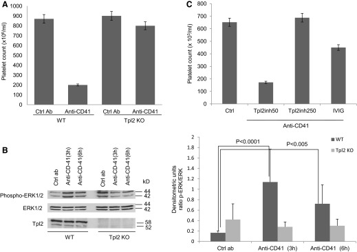
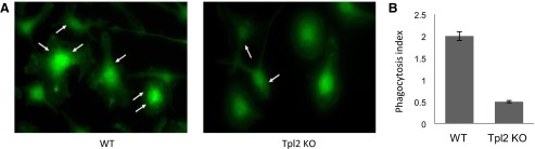
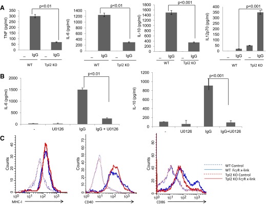
The MAPK3 Tpl2 controls innate and adaptive immunity by regulating TLR, TNF-α, and GPCR signaling in a variety of cell types. Its ablation gives rise to an anti-inflammatory phenotype characterized by resistance to LPS-induced endotoxin shock, DSS-induced colitis, and TNF-α-induced IBD. Here, we address the role of Tpl2 in autoimmunity. Our data show that the ablation and the pharmacological inhibition of Tpl2 protect mice from antiplatelet antibody-induced thrombocytopenia, a model of ITP. Thrombocytopenia in this model and in ITP is caused by phagocytosis of platelets opsonized with antiplatelet antibodies and depends on FcγR activation in splenic and hepatic myeloid cells. Further studies explained how Tpl2 inhibition protects from antibody-induced thrombocytopenia, by showing that Tpl2 is activated by FcγR signals in macrophages and that its activation by these signals is required for ERK activation, cytoplasmic Ca(2+) influx, the induction of cytokine and coreceptor gene expression, and phagocytosis.

Keywords: MAPK; immune complex; macrophages.


Figures

Figure 1.
Figure 1.. Tpl2 inhibition protects mice from antiplatelet, antibody-induced thrombocytopenia.
(A) WT and Tpl2 KO mice were injected i.p. with an anti-CD41 IgG1 antibody or the isotype control IgG1 (2 μg/mouse). Platelet counts were evaluated 24 h later. Cumulative results from three independent experiments (total number of mice/group, n=15; bars are sem) are shown. Ctrl Ab, Control antibody. (B) ERK1/2 phosphorylation in spleen protein extracts from mice injected with the anti-CD41 antibody or the isotype control IgG1. Western blots of the cell lysates were probed with antibodies against the phosphorylated or total ERK1, ERK2. To confirm the ablation of Tpl2 in Tpl2 KO mice, the same lysates were also probed with the anti-Tpl2 antibody. (Right) Densitometry analysis of the bands at left [ratio of phosphorylated (p)-ERK1/2:total ERK1/2]. Data are representative of three experiments (error bars, sd of five mice/group). (C) WT mice were injected i.p. with escalating doses of the Tpl2 inhibitor (inh; 2 mg/kg and 10 mg/kg). Each mouse received one injection/day for 2 sequential days. Alternatively, the mice were injected with IVIg (2 g/kg). Twenty-four hours after the pretreatment with the Tpl2 inhibitor or IVIg, the mice were injected i.p. with the anti-CD41 antibody or with the isotype antibody control. Platelet counts were determined 24 h later. The results shown are cumulative results of three independent experiments (total number of mice/group; n=15; bars are sem).
Figure 2.
Figure 2.. Tpl2 is activated by FcγR signals and is required for FcγR-induced MEK/ERK1/2 MAPK phosphorylation and cytoplasmic Ca2+ influx.
(A) FcγR cross-linking activates Tpl2, which was immunoprecipitated (IP) from WT BMDMs after FcγR cross-linking (x-link), and the immunoprecipitates were used in an in vitro kinase reaction, using a GST-MEK peptide as the substrate. Phosphorylation (p) of the GST-MEK peptide was determined by probing the products of the in vitro kinase reaction with an antiphospho-MEK and an anti-MEK antibody. (B) Phosphorylation of MEK, ERK1 and ERK2, p38MAPK, and JNK upon FcγR cross-linking in WT and Tpl2 KO BMDMs. Phosphorylation was monitored by probing Western blots of cell lysates harvested 30 min after the start of the stimulation with IgG1. (C) Tpl2 is not required for ERK phosphorylation in cells stimulated with MCSF, GM-CSF, or PMA. Phosphorylation was monitored by probing Western blots of cell lysates harvested 30 min after the start of the stimulation with the indicated cytokines. (D) WT and Tpl2 KO BMDMs were loaded with the fluorescent Ca2+ indicator, Fluo-4 NW. Following incubation with mouse IgG for 30 min, FcγRs were cross-linked with a goat anti-mouse IgG (Fab-specific). Cytoplasmic Ca2+ influx was monitored fluorimetrically (excitation at 494 nm and emission at 516 nm). Results are expressed as mean fluorescence ± se of the mean, and they are calculated by combining the results of two independent experiments. Each experiment included triplicate measurements for each time-point (n=6). (E) Degradation of IκBα upon FcγR cross-linking in WT and Tpl2 KO BMDMs. Molecular mass values represent the estimated molecular mass of the proteins. Each panel is representative of three to six independent experiments, all of which gave similar results.
Figure 3.
Figure 3.. Tpl2 regulates FcγR-induced phagocytosis.
(A) WT and Tpl2 KO macrophages were seeded on glass coverslips, and they were incubated with IgG-opsonized polystyrene latex beads for 1.5 h. Cells were fixed, and actin was visualized with phalloidin-FITC. Representative pictures of two independent experiments. Original magnification scale, ×40. Arrows indicate phagocytosed opsonized latex beads. (B) The phagocytosis index was calculated as the mean number of engulfed IgG-opsonized latex beads/macrophage.
Figure 4.
Figure 4.. Tpl2 regulates FcγR-induced cytokine production and costimulatory molecule expression.
(A) WT and Tpl2 KO BMDMs were stimulated with plate-bound IgG (50 μg/ml). Cytokine levels were determined by ELISA in culture supernatants from cells stimulated for 3 h for TNF and for 24 h for the other cytokines. (B) Before stimulation with plate-bound IgG (50 μg/ml), WT BMDMs were incubated for 30 min with 2.5 μΜ U0126 or vehicle control (DMSO). Cytokine levels were determined by ELISA in culture supernatants form cells stimulated for 24 h. Results are representative of triplicate cultures from at least three independent experiments. (C) BMDMs isolated from WT and Tpl2 KO mice were stimulated with plate-bound IgG (50 μg/ml). Up-regulation of MHC-I, CD40, and CD86 was monitored by flow cytometry.

Similar articles

Cited by

References

    1. Nimmerjahn F., Ravetch J. V. (2008) Fcγ receptors as regulators of immune responses. Nat. Rev. Immunol. 8, 34–47 - PubMed
    1. Salmon J. E., Millard S., Schachter L. A., Arnett F. C., Ginzler E. M., Gourley M. F., Ramsey-Goldman R., Peterson M. G., Kimberly R. P. (1996) Fc γ RIIA alleles are heritable risk factors for lupus nephritis in African Americans. J. Clin. Invest. 97, 1348–1354 - PMC - PubMed
    1. Salmon J. E., Pricop L. (2001) Human receptors for immunoglobulin G: key elements in the pathogenesis of rheumatic disease. Arthritis Rheum. 44, 739–750 - PubMed
    1. Clynes R., Dumitru C., Ravetch J. V. (1998) Uncoupling of immune complex formation and kidney damage in autoimmune glomerulonephritis. Science 279, 1052–1054 - PubMed
    1. Ji H., Ohmura K., Mahmood U., Lee D. M., Hofhuis F. M., Boackle S. A., Takahashi K., Holers V. M., Walport M., Gerard C., Ezekowitz A., Carroll M. C., Brenner M., Weissleder R., Verbeek J. S., Duchatelle V., Degott C., Benoist C., Mathis D. (2002) Arthritis critically dependent on innate immune system players. Immunity 16, 7–168 - PubMed

Publication types

MeSH terms