. 2013 Nov;94(5):953-62.
doi: 10.1189/jlb.0313174. Epub 2013 Jul 29.

Macrophage migration arrest due to a winning balance of Rac2/Sp1 repression over β-catenin-induced PLD expression

Affiliations

Macrophage migration arrest due to a winning balance of Rac2/Sp1 repression over β-catenin-induced PLD expression

Francis J Speranza et al. J Leukoc Biol. 2013 Nov.

Abstract

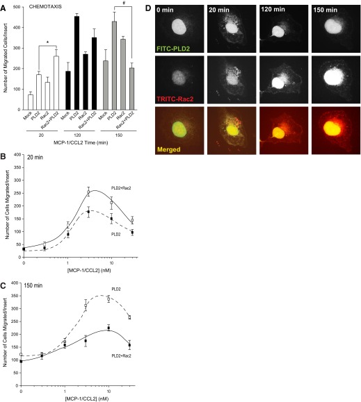
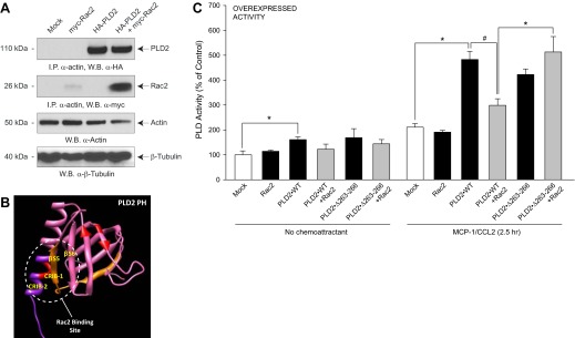
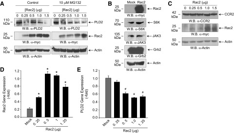
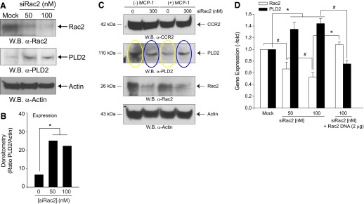
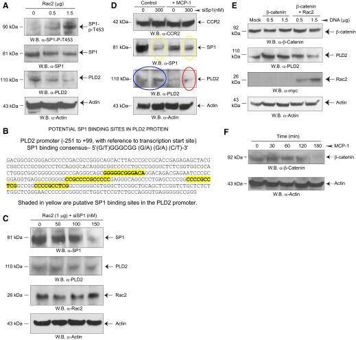
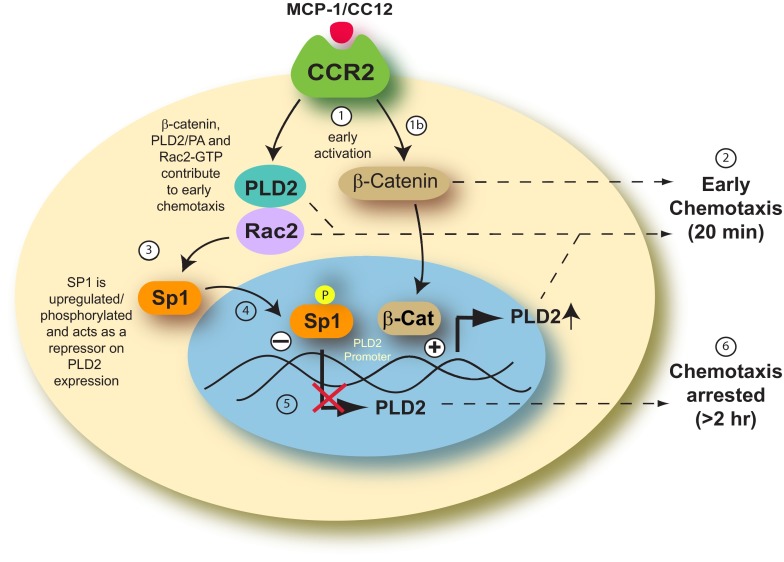
Monocytes and neutrophils infiltrate into tissues during inflammation and stay for extended periods of time until the initial insult is resolved or sometimes remain even longer in the case of chronic inflammation. The mechanism as to why phagocytes become immobilized after the initial cell migration event is not understood completely. Here, we show that overexpression or hyperactivation of Rac2 decreases sustained chemotactic responses of macrophages to MCP-1/CCL2. The resulting leukocyte arrest is not caused by a diminished availability of the cytokine receptor CCR2 that remains intact during MCP-1 stimulation. We show a novel mechanism that links the Rac2-dependent arrest of chemotaxis to decreased expression of PLD2 through the transcription regulator Sp1. Prolonged Rac2 activity leads to nuclear overactivation of Sp1, which acts as a repressor for PLD2. Also, another signaling component plays a regulatory role: β-catenin. Although early times of stimulation (≈ 20 min) with MCP-1/CCL2 resulted in activation of β-catenin with a positive effect on PLD2, after ≈ 3 h of stimulation, the levels of β-catenin were reduced and not able to prevent the negative effect of Rac2 on PLD2 activity. This is a novel molecular mechanism underlying immobilization of monocyte/macrophage migration that is important for the physiological maintenance of leukocytes at the site of inflammation. If this immobilization is prolonged enough, it could lead to chronic inflammation.

Keywords: cell migration; chemotaxis; gene expression regulation; granulocyte; monocyte signaling cascade.


Figures

Figure 1.
Figure 1.. Rac2 reduces PLD lipase activity at late times of cell migration.
(A) Migration assay of RAW264.7/LR5 cells as a function of time. Cells were transfected with PLD2-WT, Rac2-WT, or PLD2-WT + Rac2-WT, respectively. The number of cells that migrated was measured in the presence of 3 nM MCP-1. The * symbol is higher; # is lower statistically significant (P < 0.05) differences than controls at zero time by ANOVA. (B and C) Migration assay of RAW264.7/LR5 cells as a function of MCP-1 concentration. Cells were transfected with 2 μg PLD2-WT + 0.5 μg Rac2-WT. The number of cells that migrated was measured at 20 (early time; B) and 150 (later time; C) min toward MCP-1. (D) PLD2 and Rac2 localize in the nucleus, as well as in the cytoplasm, but only at short times after cell stimulation. When ready for immunofluorescence microscopy, cells were treated with 3 nM MCP-1 that was added to a localized region on the coverslip with a micropipette, which forms a temporary chemoattractant gradient.
Figure 2.
Figure 2.. A Rac2:PLD2-binding interaction negatively affects PLD activity.
(A) Rac2 and PLD2 form a protein:protein complex with actin. RAW264.7/LR5 cells were transfected with myc-Rac2 or HA-PLD2 alone or in combination, and samples were taken for immunoprecipitation (I.P.) and Western blots (W.B.). (B) Schematic of 3D modeling of the PH domain, a β-sheet composed of four anti-parallel β-strands and one single α-helix, which comprises CRIB domains. This structure was predicted using I-TASSER and viewed using the structure viewer “Chimera” http://www.cgl.ucsf.edu/chimera/download.html. (C) Effect of overexpressed Rac2 on PLD2 lipase activity assay in the presence of MCP-1 and lysates was prepared and then used for the PLD assay. Error bars are sem. Symbols */# denote statistically significant (p < 0.05) differences between means by ANOVA, higher (*) or lower (#) than the indicated controls.
Figure 3.
Figure 3.. Effect of Rac2 overexpression on PLD2.
(A) RAW264.7/LR5 cells were transfected with increasing amounts of Rac2 DNA. Twenty-four hours post-transfection, a proteasome inhibitor (MG132) was added to separate sets of cells, and 48 h post-transfection lysates were prepared, and Western blots were performed. (B) Effect of Rac2 overexpression on several signaling proteins. Protein loading control using anti-actin staining (bottom blot) as well as the effective expression of Rac2 (top blot) are included. (C) Effect of Rac2 overexpression on MCP-1/CCL2 receptor, CCR2. Protein loading control using anti-actin staining is included. (D and E) qRT-PCR gene expression of overexpressing Rac2 (D) or endogenous PLD2 (E) in RAW macrophages. Error bars are sem. Symbols */# denote statistically significant (p < 0.05) differences between means by ANOVA, higher (*) or lower (#) than Mock controls.
Figure 4.
Figure 4.. Effect of silencing Rac2 on PLD2 protein and gene expression.
(A) RAW264.7/LR5 cells were silenced with increasing Rac2 siRNA for 3 days, and lysates containing proteins were obtained. Western blots were probed with anti-Rac2 or anti-PLD2 antibodies. (B) Quantification of Western blot results of the ratio of endogenous PLD2 relative to silenced Rac2 expression. Results in this figure are the means ± sem from at least three independent experiments conducted in duplicate. (C) Sustained MCP-1 stimulation decreases endogenous PLD2 protein levels via a mechanism that is rescued by Rac2 siRNA. RAW264.7/LR5 cells were mock-silenced or silenced with increasing Rac2 siRNA for 3 days and simultaneously mock-stimulated or stimulated with 5 nM MCP-1. (D) qRT-PCR RNA expression of endogenous PLD2 and Rac2 in the presence of increasing siRNA, specific for Rac2 and probed with FAM-tagged PLD2 or Rac2 primers. Error bars are sem. Symbols */# denote statistically significant (p < 0.05) differences between means by ANOVA, higher (*) or lower (#) than the indicated controls.
Figure 5.
Figure 5.. Effect of Rac2 on Sp1 phosphorylation.
(A) Effect of Rac2 overexpression on Sp1 activation/phosphorylation. RAW264.7/LR5 cells were transfected with increasing Rac2 DNA, and resulting Western blots were performed that were stained with the indicated antibodies. (B) Schematic for potential Sp1 binding to PLD2 promoter. Shaded in yellow are putative Sp1-binding sites in the PLD2 promoter. (C) Effect of silencing Sp1 on PLD2 expression. (D) Effect of prolonged MCP-1 stimulation on Sp1-mediated rescue of PLD2 protein expression. (E) Overexpression of β-catenin reverses the negative effect of Rac2 on PLD2 expression. (F) β-Catenin is activated at early times of incubation with MCP-1/CCL2 but not at later (>1 h) times. RAW264.7/LR5 cells were stimulated with 5 nM MCP-1.
Figure 6.
Figure 6.. Model of proposed mechanism for monocyte arresting.
MCP-1/CC12 binds to the CCR2 receptor and stimulates the cell (1). Rac2 alone and Rac2 complexed with PLD2 activate chemotaxis at early times of inflammation (2). At longer times after stimulation, the following mechanism takes place: Rac2 influences Sp1 (3) in a yet-unknown way that makes it translocate from the cytoplasm to the nucleus (4), where it becomes phosphorylated (P in yellow circle) and down-regulates PLD2 expression (5), thus mediating the negative effect of Rac2 on PLD expression. This down-regulation leads to suppressed chemotaxis at later times of cell migration (6). β-Catenin is a regulator of this mechanism, in that initially, and as activated after MCP-1 stimulation, it is a positive effector of early chemotaxis (1b) and can also be a positive PLD2 gene enhancer. However, at later times, as Rac2 and Sp1 accumulate, they overcome the positive action of β-catenin, and the balance is tipped toward leukocyte immobilization (6).

Similar articles

Cited by

References

    1. Koshland D. E. (1982) Special topic: chemotaxis and motility. Annu. Rev. Physiol. 44, 499–500
    1. Ridley A. J., Schwartz M. A., Burridge K., Firtel R. A., Ginsberg M. H., Borisy G., Parsons J. T., Horwitz A. R. (2003) Cell migration: integrating signals from front to back. Science 302, 1704–1709 - PubMed
    1. Vicente-Manzanares M., Webb D. J., Horwitz A. R. (2005) Cell migration at a glance. J. Cell Sci. 118, 4917–4919 - PubMed
    1. Luster A. D., Alon R., von Andrian U. H. (2005) Immune cell migration in inflammation: present and future therapeutic targets. Nat. Immunol. 6, 1182–1190 - PubMed
    1. Capoccia B. J., Gregory A. D., Link D. C. (2008) Recruitment of the inflammatory subset of monocytes to sites of ischemia induces angiogenesis in a monocyte chemoattractant protein-1-dependent fashion. J. Leukoc. Biol. 84, 760–768 - PMC - PubMed

Publication types