

The ascending reticular activating system from pontine reticular formation to the thalamus in the human brain
- PMID: 23898258
- PMCID: PMC3722571
- DOI: 10.3389/fnhum.2013.00416
The ascending reticular activating system from pontine reticular formation to the thalamus in the human brain
Abstract
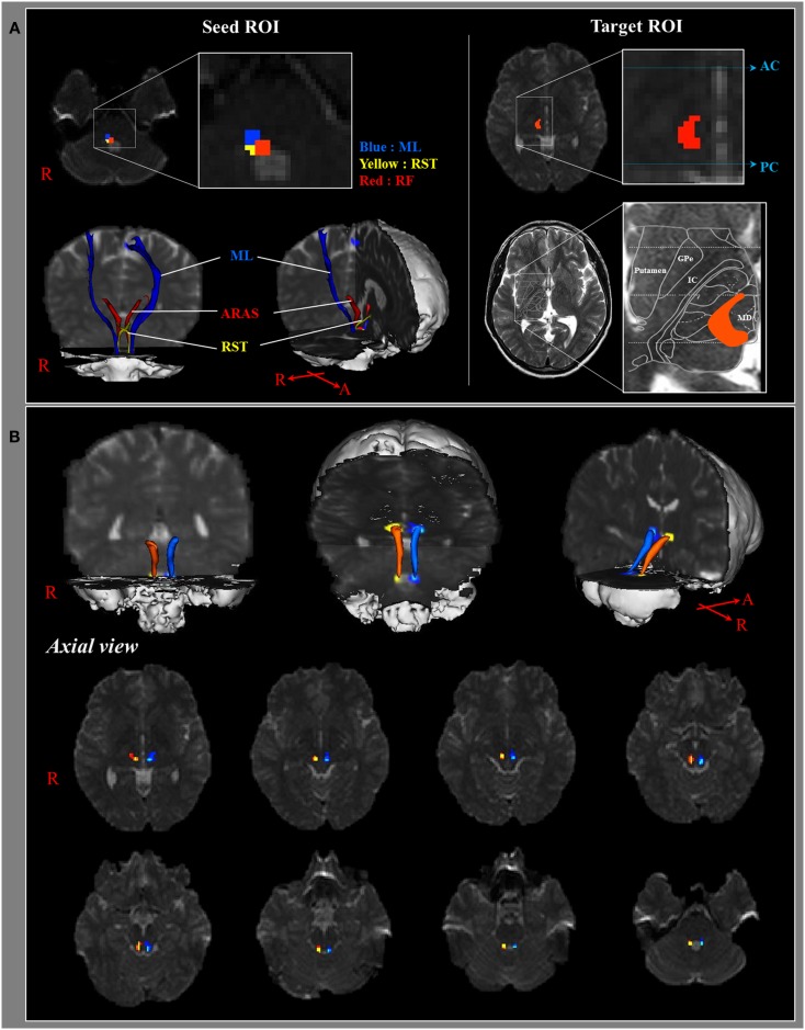
Introduction: Action of the ascending reticular activating system (ARAS) on the cerebral cortex is responsible for achievement of consciousness. In this study, we attempted to reconstruct the lower single component of the ARAS from the reticular formation (RF) to the thalamus in the normal human brain using diffusion tensor imaging (DTI).
Methods: Twenty six normal healthy subjects were recruited for this study. A 1.5-T scanner was used for scanning of diffusion tensor images, and the lower single component of the ARAS was reconstructed using FMRIB software. We utilized two ROIs for reconstruction of the lower single component of the ARAS: the seed ROI - the RF of the pons at the level of the trigeminal nerve entry zone, the target ROI - the intralaminar nuclei of the thalamus at the level of the commissural plane.
Results: The reconstructed ARAS originated from the pontine RF, ascended through the mesencephalic tegmentum just posterior to the red nucleus, and then terminated on the intralaminar nuclei of the thalamus. No significant differences in fractional anisotropy, mean diffusivity, and tract number were observed between hemispheres (p > 0.05).
Conclusion: We reconstructed the lower single component of the ARAS from the RF to the thalamus in the human brain using DTI. The results of this study might be of value for the diagnosis and prognosis of patients with impaired consciousness.
Keywords: ascending reticular activating system; consciousness; diffusion tensor imaging; reticular formation.
Figures
References
-
- Afifi A. K., Bergman R. A. (2005). Functional Neuroanatomy: Text and Atlas. New York: Lange Medical Books/McGraw-Hill
LinkOut - more resources
Full Text Sources
Other Literature Sources