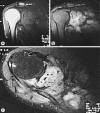
Primary alveolar soft part sarcoma of the scapula
- PMID: 23898281
- PMCID: PMC3724132
- DOI: 10.1159/000353927
Primary alveolar soft part sarcoma of the scapula
Abstract
Alveolar soft part sarcoma (ASPS) is an unusual soft tissue malignity, occurring in less than 1% of sarcomas and typically found in the head and neck tissues in children or, in adults, in the deep soft tissues of the lower extremities. In this report, we present a 33-year-old male with primary ASPS in the right scapular bone and discuss the radiologic features of this tumor in the context of the current literature.
Keywords: Alveolar soft part sarcoma; Lung metastasis; Scapula.
Figures


Similar articles
-
Alveolar soft part sarcoma metastatic to the mandible: A report and review of literature.J Stomatol Oral Maxillofac Surg. 2017 Dec;118(6):379-382. doi: 10.1016/j.jormas.2017.07.004. Epub 2017 Jul 31. J Stomatol Oral Maxillofac Surg. 2017. PMID: 28774857
-
Primary intraosseous alveolar soft part sarcoma: Report of two cases with radiologic-pathologic correlation.Ann Diagn Pathol. 2023 Feb;62:152078. doi: 10.1016/j.anndiagpath.2022.152078. Epub 2022 Dec 13. Ann Diagn Pathol. 2023. PMID: 36543620
-
Alveolar soft part sarcoma of the tongue: a case report and review of the literature.Int J Clin Exp Pathol. 2020 May 1;13(5):1275-1282. eCollection 2020. Int J Clin Exp Pathol. 2020. PMID: 32509104 Free PMC article.
-
Alveolar soft part sarcoma of the uterine cervix.Mod Pathol. 1989 Nov;2(6):676-80. Mod Pathol. 1989. PMID: 2479947 Review.
-
Primary Intracranial Alveolar Soft-Part Sarcoma: Report of Two Cases and a Review of the Literature.World Neurosurg. 2016 Jun;90:699.e1-699.e6. doi: 10.1016/j.wneu.2016.02.005. Epub 2016 Feb 6. World Neurosurg. 2016. PMID: 26862023 Review.
Cited by
-
Primary alveolar soft part sarcoma of the right femur and primary lymphoma of the left femur: A case report and literature review.Oncol Lett. 2016 Jan;11(1):89-94. doi: 10.3892/ol.2015.3906. Epub 2015 Nov 10. Oncol Lett. 2016. PMID: 26870173 Free PMC article.
-
Alveolar soft-part sarcoma in the left forearm with cardiac metastasis: A case report and literature review.Oncol Lett. 2016 Jan;11(1):81-84. doi: 10.3892/ol.2015.3889. Epub 2015 Nov 6. Oncol Lett. 2016. PMID: 26870171 Free PMC article.
-
Brain metastatic alveolar soft-part sarcoma: Clinicopathological profiles, management and outcomes.Oncol Lett. 2017 Nov;14(5):5779-5784. doi: 10.3892/ol.2017.6941. Epub 2017 Sep 14. Oncol Lett. 2017. PMID: 29113207 Free PMC article.
-
Alveolar Soft Part Sarcoma with Unusual Cardiac Metastasis: A Case Report and Review of the Literature.Case Rep Cardiol. 2017;2017:7248727. doi: 10.1155/2017/7248727. Epub 2017 Aug 6. Case Rep Cardiol. 2017. PMID: 28845314 Free PMC article.
-
Adult alveolar soft part sarcoma of the head and neck: a report of two cases and literature review.Case Rep Oncol Med. 2014;2014:597291. doi: 10.1155/2014/597291. Epub 2014 Dec 23. Case Rep Oncol Med. 2014. PMID: 25587475 Free PMC article.
References
-
- Smetana HF, Scott WF., Jr Malignant tumors of nonchromaffin paraganglia. Mil Surg. 1951;109:330–349. - PubMed
-
- Christopherson WM, Foote FW, Stewart FW. Alveolar soft part sarcomas: structurally characteristic tumors of uncertain histogenesis. Cancer. 1952;5:100–111. - PubMed
-
- Lieberman PH, Foote FW, Jr, Stewart FW, Berg JW. Alveolar soft-part sarcoma. JAMA. 1966;198:1047–1051. - PubMed
-
- Salvati M, Cervoni L, Caruso R, Gagliardi FM, Delfini R. Sarcoma metastatic to the brain: a series of 15 cases. Surg Neurol. 1998;49:441–444. - PubMed
Publication types
LinkOut - more resources
Full Text Sources
Other Literature Sources

