

Effect of dichloromethylene diphosphonate on liver regeneration following thioacetamide-induced necrosis in rats
- PMID: 23898371
- PMCID: PMC3724966
- DOI: 10.4254/wjh.v5.i7.379
Effect of dichloromethylene diphosphonate on liver regeneration following thioacetamide-induced necrosis in rats
Abstract
Aim: To study the effect of dichloromethylene diphosphonate (DMDP), a selective Kupffer cell toxicant in reference to liver damage and postnecrotic liver regeneration in rats induced by sublethal dose thioacetamide (TA).
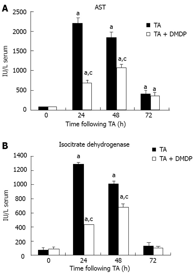
Methods: Rats, intravenously (iv) pre-treated with a single dose of DMDP (10 mg/kg), were intraperitoneally (ip) injected with TA 6.6 mmol/kg (per 500 mg/kg body weight). Hepatocytes were isolated from rats at 0, 24, 48 and 72 h following TA intoxication and blood and liver samples were obtained. To evaluate the mechanisms involved in the postnecrotic regenerative state, DNA distribution and ploidy time course were assayed in isolated hepatocytes. Circulating cytokine tumor necrosis factor-alpha (TNF-α) was assayed in serum and determined by reverse transcriptase-polymerase chain reaction in liver extract.
Results: The effect of DMDP induced noticeable changes in postnecrotic regeneration, causing an increased percentage of hepatocytes in the cell cycle S phase. The increase at 24 h in S1 population in rats pretreated with DMDP + TA was significantly (P < 0.05) different compared with that of the TA group (18.07% vs 8.57%). Hepatocytes increased their proliferation as a result of these changes. Also, TNF-α expression and serum level were increased in rats pre-treated with DMDP. Thus, DMDP pre-treatment reduced TA-induced liver injury and accelerated postnecrotic liver regeneration.
Conclusion: These results demonstrate that Kupffer cells are involved in TA-induced liver, as well as in postnecrotic proliferative liver states.
Keywords: Cell cycle; Dichloromethylene diphosphonate; Hepatotoxicity; Kupffer cells; Thioacetamide.
Figures

References
-
- Schiedner G, Hertel S, Johnston M, Dries V, van Rooijen N, Kochanek S. Selective depletion or blockade of Kupffer cells leads to enhanced and prolonged hepatic transgene expression using high-capacity adenoviral vectors. Mol Ther. 2003;7:35–43. - PubMed
-
- Laskin DL. Nonparenchymal cells and hepatotoxicity. Semin Liver Dis. 1990;10:293–304. - PubMed
-
- Bautista AP, Skrepnik N, Niesman MR, Bagby GJ. Elimination of macrophages by liposome-encapsulated dichloromethylene diphosphonate suppresses the endotoxin-induced priming of Kupffer cells. J Leukoc Biol. 1994;55:321–327. - PubMed
-
- Ishiyama H, Sato M, Matsumura K, Sento M, Ogino K, Hobara T. Proliferation of hepatocytes and attenuation from carbon tetrachloride hepatotoxicity by gadolinium chloride in rats. Pharmacol Toxicol. 1995;77:293–298. - PubMed
-
- Iimuro Y, Yamamoto M, Kohno H, Itakura J, Fujii H, Matsumoto Y. Blockade of liver macrophages by gadolinium chloride reduces lethality in endotoxemic rats--analysis of mechanisms of lethality in endotoxemia. J Leukoc Biol. 1994;55:723–728. - PubMed
LinkOut - more resources
Full Text Sources
Other Literature Sources
