. 2013 Jul 23:2:e00762.
doi: 10.7554/eLife.00762.

A mammalian pseudogene lncRNA at the interface of inflammation and anti-inflammatory therapeutics

Affiliations

A mammalian pseudogene lncRNA at the interface of inflammation and anti-inflammatory therapeutics

Nicole A Rapicavoli et al. Elife. .

Abstract

Pseudogenes are thought to be inactive gene sequences, but recent evidence of extensive pseudogene transcription raised the question of potential function. Here we discover and characterize the sets of mouse lncRNAs induced by inflammatory signaling via TNFα. TNFα regulates hundreds of lncRNAs, including 54 pseudogene lncRNAs, several of which show exquisitely selective expression in response to specific cytokines and microbial components in a NF-κB-dependent manner. Lethe, a pseudogene lncRNA, is selectively induced by proinflammatory cytokines via NF-κB or glucocorticoid receptor agonist, and functions in negative feedback signaling to NF-κB. Lethe interacts with NF-κB subunit RelA to inhibit RelA DNA binding and target gene activation. Lethe level decreases with organismal age, a physiological state associated with increased NF-κB activity. These findings suggest that expression of pseudogenes lncRNAs are actively regulated and constitute functional regulators of inflammatory signaling. DOI:http://dx.doi.org/10.7554/eLife.00762.001.

Keywords: Mouse; NF-κB; genomics; noncoding RNA.


Conflict of interest statement

The authors declare that no competing interests exist.

Figures
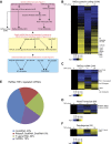
Figure 1.
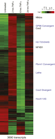
Figure 1.. TNFα regulates the transcription of many coding and noncoding genes.
(A) Workflow for strategy for discovery of NF-κB regulated lncRNAs. (B) 3596 RefSeq protein coding genes are regulated by TNFα. Values are normalized to the 0 hr time point. (C) 244 RefSeq ncRNAs are regulated by TNFα. (D) 64 de novo lncRNAs are regulated by TNFα. (E) The fraction of all RefSeq ncRNAs for each class of transcript. (F) 54 pseudogene lncRNAs are regulated by TNFα. DOI: http://dx.doi.org/10.7554/eLife.00762.003
Figure 1—Figure supplement 1.
Figure 1—Figure supplement 1.. Heatmap of RefSeq genes.
Mean centered heatmap of RefSeq protein coding genes and RefSeq lncRNAs. DOI: http://dx.doi.org/10.7554/eLife.00762.004
Figure 2.
Figure 2.. LncRNAs distinguish between different stimuli and are regulated by NF-κB.
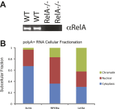
(A) Validation of lncRNAs expression alongside the closest protein coding gene under a variety of different stimuli by qRT-PCR. Genomic organization is shown below. MEFs were treated with 20 ng/ml TNFα for 0 and 6 hr. Quantitative Taqman real time RT-PCR of the indicated RNAs is normalized to Actin levels (mean ± SD). (B) LncRNAs are regulated by RelA. qRT-PCR in WT and RelA−/− littermate cells under a variety of different stimuli. MEFs were treated with 20 ng/ml TNFα for 0 and 6 hr. Quantitative Taqman real time RT-PCR of the indicated RNAs is normalized to Actin levels (mean ± SD). (C) Endogenous RelA is recruited to the promoters of lncRNAs. MEFs were treated with 20 ng/ml TNFα for 0 and 15 min. ChIP with α-RelA antibodies was performed and RelA percent enrichment relative to input is shown (mean ± SD, Nkfbia, p<0.0518; Gp96 Convergent, p<0.007; Lethe, p<0.002). (D) LncRNAs are found throughout the cell. Cellular fractionation was performed and fraction found in the chromatin, nucleus and cytoplasm is shown. MEFs were treated with 20 ng/ml TNFα for 6 hr. Quantitative Taqman real time RT-PCR of the indicated RNAs is shown (mean ± SD is shown). (E) LncRNAs are found on the chromatin. MEFs were treated with 20 ng/ml TNFα for 6 hr. RNA-IP with α-H3 antibodies was performed. RNA was isolated and Quantitative Taqman real time RT-PCR of the indicated RNAs is shown (mean ± SD, Lethe p<0.004). DOI: http://dx.doi.org/10.7554/eLife.00762.005
Figure 2—figure supplement 1.
Figure 2—figure supplement 1.. (A) Western analysis of RelA protein levels. (B) PolyA+ Lethe is found on the chromatin.
Immunoblot of wildtype and RelA−/− MEFs. Cellular fractionation was performed, total RNA was purified and polyA+ selection was performed. The fraction polyA+ RNA found in the chromatin, nucleus and cytoplasm is shown. MEFs were treated with 20 ng/ml TNFα for 6 hr. Quantitative Taqman real time RT-PCR of the indicated RNAs is shown (mean ± SD is shown). DOI: http://dx.doi.org/10.7554/eLife.00762.006
Figure 3.
Figure 3.. Lethe is a pseudogene lncRNAs that is regulated by NF-κB, Glucocorticoid Receptor and in aging.
(A) Gene structure, homology and Taqman probe design of Rps15a and pseudogene family members. (B) Lethe is induced by TNFα, but other family members are not. MEFs were treated with 20 ng/ml TNFα for 0 and 1.5 hr. Quantitative Taqman real time RT-PCR of the indicated RNAs is shown normalized to Actin levels (mean ± SD, p<0.012). (C) Genomic organization of Lethe with RNA-Seq data at time 0, 1.5, 6 and 24 hr post TNFα treatment. Lethe is located on mouse chromosome 4 between Gmeb1 and Ythd2. Gmeb1 and Ythd2 are not induced by TNFα stimulation. (D) Lethe is induced by dexamethasone treatment, but not other nuclear hormone receptor agonists. MEFs were treated with either 10 nM vitamin D, 100 nM methyltrienolone, 100 nM estradiol, or 1 μM dexamethasone for 0 and 6 hr. Quantitative Taqman real time RT-PCR of the indicated RNAs is shown normalized to Actin levels (mean ± SD, p<0.003). (E) Lethe is down-regulated in aged mice. Lethe is expressed in young spleen from male and female mice. Five mice were used for each sex and time point. Quantitative Taqman real time RT-PCR of the indicated RNAs is shown normalized to Actin levels (mean ± SD, Lethe p<0.001, Gmeb1 p<0.003). ANOVA analysis was performed to determine significance. DOI: http://dx.doi.org/10.7554/eLife.00762.008
Figure 3—figure supplement 1.
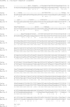
Figure 3—figure supplement 1.. Alignment of Lethe with Rps15a-ps6.
ClustalW2 alignment was performed on Lethe (Rps15a-ps4) and Rps15a-ps6. DOI: http://dx.doi.org/10.7554/eLife.00762.009
Figure 4.
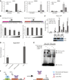
Figure 4.. Lethe Binds to RelA and inhibits RelA occupancy of DNA.
(A) Increased expression of NF-κB regulated genes in Lethe knockdown cells. Quantitative Taqman real time RT-PCR of the indicated RNAs is shown normalized to Actin levels (mean ± SD, p<0.05 is shown) (B) Lethe inhibits TNFα induced reporter gene expression. RLU of 3x-κB reporter activity and mutant reporter activity (mean ± SD, p<0.05 is shown) in CMV_Lethe transfected 293T cells. Reporter constructs are diagrammed above. (C) Endogenous RelA recruitment to the promoters of target genes is reduced in the presence of Lethe. 293T expressing CMV_GFP or CMV_Lethe were treated with 20 ng/ml TNFα for 15 min. ChIP with α-RelA antibodies was performed and RelA percent enrichment relative to input is shown (mean ± SD; Il6, p<0.033; Sod2, p<0.001; Il8, p<0.003; Nfkbia, p<0.015). (D) Lethe binds to RelA. MEFs were treated with 20 ng/ml TNFα for 6 hr. RNA-IP with α-RelA antibodies was performed. RNA was isolated and Quantitative Taqman real time RT-PCR of the indicated RNAs is shown (mean ± SD, p<0.020). (E) Lethe expression blocks DNA binding of the RelA homodimer to its target. NF-κB EMSA of GFP or Lethe transfected 293T nuclear extracts treated with 20 ng/ml TNFα for 15 min. Extracts were pretreated with unlabeled NF-κB (specific) or CREB (nonspecific competitor), or α-RelA antibodies for 15 min prior to incubation with probe. (F) Model for Lethe regulation of gene expression. Upon addition of TNFα or dexamthasone, Lethe is transcribed. Lethe can then bind to RelA–RelA homodimers and block binding to other NF-κB response elements, inhibiting NF-κB. DOI: http://dx.doi.org/10.7554/eLife.00762.010

References

    1. Adler AS, Sinha S, Kawahara TLA, Zhang JY, Segal E, Chang HY. 2007. Motif module map reveals enforcement of aging by continual NF-kappaB activity. Genes Dev 21:3244–57. 10.1101/gad.1588507 - DOI - PMC - PubMed
    1. Boldin MP, Baltimore D. 2012. MicroRNAs, new effectors and regulators of NF-kappaB. Immunol Rev 246:205–20. 10.1111/j.1600-065X.2011.01089.x - DOI - PubMed
    1. Bond AM, Van Gompel MJ, Sametsky EA, Clark MF, Savage JC, Disterhoft JF, et al. 2009. Balanced gene regulation by an embryonic brain ncRNA is critical for adult hippocampal GABA circuitry. Nat Neurosci 12:1020–7. 10.1038/nn.2371 - DOI - PMC - PubMed
    1. Cabili MN, Trapnell C, Goff L, Koziol M, Tazon-Vega B, Regev A, et al. 2011. Integrative annotation of human large intergenic noncoding RNAs reveals global properties and specific subclasses. Genes Dev 25:1915–27. 10.1101/gad.17446611 - DOI - PMC - PubMed
    1. Chang AL, Bitter PH, Jnr, Qu K, Lin M, Rapicavoli NA, Chang HY. 2013. Rejuvenation of gene expression pattern of aged human skin by broadband light treatment: a pilot study. J Invest Dermatol 133:394–402. 10.1038/jid.2012.287 - DOI - PMC - PubMed

Publication types

MeSH terms

Substances