. 2014 Feb;13(1):19-28.
doi: 10.1111/acel.12145. Epub 2013 Sep 11.

Longevity effect of IGF-1R(+/-) mutation depends on genetic background-specific receptor activation

Affiliations

Longevity effect of IGF-1R(+/-) mutation depends on genetic background-specific receptor activation

Jie Xu et al. Aging Cell. 2014 Feb.

Abstract

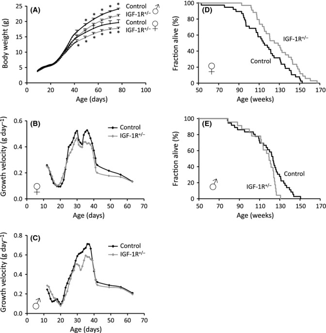
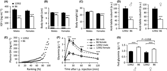
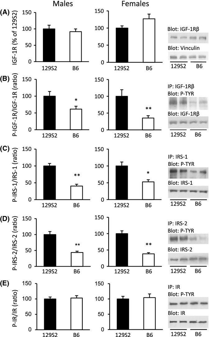
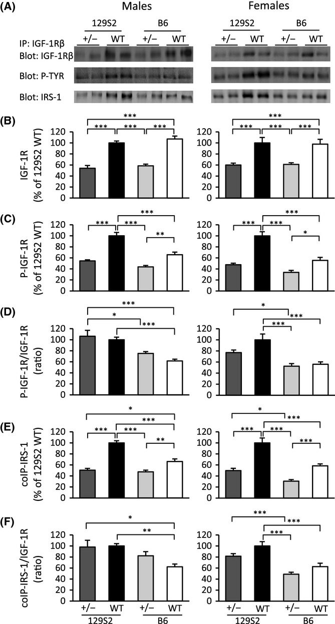
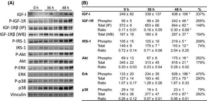
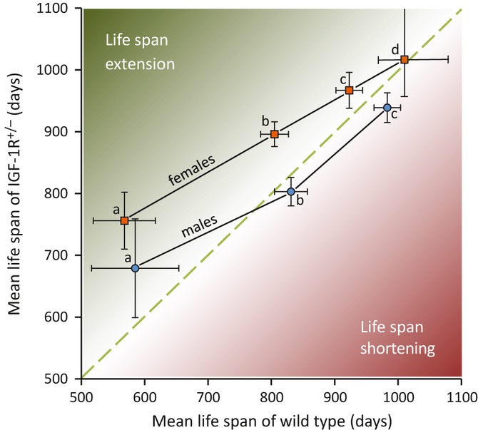
Growth hormone (GH) and insulin-like growth factor (IGF) signaling regulates lifespan in mice. The modulating effects of genetic background gained much attention because it was shown that life-prolonging effects in Snell dwarf and GH receptor knockout vary between mouse strains. We previously reported that heterozygous IGF-1R inactivation (IGF-1R(+/-) ) extends lifespan in female mice on 129/SvPas background, but it remained unclear whether this mutation produces a similar effect in other genetic backgrounds and which molecules possibly modify this effect. Here, we measured the life-prolonging effect of IGF-1R(+/-) mutation in C57BL/6J background and investigated the role of insulin/IGF signaling molecules in strain-dependent differences. We found significant lifespan extension in female IGF-1R(+/-) mutants on C57BL/6J background, but the effect was smaller than in 129/SvPas, suggesting strain-specific penetrance of longevity phenotypes. Comparing GH/IGF pathways between wild-type 129/SvPas and C57BL/6J mice, we found that circulating IGF-I and activation of IGF-1R, IRS-1, and IRS-2 were markedly elevated in 129/SvPas, while activation of IGF pathways was constitutively low in spontaneously long-lived C57BL/6J mice. Importantly, we demonstrated that loss of one IGF-1R allele diminished the level of activated IGF-1R and IRS more profoundly and triggered stronger endocrine feedback in 129/SvPas background than in C57BL/6J. We also revealed that acute oxidative stress entails robust IGF-1R pathway activation, which could account for the fact that IGF-1R(+/-) stress resistance phenotypes are fully penetrant in both backgrounds. Together, these results provide a possible explanation why IGF-1R(+/-) was less efficient in extending lifespan in C57BL/6J compared with 129/SvPas.

Keywords: Genetic background; IGF-I; IRS; gene knockout; lifespan; stress resistance.


Figures

Figure 1
Figure 1
Heterozygous knockout of IGF-1R in C57BL/6J genetic background inhibits growth in both sexes and extends lifespan in females. (A) Postnatal growth in male and female IGF-1R+/− mice. Significant differences existed from 6 weeks of age onwards in females (P < 0.05) and from 7 weeks onwards in males (P < 0.01; Student’s t-test, N = 18–24, error bars represent SEM). A star indicates difference between genotypes within the same sex (P values between 0.036 and < 0.001). (B and C) Male and female growth velocity was calculated as weight gain per day using data from (A). Peaks of growth velocity at 4 weeks of age are blunted in IGF-1R+/− mutants. (D) Heterozygous knockout of IGF-1R extended lifespan in female mice. IGF-1R+/− females (gray line) lived 11% longer than controls (black) (896 ± 23 vs. 805 ± 26 day; P = 0.023, Cox regression test; P = 0.021, log-rank test). (E) IGF-1R+/− males (gray line) showed reduced maximum lifespan (803 ± 20 vs. 831 ± 22 day; P = 0.027, Cox regression test; P = 0.025, log-rank test). Detailed lifespan data and descriptive statistics are provided in Tables S1 and S2. For comparison, mean lifespan in B6 control groups from the recent literature is on average 811 days in females and 822 days in males (Table S3).
Figure 2
Figure 2
Comparing somatotropic hormones, adult body size and glucose homeostasis between wild-type (WT) B6 and 129S2 mice. (A) Plasma IGF-I concentration. ***P < 0.001, Student’s t-test; N = 18–19 per group. (B) Body length (naso-anal distance). (C) Body weight. N = 11–13 per group. (D) Plasma acid-labile subunit (ALS). ***P < 0.001, Student’s t-test; N = 9 per group. (E) Comparing plasma GH concentration by rank plot analysis (Xu et al., 2011) between WT females from B6 (open marks) and 129S2 (black marks) background (P = 0.012, log-rank test). (F) Glucose tolerance test (GTT) in male and female mice. Glucose (2 g kg−1 body weight) was intraperitoneally (i.p.) injected at T0 and glycemia measured at T0, 15, 30, 60, and 120 min. Note that all groups rapidly regained control over glycemia. ***P < 0.002 (N = 7–8 per group) comparing B6 with 129S2. Individual area under curve (AUC) between both strains is significantly different (P < 0.001, one-way ANOVA with Bonferroni’s post hoc test). (G) Glycemia in the fed state. ***P < 0.001 (N = 6–10 per group) (one-way ANOVA with Bonferroni’s post hoc test). A-E and G were performed in 11- to 13-week-old mice. Tests in panel F were performed in 2-month-old mice. Error bars indicate SEM.
Figure 3
Figure 3
Activation of IGF-1R and substrates IRS-1 and IRS-2 is higher in WT mice of 129S2 than in WT mice of B6 genetic background, in males and females. Ad libitum-fed mice (11–13 week old) were used to recapitulate pathway activation under physiological conditions. Results shown are from skeletal muscle. Representative Western blots (WB) are displayed on the right. (A) Total IGF-1R; vinculin was used as loading control. (B) Phospho-IGF-1R (P-IGF-1R), (C) P-IRS-1, (D) P-IRS-2, and (E) P-IR were detected by immunoprecipitation (IP) using specific antibodies, followed by WB using an anti-phosphotyrosine (P-TYR) antibody. Signals of activated protein were expressed relative to total protein. N = 6 per group, in males and females; *P < 0.05, **P < 0.01, Student’s t-test; error bars represent SEM.
Figure 4
Figure 4
IGF-1R phosphotyrosine activation and IRS-1 recruitment differ significantly between 129S2 and B6 mice of IGF-1R+/− (+/−) and WT genotype. Ad libitum-fed 11- to 13-week-old mice were used. (A) Representative WB results. (B) Prevalence of IGF-1R revealed by IP and subsequent WB. (C) Phospho-IGF-1R (P-IGF-1R) detected from total immunoprecipitated IGF-1R using phosphotyrosine-specific antibody (P-TYR). (D) P-IGF-1R relative to total immunoprecipitated IGF-1R. (E) IRS-1 co-immunoprecipitating with IGF-1R shows a profile similar to (C). (F) Expressing co-immunoprecipitated IRS-1 relative to IGF-1R shows a profile similar to (D). In each graph, the mean of WT 129S2 mice was arbitrarily set to 100, and results from other groups expressed relative to that. N = 7–12 per group; *P < 0.05, **P < 0.01, ***P < 0.001, in one-way ANOVA with Bonferroni’s post hoc test; error bars represent SEM.
Figure 5
Figure 5
Activation of IGF signaling pathways 36 and 48 h after i.p. injection of paraquat. (A) WB detection of IGF-I from blood and of key signal transduction proteins in IGF pathways (P-tyrosine-activated forms and total protein) from lung tissue. Gel loading was controlled by vinculin. (B) Quantification of WB by chemiluminescence. Phosphotyrosine-IGF-1R (P-IGF-1R) and P-IRS-1 were detected using a phosphotyrosine-specific antibody after IP. Note that increase in IGF-1R abundance over time was very similar whether detected from IP samples or by direct WB. Tests were performed in 11- to 13-week-old mice. N = 3 per group; mean ± SEM, expressed in arbitrary units. *P < 0.05, Mann–Whitney U-test.
Figure 6
Figure 6
Schematic illustration suggesting that heterozygous IGF-1R knockout extends lifespan depending on longevity of genetic background and on sex. Data are from Holzenberger et al., (a, 129S2), Xu et al. (b, B6; this paper), and Bokov et al. (2011) (c, B6; d, hybrid B6/129). Data are used to compare mean lifespan of IGF-1R+/− mutant populations (y-axis) with mean lifespan of corresponding WT control populations (x-axis). In d, median lifespan was used. Error bars represent SEM in a, b, and c, and 95% confidence interval in d. In females, lifespan-extending effect of heterozygous IGF-1R knockout tends to diminish with increasing mean lifespan of WT control. Male populations show a similar trend, but seem to be exposed to adverse, life-shortening effects of IGF receptor inactivation as mean lifespan increases. This schematic representation raises the question whether stronger IGF-1R gene inactivation further extends longevity, in spontaneously long-lived strains or under environmental conditions that already favor long lifespan. It also suggests that DR may extend lifespan more efficiently in 129S2 than in B6 (see also Swindell, 2012).

References

    1. Ahamed K, Epaud R, Holzenberger M, Bonora M, Flejou JF, Puard J, Clement A, Henrion-Caude A. Deficiency in type 1 insulin-like growth factor receptor in mice protects against oxygen-induced lung injury. Respir. Res. 2005;6:31. - PMC - PubMed
    1. Ahluwalia A, Clodfelter KH, Waxman DJ. Sexual dimorphism of rat liver gene expression: regulatory role of growth hormone revealed by deoxyribonucleic acid microarray analysis. Mol. Endocrinol. 2004;18:747–760. - PubMed
    1. Berryman DE, Christiansen JS, Johannsson G, Thorner MO, Kopchick JJ. Role of the GH/IGF-1 axis in lifespan and healthspan: lessons from animal models. Growth Horm. IGF Res. 2008;18:455–471. - PMC - PubMed
    1. Blüher M, Kahn B, Kahn CR. Extended longevity in mice lacking the insulin receptor in adipose tissue. Science. 2003;299:572–574. - PubMed
    1. Bokov AF, Garg N, Ikeno Y, Thakur S, Musi N, DeFronzo RA, Zhang N, Erickson RC, Gelfond J, Hubbard GB, Adamo ML, Richardson A. Does reduced IGF-1R signaling in Igf1r+/− mice alter aging? PLoS ONE. 2011;6:e26891. - PMC - PubMed

Publication types

MeSH terms