. 2013 Aug 14;135(32):11869-78.
doi: 10.1021/ja4041779. Epub 2013 Jul 30.

Multiplexed electrochemistry of DNA-bound metalloproteins

Affiliations

Multiplexed electrochemistry of DNA-bound metalloproteins

Catrina G Pheeney et al. J Am Chem Soc. .

Abstract

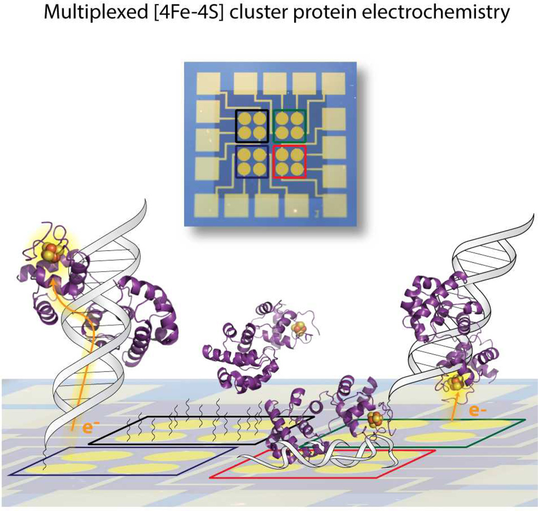
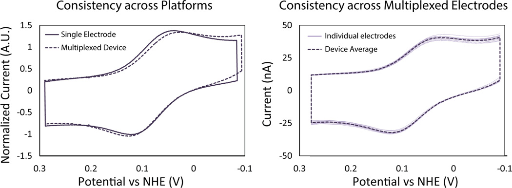
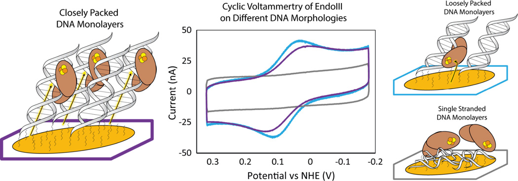
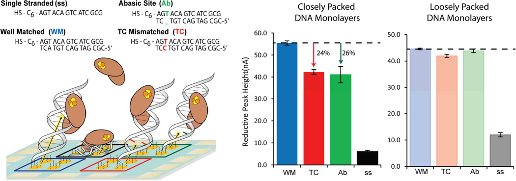
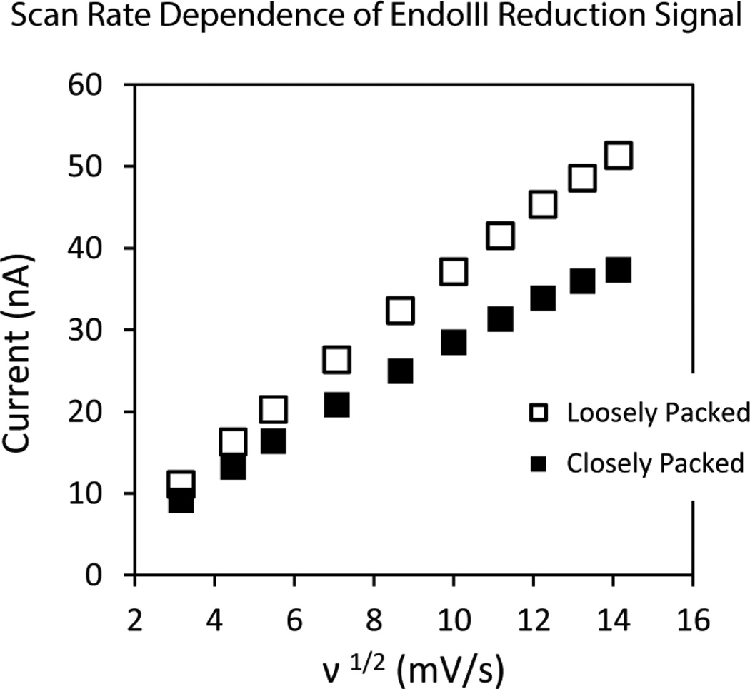
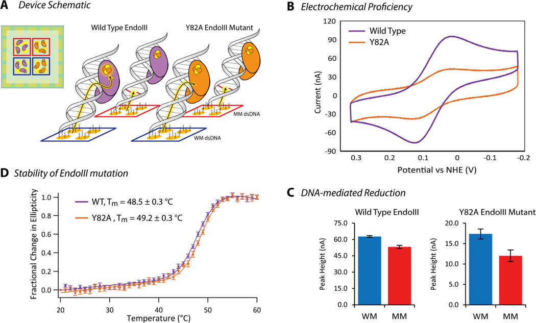
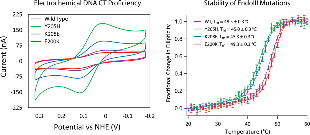
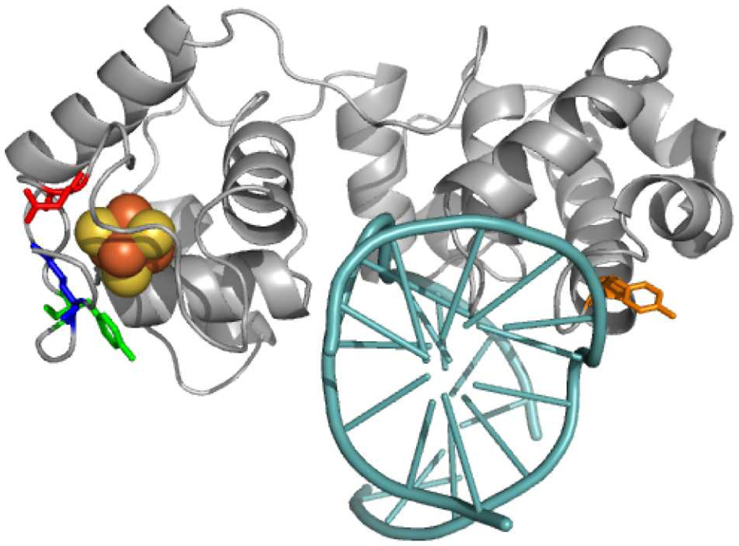
Here we describe a multiplexed electrochemical characterization of DNA-bound proteins containing [4Fe-4S] clusters. DNA-modified electrodes have become an essential tool for the characterization of the redox chemistry of DNA repair proteins containing redox cofactors, and multiplexing offers a means to probe different complex samples and substrates in parallel to elucidate this chemistry. Multiplexed analysis of endonuclease III (EndoIII), a DNA repair protein containing a [4Fe-4S] cluster known to be accessible via DNA-mediated charge transport, shows subtle differences in the electrochemical behavior as a function of DNA morphology. The peak splitting, signal broadness, sensitivity to π-stack perturbations, and kinetics were all characterized for the DNA-bound reduction of EndoIII on both closely and loosely packed DNA films. DNA-bound EndoIII is seen to have two different electron transfer pathways for reduction, either through the DNA base stack or through direct surface reduction; closely packed DNA films, where the protein has limited surface accessibility, produce electrochemical signals reflecting electron transfer that is DNA-mediated. Multiplexing furthermore permits the comparison of the electrochemistry of EndoIII mutants, including a new family of mutations altering the electrostatics surrounding the [4Fe-4S] cluster. While little change in the midpoint potential was found for this family of mutants, significant variations in the efficiency of DNA-mediated electron transfer were apparent. On the basis of the stability of these proteins, examined by circular dichroism, we propose that the electron transfer pathway can be perturbed not only by the removal of aromatic residues but also through changes in solvation near the cluster.


Figures

Figure 1
Figure 1
Schematic depicting the versatility of multiplexed analysis for the investigation of metalloprotein electrochemistry. Our multiplexed devices are composed of 16 electrodes that are divisible into 4 separate quadrants of 4 electrodes, having the capability of producing 4 distinct experimental conditions on a single Au surface. This assembly allows for facile comparisons of the electrochemical signal from various DNA-bound proteins across varying DNA substrates and morphologies.
Figure 2
Figure 2
Consistency of DNA-modified electrodes. (Left) The signals generated after incubation of loosely packed DNA-modified electrodes with EndoIII (30 µM) in phosphate buffer (20 mM sodium phosphate, 100 mM NaCl, 0.5 mM EDTA, 20 % glycerol, pH 7.4) were used to directly compared the single (solid) and multiplexed (dashed) electrochemical assemblies. The cyclic voltammetry (scan rate = 100 mV/s) was normalized, based on the capacitance at 0.3 mV vs NHE, so relative signal sizes could be compared across platforms. (Right) The variability of the EndoIII signal, under the same conditions, across all 16 electrodes (light solid line) of a single multiplexed device was within 3.5% of the average CV for the device (dark dashed line).
Figure 3
Figure 3
The electrochemistry of EndoIII on DNA-modified electrodes was determined as a function of the underlying DNA film morphology. DNA monolayers were allowed to self-assemble over 16–24 hours either with or without 100 mM MgCl2 to form either closely (purple) or loosely (blue) packed DNA monolayers. All morphologies were directly compared on the same multiplexed device so the differences in the EndoIII (60 µM) redox signal could be resolved. Cyclic voltammetry scans (scan rate = 100 mV/s) were compared in phosphate buffer (20 mM sodium phosphate, 100 mM NaCl, 0.5 mM EDTA, 20 % glycerol, pH 7.4) and the peak splitting and signal size were both quantified. Single-stranded DNA monolayers (grey) were prepared and shown to not produce a DNA-bound EndoIII signal.
Figure 4
Figure 4
The degree of signal attenuation induced by a single perturbation to the π-stack, for both closely and loosely packed DNA monolayers was investigated. (Left) Schematic of multiplexed devices prepared with well-matched (blue), TC mismatched (red), and abasic site (green) duplex DNA, and a single-stranded control (black).The sequences are indicated above. (Right) The reductive signals from DNA-bound EndoIII in phosphate buffer (20 mM sodium phosphate, 100 mM NaCl, 0.5 mM EDTA, 20 % glycerol, pH 7.4) were quantified for DNA monolayers assembled in both the presence (dark) and absence (light) of 100 mM MgCl2 yielding closely and loosely packed DNA, respectively. The percent signal attenuations of the TC mismatch and abasic site were determined based on the average signal size, across all four electrodes in a quadrant, compared to that of well-matched DNA. The signals generated from closely packed DNA-films displayed distinct attenuation upon introducing either a mismatch or abasic site, while the signals from loosely packed DNA-films did not display this sequence dependence.
Figure 5
Figure 5
Kinetic analysis of the signal generated from EndoIII on differing DNA film morphologies is indicated. Cyclic voltammetry (scan rates ranging from 10 mV/s to 200 mV/s) of EndoIII were obtained in phosphate buffer (20 mM sodium phosphate, 100 mM NaCl, 0.5 mM EDTA, 20 % glycerol, pH 7.4) for both closely (solid) and loosely (outlined) packed DNA monolayers. The reductive peak height for both morphologies, on the same multiplexed device, was quantified and plotted as a function of the square root of the scan rate, v½. The non-linearity of the signal from closely packed DNA films indicates that the signal is not diffusion rate limited.
Figure 6
Figure 6
Comparison of the electrochemical properties and stability of wild type EndoIII and a Y82A mutant. (A) Multiplexed electrode assembly schematic where electrodes are assembled with 100 mM MgCl2, with either well-matched (blue) or TC mismatched (red) duplex DNA, and then incubated with either wild type (purple) or Y82A (orange) EndoIII (70 µM, based on absorbance at 410 nm). (B) Cyclic voltammetry (scan rate = 100 mV/s) in phosphate buffer (20 mM sodium phosphate, 100 mM NaCl, 0.5 mM EDTA, 20 % glycerol, pH 7.4) are indicated for both wild type and Y82A EndoIII on closely packed, well-matched DNA monolayers. (C) The reductive signal upon introducing a TC mismatch (red) compared to well-matched (blue) validates the mechanism of reduction to be DNA-mediated for both proteins. (D) Circular dichroism thermal denaturation (5 µM protein) validates that the Y82A mutation does not significantly alter the stability of the protein.
Figure 7
Figure 7
Electrochemical and stability comparison of a new family of electrostatic EndoIII mutations, Y205H (green), K208E (blue), and E200K (red), with wild type EndoIII (purple). (Left) Cyclic voltammetry (scan rate = 100 mV/s) in phosphate buffer (20 mM sodium phosphate, 100 mM NaCl, 0.5 mM EDTA, 20 % glycerol, pH 7.4) is displayed for all four proteins on closely packed, (assembled with 100 mM Mg Cl2) well-matched DNA monolayers. Protein samples had equivalent concentrations of [4Fe-4S] (70 µM based on the 410 nm absorbance). (Right) Circular dichroism thermal denaturation (5 µM protein) was performed to correlate the altered electronic coupling of these mutations in close proximity of the [4Fe-4S] cluster with the differential stability of the proteins.
Figure 8
Figure 8
Crystal structure of EndoIII with the location of mutants shown relative to the DNA (cyan) and [4Fe-4S] cluster: Y205 (green), K208 (blue), E200 (red), and Y82 (orange). PDB: 2ABK with DNA from 1ORN.

References

    1. Zheng G, Patolsky F, Cui Y, Wang UW, Lieber CM. Nature Biotechnol. 2005;23:1294. - PubMed
    1. Morrow TJ, Li M, Kim J, Mayer TS, Keating CD. Science. 2009;323:352. - PMC - PubMed
    1. Swensen JS, Xiao Y, Ferguson BS, Lubin AA, Lai RY, Heeger AJ, Plaxco KW, Soh HT. J. Am. Chem. Soc. 2009;131:4262. - PMC - PubMed
    1. Plaxco KW, Soh HT. Trends in Biotechnology. 2011;29:1. - PMC - PubMed
    1. Bonham AJ, Hsieh K, Ferguson BS, Vallee-Belisle A, Ricci F, Soh HT, Plaxco KW. J. Am. Chem. Soc. 2012;134:3346. - PMC - PubMed

Publication types

MeSH terms