

The role of fundus autofluorescence in late-onset retinitis pigmentosa (LORP) diagnosis
- PMID: 23899229
- PMCID: PMC4377133
- DOI: 10.3109/13816810.2013.800891
The role of fundus autofluorescence in late-onset retinitis pigmentosa (LORP) diagnosis
Abstract
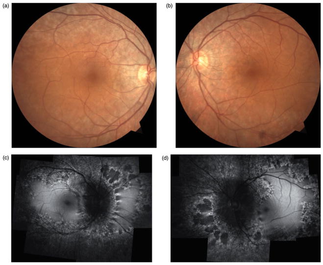
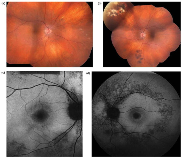
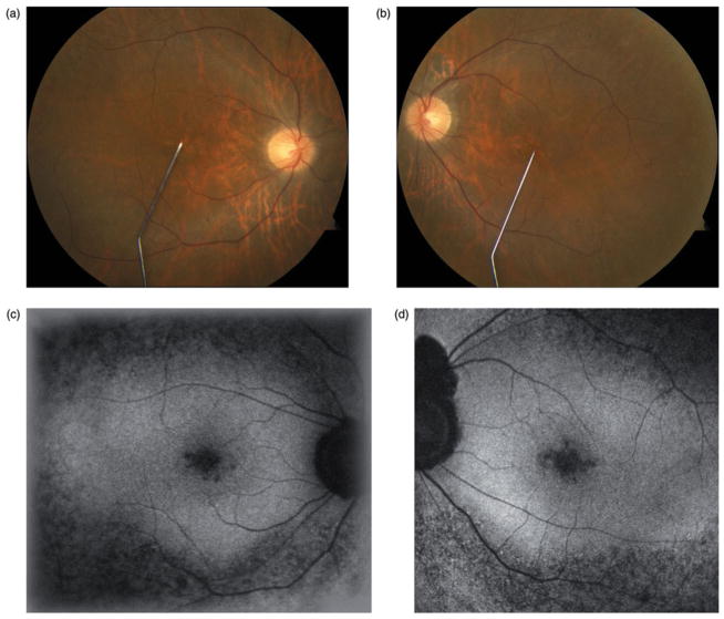
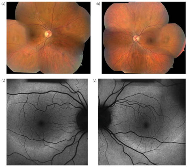
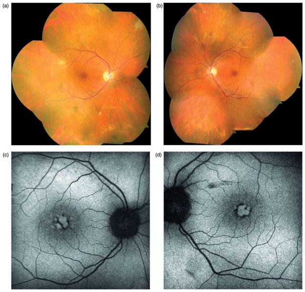
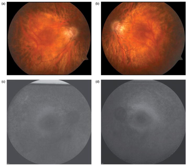
Purpose: To demonstrate the utility and characteristics of fundus autofluorescence in late-onset retinitis pigmentosa.
Methods: Observational case series. Patients diagnosed with late-onset retinitis pigmentosa were identified retrospectively in an institutional setting. Twelve eyes of six patients were identified and medical records were reviewed.
Results: All patients presented with slowly progressive peripheral field loss and initial clinical examination revealed only subtle retinal changes. There was a notable lack of intraretinal pigment migration in all patients. Five out of six patients underwent magnetic resonance imaging of the brain to rule out intracranial processes and all were referred from another ophthalmologist for further evaluation. Fundus autofluorescence was ultimately employed in all patients and revealed more extensive retinal pathology than initially appreciated on clinical examination. Fundus autofluorescence directed the workup toward a retinal etiology in all cases and led to the eventual diagnosis of late-onset retinitis pigmentosa through electroretinogram testing.
Conclusion: Fundus autofluorescence may be a more sensitive marker for retinal pathology than stereo fundus biomicroscopy alone in late-onset retinitis pigmentosa. Early use of fundus autofluorescence imaging in the evaluation of patients with subtle retinal lesions and complaints of peripheral field loss may be an effective strategy for timely and cost-efficient diagnosis.
Keywords: Fundus autofluorescence; late-onset retinitis pigmentosa; magnetic resonance imaging.
Conflict of interest statement
The authors report no conflicts of interest. The authors alone are responsible for the content and writing of the paper.
Figures
Similar articles
-
Fundus Autofluorescence Lifetime Patterns in Retinitis Pigmentosa.Invest Ophthalmol Vis Sci. 2018 Apr 1;59(5):1769-1778. doi: 10.1167/iovs.17-23336. Invest Ophthalmol Vis Sci. 2018. PMID: 29610860
-
Estimation of Visual Function Using Deep Learning From Ultra-Widefield Fundus Images of Eyes With Retinitis Pigmentosa.JAMA Ophthalmol. 2023 Apr 1;141(4):305-313. doi: 10.1001/jamaophthalmol.2022.6393. JAMA Ophthalmol. 2023. PMID: 36821134 Free PMC article.
-
Wide-field fundus autofluorescence imaging to evaluate retinal function in patients with retinitis pigmentosa.Am J Ophthalmol. 2014 Nov;158(5):1093-8. doi: 10.1016/j.ajo.2014.07.021. Epub 2014 Jul 22. Am J Ophthalmol. 2014. PMID: 25062603 Clinical Trial.
-
Wide-Field Fundus Autofluorescence for Retinitis Pigmentosa and Cone/Cone-Rod Dystrophy.Adv Exp Med Biol. 2016;854:307-13. doi: 10.1007/978-3-319-17121-0_41. Adv Exp Med Biol. 2016. PMID: 26427426 Review.
-
[Long-term follow-up of a case of unilateral retinitis pigmentosa].Nippon Ganka Gakkai Zasshi. 2012 Nov;116(11):1086-93. Nippon Ganka Gakkai Zasshi. 2012. PMID: 23316657 Review. Japanese.
Cited by
-
Next-generation sequencing revealed a novel mutation in the gene encoding the beta subunit of rod phosphodiesterase.Ophthalmic Genet. 2014 Sep;35(3):142-50. doi: 10.3109/13816810.2014.915328. Epub 2014 May 14. Ophthalmic Genet. 2014. PMID: 24828262 Free PMC article.
-
Spotlight on fundus autofluorescence.Clin Optom (Auckl). 2018 Mar 27;10:25-32. doi: 10.2147/OPTO.S134637. eCollection 2018. Clin Optom (Auckl). 2018. PMID: 30214339 Free PMC article. Review.
-
Occult inflammation detected by autofluorescence May Be the cause of idiopathic choroidal neovascularization.Am J Ophthalmol Case Rep. 2020 Oct 17;20:100965. doi: 10.1016/j.ajoc.2020.100965. eCollection 2020 Dec. Am J Ophthalmol Case Rep. 2020. PMID: 33117916 Free PMC article.
-
Structural evaluation in inherited retinal diseases.Br J Ophthalmol. 2021 Dec;105(12):1623-1631. doi: 10.1136/bjophthalmol-2021-319228. Epub 2021 May 12. Br J Ophthalmol. 2021. PMID: 33980508 Free PMC article. Review.
-
Bilateral Concordance of the Fundus Hyperautofluorescent Ring in Typical Retinitis Pigmentosa Patients.Ophthalmic Genet. 2015 Jun;36(2):113-22. doi: 10.3109/13816810.2013.841962. Epub 2013 Oct 10. Ophthalmic Genet. 2015. PMID: 24111858 Free PMC article.
References
-
- Berson EL. Retinitis pigmentosa. The Friedenwald Lecture. Invest Ophthalmol Vis Sci. 1993;34:1659–1676. - PubMed
-
- Haim M, Holm NV, Rosenberg T. Prevalence of retinitis pigmentosa and allied disorders in Denmark. I. Main results. Acta Ophthalmol (Copenh) 1992;70:178–186. - PubMed
-
- Dryja TP, Li T. Molecular genetics of retinitis pigmentosa. Hum Mol Genet. 1995;4:1739–1743. - PubMed
-
- Haim M. Prevalence of retinitis pigmentosa and allied disorders in Denmark. II. Systemic involvement and age at onset. Acta Ophthalmol (Copenh) 1992;70:417–426. - PubMed
-
- Phelan JK, Bok D. A brief review of retinitis pigmentosa and the identified retinitis pigmentosa genes. Mol Vis. 2000;6:116–124. - PubMed
Publication types
MeSH terms
Grants and funding
LinkOut - more resources
Full Text Sources
Other Literature Sources