. 2013 Jul 30:10:80.
doi: 10.1186/1742-4690-10-80.

HIV-1 Nef promotes the localization of Gag to the cell membrane and facilitates viral cell-to-cell transfer

Affiliations

HIV-1 Nef promotes the localization of Gag to the cell membrane and facilitates viral cell-to-cell transfer

Marine Malbec et al. Retrovirology. .

Abstract

Background: Newly synthesized HIV-1 particles assemble at the plasma membrane of infected cells, before being released as free virions or being transferred through direct cell-to-cell contacts to neighboring cells. Localization of HIV-1 Gag precursor at the cell membrane is necessary and sufficient to trigger viral assembly, whereas the GagPol precursor is additionally required to generate a fully matured virion. HIV-1 Nef is an accessory protein that optimizes viral replication through partly defined mechanisms. Whether Nef modulates Gag and/or GagPol localization and assembly at the membrane and facilitates viral cell-to-cell transfer has not been extensively characterized so far.

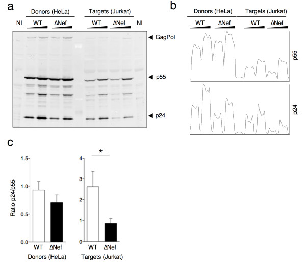
Results: We report that Nef increases the total amount of Gag proteins present in infected cells, and promotes Gag localization at the cell membrane. Moreover, the processing of p55 into p24 is improved in the presence of Nef. We also examined the effect of Nef during HIV-1 cell-to-cell transfer. We show that without Nef, viral transfer through direct contacts between infected cells and target cells is impaired. With a nef-deleted virus, the number of HIV-1 positive target cells after a short 2h co-culture is reduced, and viral material transferred to uninfected cells is less matured. At later time points, this defect is associated with a reduction in the productive infection of new target cells.

Conclusions: Our results highlight a previously unappreciated role of Nef during the viral replication cycle. Nef promotes HIV-1 Gag membrane localization and processing, and facilitates viral cell-to-cell transfer.


Figures

Figure 1
Figure 1
Nef increases the amount of Gag proteins in HeLa cells. HeLa cells were infected with VSV-G-pseudotyped wild type (WT) or nef-deleted (∆Nef) viruses. At day 2 post-infection cells were stained with the anti HIV-1 p24 KC57 antibody and the percentage of KC57 positive cells and their relative mean fluorescence intensity (MFI) were analyzed by flow cytometry. (a) Representative dot plots of infected HeLa cells (left panels). Percentage of KC57 positive cells is indicated in the top right corner of the gated population. MFI for the gated population is also indicated. A compilation of 10 independent experiments (Mean+SEM) of the percentage of KC57 positive cells and the Gag (KC57) MFI are shown (right panels). (b). Levels of HIV-1 p24 (in ng/ml) in the supernatants and cell lysates derived from cells described in (a) and measured by ELISA using the anti HIV-1 p24 183-H12-5C antibody. (c) The efficiency of viral release was calculated as the ratio between the levels of HIV-1 p24 in the supernatants and the total antigen HIV-1 p24 (supernatant + cell associated). Mean+SEM is shown. (d). HeLa cells were infected with VSV-G-pseudotyped WT- or ∆Nef viruses in which the green fluorescent protein (GFP) was inserted in frame with Gag-p17 (Gag-GFP). Cells were harvested at day 2 post-infection and analyzed by flow cytometry. Of note GFP signal does not distinguish between the mature and immature forms of Gag-GFP. The Gag-GFP MFI was measured on gated Gag-GFP positive HeLa cells. Representative dot plot analysis (left panel) in which the percentage of Gag-GFP positive cells is indicated in the top right corner of the gated population together with the MFI. Mean+SEM of 3 independent experiments of Gag-GFP MFI (right panel). *p<0.05; **p<0.01 (Mann Whitney test).
Figure 2
Figure 2
Nef increases Gag proteins levels in infected primary CD4+ T cells. PHA-activated primary CD4+ T cells were exposed to VSV-G-pseudotyped WT or ∆Nef (50–150 ng of p24/ml) for 3 h. The virus was washed off and the infected cells were cultured for up to 3 days. Productive infection was followed by flow cytometry of intracellular HIV-1 Gag using the KC57 anti-HIV-1 p24 monoclonal antibody. (a) Evolution of the fraction of Gag (KC57) positive cells at the indicated days post infection. Data are Mean±SEM of cells from three independent donors. (b) MFI of intracellular Gag (KC57) staining calculated on the fraction of Gag (KC57) positive cells. Maximum, Minimum and Mean of results obtained in cells described in Figure 1a are indicated. (c) Representative dot-plot analysis of Gag (KC57) staining of primary CD4+T cells infected with WT or ∆Nef (at day 2 post-infection). Cells were exposed to a higher viral input of ∆Nef than WT, in order to obtain similar fraction of infected cells. The percentage of Gag (KC57) positive cells is indicated in the top right corner of the gated population. MFI is also indicated. (d) Analysis of infected cells from 11 independent infections (8 donors), selected for the same fraction of Gag(KC57) positive cells at day 2 post-infection (left panel). The MFI of Gag (KC57) is reduced in absence of Nef (right panel). Each infection has been symbol-coded. *p<0.05 (Mann Whitney test)
Figure 3
Figure 3
Nef enhances viral cell-to-cell transfer in primary CD4+ T cells. Primary CD4+ T cells were infected with VSV-G-pseudotyped WT- or ∆Nef in order to get similar levels of Gag (KC57) positive cells, or, as a negative control, left uninfected (NI). These cells were then co-cultivated with target lymphocytes pre-stained with carboxyfluorescein succinimidyl ester (CFSE) for 2h, and analyzed by flow cytometry. (a) Dot plot analysis of donors (upper panels) and targets (lower panels) in one representative experiment. The percentage of Gag (KC57) positive cells is indicated in the top right corner of the gated population. MFI is also indicated. (b) Percentages of Gag (KC57) positive primary CD4+ target cells in 5 independent experiments. (c) Contacts and virological synapses between infected donors (D) and uninfected CD4+ lymphocytes targets (T), visualized by immunofluorescence. Donor cells were co-cultivated with CFSE-labeled (green) target cells for 1h and stained for HIV-1 Gag proteins (red) using a polyclonal rabbit anti-Gag antiserum. A contact was defined as a tight interaction between the cells (upper panel). A virological synapse was defined as a cell conjugate in which a polarization of Gag proteins was visible at the contact zone (lower panel). (c, d, e). Quantification of the percentage of conjugates (d) and virological synapses (e) formed between donor and target cells. *p<0.05 (Mann Whitney test).
Figure 4
Figure 4
Nef increases viral transfer from HeLa to Jurkat cells. (a) HeLa cells infected with VSV-G-pseudotyped WT- or ∆Nef for two days and having similar levels of Gag (KC57) positive cells by flow cytometry were co-cultivated with target Jurkat cells for 2h. Jurkat cells were then harvested, and the percentage of Gag (KC57) positive cells was measured by flow-cytometry at the indicated time points. Co-cultures were performed either in static conditions, to allow cell-to-cell contacts, in the presence of reverse transcriptase inhibitor nevirapine (NVP) to evaluate productive transmission, or under gentle shaking to limit cell-to-cell contacts. A compilation of at least 4 independent experiments per condition (mean + SEM), at 2h and 24h, is depicted. (b). Effect of Nef proteins from different alleles on viral cell-to-cell transfer. HeLa cells were co-transfected with HIV-1 ∆Nef and with plasmids expressing Nef from HIV-1 (LAI, NA7 and CFA-1), HIV-2 or SIVmac. Cells were then co-cultivated with Jurkat target cells, which were harvested after 2h. The percentage of Gag (KC57) positive Jurkat cells was measured at 2h and set up at 1 for WT. One representative experiment out of 2 is presented. *p<0.05; **p<0.01 (Mann Whitney test).
Figure 5
Figure 5
Nef increases HIV-1 p24 localization at the plasma membrane of HeLa cells. (a) HeLa cells were infected with VSV-G-pseudotyped WT- or ∆Nef-Gag-GFP viruses and plated on coverslips. 48h after infection cells were fixed and analyzed by confocal microscopy. Representative images for WT-GagGFP infected (A-D) and ∆Nef-GagGFP (E-H) are shown. White arrows indicate Gag-GFP proteins localized at the plasma membrane. Red arrows point to Gag-GFP proteins accumulating intracellulary. (b, c) HeLa cells were transfected with a plasmid coding for Gag (pGag) or GagPol (pGagPol) together with a plasmid coding for HIV-1Lai Nef protein (pCMV-Nef) or a control plasmid in which the Nef sequence was in antisense (pCMV-NefAS). 48h after transfection, dounce-homogenized cells were subjected to membrane flotation analysis. Panels (b) and (c) show representative western blots probed with an HIV-1 p24 monoclonal antibody (25A). Numbers on top of each lane indicate the loaded fractions. Fractions 2–4 and 8–10 correspond to membranes and cytoplasm, respectively. The immature (p55) and mature (p24) forms of Gag/GagPol are indicated. The blots were also probed with an anti-caveolin-1 antibody. (d) The percentages of the different Gag proteins in the membrane fractions were calculated and the mean+SEM of 3 independent experiments is shown in **p<0.01 (Mann Whitney test).
Figure 6
Figure 6
Nef induces HIV-1 p55 and p24 localization in membranes of infected primary CD4+T cells. (a) Primary CD4+T cells were infected with VSV-G-pseudotyped WT or ∆Nef. Two days post-infection cells were collected, dounce-homogenized and subjected to membrane flotation analysis as in Figure 5. Representative western blots of WT- (left) or ∆Nef-infected (right) primary CD4+T cells are shown. (b). Quantitative densitometry analysis of the western blots for p55 and p24. The x-axis shows the pixel location in each fraction and y-axis indicates the pixel intensity. (c). The percentages of p55 and p24 found in the membrane fractions was calculated and the mean+SEM of experiments performed with cells from 4 independent donors is shown. *p<0.05 (Mann Whitney test).
Figure 7
Figure 7
∆Nef-infected donor cells mostly transfer immature viral material to target cells. (a) HeLa cells infected with two doses of VSV-G-pseudotyped WT or ∆Nef viruses were used as donors to transfer the infection to Jurkat target cells as described in Figure 4. After a co-culture of 2h, donors and targets were separately harvested and cell lysates were analyzed by western blotting with a monoclonal anti-HIV-1 p24 antibody (25A). A representative experiment is shown. (b) Quantitative densitometry analysis of the western blots for p55 and p24. The x-axis shows the pixel location in each fraction and y-axis indicates the pixel intensity. (c) The ratio between mature (p24) and immature (p55) forms of Gag proteins in HeLa donors and Jurkat target cells was calculated and the mean+SEM of 3 independent experiments is shown. *p<0.05 (Mann Whitney test).

Similar articles

Cited by

References

    1. Jacks T, Power M, Masiarz F, Luciw P, Barr P, Varmus H. Characterization of ribosomal frameshifting in HIV-1 gag-pol expression. Nature. 1988;331:280–283. doi: 10.1038/331280a0. - DOI - PubMed
    1. Morita E, Sundquist W. Retrovirus budding. Annu Rev Cell Dev Biol. 2004;20:395–425. doi: 10.1146/annurev.cellbio.20.010403.102350. - DOI - PubMed
    1. Smith AJ, Srinivasakumar N, Hammarskjold ML, Rekosh D. Requirements for incorporation of Pr160gag-pol from human immunodeficiency virus type 1 into virus-like particles. J Virol. 1993;67:2266–2275. - PMC - PubMed
    1. Park J, Morrow CD. The nonmyristylated Pr160gag-pol polyprotein of human immunodeficiency virus type 1 interacts with Pr55gag and is incorporated into viruslike particles. J Virol. 1992;66:6304–6313. - PMC - PubMed
    1. Adamson CS, Salzwedel K, Freed EO. Virus maturation as a new HIV-1 therapeutic target. Expert Opin Ther Targets. 2009;13:895–908. doi: 10.1517/14728220903039714. - DOI - PMC - PubMed

Publication types

MeSH terms

Substances

LinkOut - more resources