Review
. 2014 Apr;10(4):1571-1580.
doi: 10.1016/j.actbio.2013.07.025. Epub 2013 Jul 27.

Design of cell-matrix interactions in hyaluronic acid hydrogel scaffolds

Affiliations
Review

Design of cell-matrix interactions in hyaluronic acid hydrogel scaffolds

Jonathan Lam et al. Acta Biomater. 2014 Apr.

Abstract

The design of hyaluronic acid (HA)-based hydrogel scaffolds to elicit highly controlled and tunable cell response and behavior is a major field of interest in developing tissue engineering and regenerative medicine applications. This review will begin with an overview of the biological context of HA, which is needed to better understand how to engineer cell-matrix interactions in the scaffolds via the incorporation of different types of signals in order to direct and control cell behavior. Specifically, recent methods of incorporating various bioactive, mechanical and spatial signals are reviewed, as well as novel HA modifications and crosslinking schemes with a focus on specificity.

Keywords: Hyaluronic acid; Hydrogel; Scaffold; Tissue engineering.


Figures

Figure 1
Figure 1
Hyaluronic acid scaffolds can be engineered in several ways to control encapsulated cell behavior. (A) Bioactive signals can be incorporated into the scaffold in suspension or covalently bound to the polymer. (B) Tuning the mechanical properties by utilizing degradable crosslinkers and/or degree of crosslinking can regulate cell mechanosensing. (C) Spatial cues such as patterned bioactive signals, porosity control, and topographical patterns can direct cell migration. (D) New “click chemistries” have been employed to provide new ways to covalently crosslink the hydrogel network.
Figure 2
Figure 2
Structure of hyaluronic acid. Chemical modifications are commonly performed at (a) and (b).
Figure 3
Figure 3
Bioactive signals are commonly incorporated into HA hydrogels to direct cell behavior. Some signals, like peptide fragments and antibodies, can be covalently linked to the HA backbone whereas other signals, like heparin and DNA, are loaded into the hydrogel without any permanent bond.
Figure 4
Figure 4
HA hydrogels were functionalized with RGD peptides or fibronectin fragments and filamentous actin of encapsulated mouse mesenchymal stem cells were stained with phalloidin 4 days after gelation. Cell spreading can be controlled by altering both the concentration and presentation of RGD. While (A) 100 μM of homogenously distributed RGD resulted in spherical morphology, (B-C) clustering 10 or 100 μM resulted in elongated cells. Hydrogels functionalized with (D) 1 μM, (E) 5 μM, and (F) 50 μM of homogenously distributed fibronectin fragments resulted in elongated cell morphologies.
Figure 5
Figure 5
HUVECs seeded on HA hydrogels in microgrooves of 100 μm and 200 μm in width covered the grooves in monolayers (a-b), while those seeded in microgrooves of 30 μm and 50 μm in width organized into 3D cord structures (c-d) [65].
Figure 6
Figure 6
RGDS within the dashed circles was uncaged and activated via photopatterning, resulting in the formation of patterns of cell populations on HA hydrogels [66].
Figure 7
Figure 7
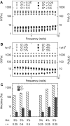
The storage and loss moduli of HA hydrogels, measured with a rheometer, can be modulated by varying (A) crosslinking densities (r-ratio) or (B) HA concentration. (C) Average G’ and G” over the frequency range tested [1].
Figure 8
Figure 8
D1 mMSC cells cultured in stiffer nonporous HA hydrogels (higher HA concentration) show less spreading (A), proliferation (B), and migration (C-D) than those cultured in softer nonporous hydrogels (lower HA concentration). The same patterns were observed in hydrogels with two different RGD concentrations [1].
Figure 9
Figure 9
Stiffness of UV-photopolymerized HA hydrogels can be controlled by varying the exposure time to UV light. Gel stiffness in this study was measured using dynamic mechanical analysis [68].

References

    1. Lei YG, Gojgini S, Lam J, Segura T. The spreading, migration and proliferation of mouse mesenchymal stem cells cultured inside hyaluronic acid hydrogels. Biomaterials. 2011;32:39–47. - PMC - PubMed
    1. Kurisawa M, Chung JE, Yang YY, Gao SJ, Uyama H. Injectable biodegradable hydrogels composed of hyaluronic acid-tyramine conjugates for drug delivery and tissue engineering. Chem Commun (Camb) 2005:4312–4. - PubMed
    1. Gojgini S, Tokatlian T, Segura T. Utilizing cell-matrix interactions to modulate gene transfer to stem cells inside hyaluronic acid hydrogels. Mol Pharm. 2011;8:1582–91. - PMC - PubMed
    1. Prehm P. Hyaluronate is synthesized at plasma membranes. Biochem J. 1984;220:597–600. - PMC - PubMed
    1. Meyer K, Palmer JW. The polysaccharide of the vitreous humor. Journal of Biological Chemistry. 1934;107:629–34.

Publication types

MeSH terms

Substances