Review
. 2013 Jul;93(3):1019-137.
doi: 10.1152/physrev.00028.2012.

Phosphoinositides: tiny lipids with giant impact on cell regulation

Affiliations
Review

Phosphoinositides: tiny lipids with giant impact on cell regulation

Tamas Balla. Physiol Rev. 2013 Jul.

Abstract

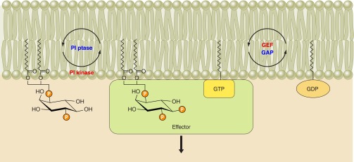
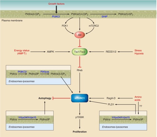
Phosphoinositides (PIs) make up only a small fraction of cellular phospholipids, yet they control almost all aspects of a cell's life and death. These lipids gained tremendous research interest as plasma membrane signaling molecules when discovered in the 1970s and 1980s. Research in the last 15 years has added a wide range of biological processes regulated by PIs, turning these lipids into one of the most universal signaling entities in eukaryotic cells. PIs control organelle biology by regulating vesicular trafficking, but they also modulate lipid distribution and metabolism via their close relationship with lipid transfer proteins. PIs regulate ion channels, pumps, and transporters and control both endocytic and exocytic processes. The nuclear phosphoinositides have grown from being an epiphenomenon to a research area of its own. As expected from such pleiotropic regulators, derangements of phosphoinositide metabolism are responsible for a number of human diseases ranging from rare genetic disorders to the most common ones such as cancer, obesity, and diabetes. Moreover, it is increasingly evident that a number of infectious agents hijack the PI regulatory systems of host cells for their intracellular movements, replication, and assembly. As a result, PI converting enzymes began to be noticed by pharmaceutical companies as potential therapeutic targets. This review is an attempt to give an overview of this enormous research field focusing on major developments in diverse areas of basic science linked to cellular physiology and disease.


Figures

Figure 1.
Figure 1.
Phosphoinositide basics. A: Agranoff's turtle demonstrating the orientation of the hydroxyl groups in myo-inositol. B: interconversions between various phosphoinositides and the enzymes catalyzing these reactions. The yeast enzymes are listed in parentheses. Where there is some ambiguity it is indicated by “??”. *It is worth pointing out that contrary to their designation, PIP5K2s are 4-kinases that act on PtdIns5P.
Figure 2.
Figure 2.
The phosphoinositide cycle as originally perceived (A) and the updated version also showing the polyphosphoinositides, PtdIns4P and PtdIns(4,5)P2 (B). The primary event in triggering the cycle is the agonist-induced PLC activation. Note that all of the products of PtdIns(4,5)P2 hydrolysis are recycled. Diacylglycerol (DG) is converted to phosphatidic acid (PtdOH) by one of many DG-kinase enzymes (DGK). PtdOH then has to be transferred from the PM to the endoplasmic reticulum by a mechanism that has not been identified. In the ER, one of two CDP-DG synthase (CDS) enzymes conjugates PtdOH with CTP, and the CDP-DG is then conjugated with myo-inositol to phosphatdylinositol (PtdIns). PtdIns synthesis takes place mainly in a highly dynamic subcompartment of the ER. Much of the inositol used for PtdIns synthesis is derived from the sequential dephosphorylation of inositol 1,4,5-trisphosphate [Ins(1,4,5)P3], the other product of PLC-mediated PtdIns(4,5)P2 hydrolysis. Several of the dephosphorylation steps are inhibited by Li+, including the final dephosphorylation of inositol monophosphates by the enzyme inositol monophosphatase (IMP). The newly synthesized PtdIns has to reach the PM by a still obscure mechanism, perhaps mediated by PtdIns/PtdCho transfer proteins (PITPs).
Figure 3.
Figure 3.
The family of PI 4-kinase enzymes. PI 4-kinases (PI4Ks) have two major types, the type III and type II enzymes [the type I enzyme(s) turned out to be the PI 3-kinases]. The type III enzymes are comprised of two proteins: the larger (∼210–230 kDa) PI4KA (Stt4p in yeast) and the smaller (∼92–110 kDa) PI4KB (Pik1p in yeast). These enzymes are relatives of PI 3-kinases and the PIK-related protein kinases, with a highly conserved COOH-terminal catalytic domain. They also have lipid-kinase unique (LKU) domains also found in PI 3-kinases. Other domains include proline-rich sequences (PR) and a frequennin-binding (Fq) domain in the PI4KB form. The smaller sized (∼56 kDa) type II PI4Ks exist in two forms in vertebrates: PI4K2A and PI4K2B that are highly homologous except at their very NH2 termini. [Only one form is found in S. cerevisiae (Lsb6) and in D. melanogaster]. The signature feature of these enzymes is a cysteine-rich (CR) sequence that is palmitoylated in the vertebrate enzymes providing stronger membrane association.
Figure 4.
Figure 4.
The family of PIP kinase enzymes. Type I PIP kinases phosphorylate PtdIns4P to PtdIns(4,5)P2. They have three forms: α, β, and γ; the latter has five splice variants in humans that differ in their very COOH termini (alternative but still commonly used names are shown in parentheses). The type II PIP kinases phosphorylate PtdIns5P to PtdIns(4,5)P2 and also exist in three forms, of which the γ form has very low catalytic activity. Yeast only has one enzyme, Mss4p, that makes PtdIns(4,5)P2 from PtdIns4P. A third group of PIP kinases are called type III PIPkins, and they phosphorylate PtdIns3P to PtdIns(3,5)P2. The mammalian enzyme is called PIKfyve, while the yeast ortholog is Fab1p. The typical feature of these enzymes is the presence of a FYVE domain close to their NH2 termini that binds PtdIns3P and a Cpn60/TCP-1 chaperonin family domain (TCP-1).
Figure 5.
Figure 5.
The family of PI 3-kinase enzymes. Class I PI 3-kinases phosphorylate PtdIns(4,5)P2 to PtdIns(3,4,5)P3. There are four different genes coding for the catalytic subunits of PI3Ks, called p110α, -β, -γ, and -δ. They all have a conserved COOH-terminal catalytic domain preceded by lipid kinase unique (LKU, also called “helical”), C2 and Ras binding domains (Ras-BD). These enzymes have tightly associated regulatory subunits: p110α, -β, and -δ associate with p85 and p55/50 regulatory subunits encoded by three different genes. Their association is mediated by an interaction between the very NH2-terminal p85-binding region (p85BR) of the catalytic chains with the inter-SH2 (iSH2) region of the regulatory subunits. These enzymes are also called class IA enzymes. The catalytic p110γ differs from the previous forms (hence it is called class IB) in that it associates with either of two adaptors, p101 and p84/p87. These different adaptors interact with NH2-terminal adaptor binding region (AdBR) of p110γ and lend regulation by G protein βγ subunits (p101) and perhaps by Ras (p84/p87) to the enzyme. Class II PI3Ks also come in three forms (α, β, and γ) and contain phox-homology (PX) and C2 domains placed COOH terminally to their catalytic domains. These enzymes can phosphorylate PtdIns and PtdIns4P in vitro, but their in vivo substrate preference is still debated. The enzymes most likely form PtdIns3P in the cell. The single class III PI3K and its yeast ortholog, Vps34p, phosphorylates PtdIns to PtdIns3P. These enzymes associate with a larger regulatory protein Vps15p in yeast and p150 in mammalian cells (not shown).
Figure 6.
Figure 6.
The inositol lipid 5-phosphatase family. These enzymes dephosphorylate PtdIns(4,5)P2 and/or PtdIns(3,4,5)P3 in the 5-position. There are three major subgroups called type II, III, and IV. [The type I enzyme is a smaller, 43-kDa protein that hydrolyzes the water-soluble Ins(1,4,5)P3 molecule but not the membrane-bound lipids.] The type II 5-phosphatase enzymes are encoded by six genes. Two of these, synaptojanin-1 and synaptojanin-2, exist in multiple splice forms differing in their respective COOH termini (only the two main forms are shown). The characteristic feature of these enzymes is the presence of a Sac1 homology domain upstream of their conserved 5-phosphatase domains. The NPF repeats of the longer form of SYNJ1 binds to the endocytic protein Eps15. OCRL and INPP5B are also very similar to one another, with a whole set of domains providing multiple interactions in addition to the catalytic 5-ptase domain. These include an NH2-terminal PH domain and COOH-terminal ASH and Rho-GAP domains. The ASH domain binds Rab5 and the adaptor protein APPL1, while Rho-GAP domain binds the small GTP binding proteins, Rac1 and Cdc42. OCRL1a contains two clathrin binding (CB) domains, the second of which is not present in OCRL1b. INPP5B has very similar structure, but it lacks the clathrin-binding domains and has a COOH-terminal CAAX domain. INPP5J and INPP5K are smaller proteins that have a SKICH domain downstream of their 5-phosphatase domain. This core structure is surrounded by proline-rich sequences in INPP5J. The type III 5-phosphatases are represented by the SH2-domain-containing enzymes SHIP1 and SHIP2. They have an NH2-terminal SH2 domain and C2 and proline-rich sequences downstream of the 5-phosphatase domain. SHIP2 also has a sterile alpha motif (SAM) at its very COOH terminus. The single type IV enzyme, INPP5E has a proline-rich sequence and a COOH-terminal CAAX box in addition to the 5-phosphatase domain.
Figure 7.
Figure 7.
The inositol lipid 3-phosphatases. A: the PTEN family of enzymes dephosphorylate PtdIns(3,4,5)P3 at the 3-position and hence “antagonize” the class I PI3Ks. The conserved phosphatase domain is followed by a C2 domain and a PDZ binding sequence in PTEN. The PBM is a phospholipid binding domain present in PTEN and also in the voltage-dependent phosphatases (these latter enzymes are actually 5-phosphatases). The TPIPα and -γ enzymes have an NH2-terminal putative transmembrane domain (TM) that is lacking in the β form. The TPTE enzyme is catalytically inactive. B: the myotubularin family of 3-phosphatases dephosphorylate PtdIns3P and PtdIns(3,5)P2. They fall into three subgroups. All of these enzymes (except for MTMR14) have a PHG (PH and GRAM) domain and coil-coil (CC) regions in addition to their phosphatase domain. The phosphatase domain also contains a SET interaction domain (SID). MTMR3 and -4 also has a FYVE domain in their COOH termini. No specific domains beyond the phosphatase domain have been described in MTMR14. There are additional MTM-related proteins that lack catalytic activity. They are not shown in this figure.
Figure 8.
Figure 8.
The inositol lipid 4-phosphatases. INPP4A and -B dephosphorylate PtdIns(3,4)P2 at the 4-position. They have a C2 domain in their NH2 termini and a phosphatase domain at their COOH termini. INPP4A also has a PEST sequence. The TMEM proteins have lower activity, and they act on PtdIns(4,5)P2 to generate PtdIns5P. These enzymes have putative transmembrane domains (TM) at their COOH termini. B: the Sac1 phosphatases can dephosphorylate any monophosphorylated PtdIns, but in the cell they primarily function as PtdIns4P 4-phosphatases. The yeast and human Sac1 are tail-anchored proteins with a COOH-terminal TM domain. Human Sac3 and yeast Fig4p work as PtdIns(3,5)P2 5-phosphatases. hSac2 is a 5-phosphatase acting on PtdIns(4,5)P2 and PtdIns(3,4,5)P3 yet, structurally is a homolog of the Sac1 family.
Figure 9.
Figure 9.
The phospholipase C family. These enzymes hydrolyze PtdIns(4,5)P2 to Ins(1,4,5)P3 and diacylglycerol, hence are called phosphoinositide-specific (PI)-PLCs. They should not be mistaken for the bacterial PLCs that are specific to PtdIns, unable to hydrolyze phosphorylated PtdIns, and hence called PI-specific PLC (unfortunately similarly abbreviated to PI-PLC). Mammalian PLCs have a core structure consisting of a PH domain, followed by EF hands, a catalytic domain formed from X and Y conserved regions and a C2 domain. PLCδ enzymes show this minimal domain organization. The only exemption is PLCζ that lacks the PH domain. This basic core is then extended in the various PLCs: the β enzymes have a characteristic COOH-terminal extension (CTR) that lends regulation by heterotrimeric G protein alpha subunits. The X and Y domains are separated with a long insert in the PLCγ enzymes consisting of two SH2 domains, an SH3 domain, and this whole insert is sandwiched in between two half PH domains. The PLCη enzymes have a long serine-proline-rich (S/P) segment in their COOH termini. The largest PLC enzyme is PLCε, which has a cysteine-reach region (CR) and a RAS-GEF domain at the NH2 terminus and two Ras association domains (RA) at the COOH terminus.
Figure 10.
Figure 10.
The mammalian phosphatidylinositol transfer proteins. The PITP domain is capable of transferring PtdIns and PtdCho between natural membranes and artificial liposomes and hence are functionally similar to the yeast Sec14 protein. The class I PITPs are encoded by two genes, PITPα and PITPβ, the latter having two splice forms differing at the very COOH termini. A highly similar PITP domain is found in the NH2 terminus of a group of larger proteins that were first described in Drosophila as the RdgB proteins. These proteins were later identified in mammalian cells as membrane-associated PITPs (hence the name PITPnm) or as a family of proteins that interact with the Ca2+-regulated tyrosine kinase Pyk2 and named Nir1–3. In fact, one of these family members, Nir1/PITPnm3/RdgBαIII, lacks the PITP domain, so it is not a PITP. These proteins (called class IIA proteins) have an acidic region, initially defined as a Ca2+ binding domain but now recognized as a region containing an FFAT domain that provides ER localization through interaction with the ER resident VAP proteins. The DDHD domain is similar to a domain found in some PLA1 proteins, whereas the LNS2 domain is found in lipins that are PtdOH phosphohydrolases. The Pyk2 binding site, which is also located in the COOH terminus, probably overlaps with the LNS2. A third form of PITPs, called class IIB, is similar to the PITP region of the class IIA forms but lacks all other domains and has two splice variants differing in their COOH termini.
Figure 11.
Figure 11.
Distribution of phosphoinositides in various membrane compartments in a generic cell. The majority of PtdIns(4,5)P2 is located in the plasma membrane (PM), and that is where it is converted to PtdIns(3,4,5)P3. PtdIns4P is also found in the PM, but it is also highly enriched in the Golgi and in some endosomes that are part of the TGN. PtdIns3P is formed on endosomes such as the early endosmes (EE) and their precursors and is converted to PtdIns(3,5)P2 at the level of sorting endosomes (SE) at regions that form the invaginating membranes destined to become luminal in multivesicular bodies (MVB). PtdIns is generated in ER-derived compartments that can reach every other membrane, but the mechanism of lipid transfer between these compartments is still unknown. It is important to note that this distribution does not rule out that each of these lipids can be present in small, undetected amounts in other membrane compartments.
Figure 12.
Figure 12.
The multiple functions of PtdIns(4,5)P2 in the plasma membrane. A: this lipid serves as a precursor of the second messengers, Ins(1,4,5)P3 and DG upon PLC activation, or of PtdIns(3,4,5)P3 during PI3K action. It can also be dephosphorylated by 5-phosphatases to PtdIns4P. B: in addition to this “precursor” role, PtdIns(4,5)P2 can 1) regulate the activity of enzymes such as PLCδ1 or PLD; 2) contribute to the recruitment of endocytic clathrin adaptors; 3) anchor the actin cytoskeleton to the PM; 4) help dock secretory vesicles and presynatptic vesicles to the membrane during exocytosis; and 5) maintain or regulate the active conformation of a variety of ion channels and transporters. C: polarized or localized distribution of PtdIns(4,5)P2 and PtdIns(3,4,5)P3 play important roles in establishing and/or maintaining polarity, such as in directional migration, in local events, such as in phagocytosis, or in polarized epithelial cells that form barriers between their luminal and basolateral sides.
Figure 13.
Figure 13.
The general principle of signaling by phosphoinositides. There are a large number of molecules that possess domains capable of recognizing phosphoinositides (effectors). These domains usually also detect the GTP-bound form of small GTP-binding proteins. The coincidental detection of the G protein and the inositol lipid either recruits the effector to the membrane or induces a conformational change in the molecule. The effector is usually found in a macromolecular complex that also contains the lipid kinase (and probably the phosphatase) as well as the GEF and GAP proteins of the small GTP binding protein. The “regulation” of the effector then depends on the speed of the respective activation-inactivation cycles on phosphoinositides and G proteins. In this model, the strict lipid-specificity of the effector is less important than the identity of the enzymes that are part of the protein signaling complex. This model also explains why enzymes producing or eliminating the same lipid cannot substitute for one another's functions. Note that there is no need to substantially increase the level of the lipids in the membrane where these processes take place. This model assumes some sort of “channeling” lipids to the effectors. It is important to note, however, that this may not be the sole mode of regulation by inositol lipids, and there are other situations where the overall lipid levels show larger changes, and the “free lipid” produced is available for multiple inositol lipid interacting proteins. These latter ones might be more easily detectable with our current molecular tools. The question of how to limit the diffusion of lipids is a critical one to maintain specificity and local control in either case.
Figure 14.
Figure 14.
Integration of metabolic control, cell growth and proliferation, and their regulation by phosphoinositides. One of the main effectors of PI3K activation is the Akt/PKB protein kinase that is activated by translocation to the membrane by binding via its PH domain to PtdIns(3,4,5)P3 [and PtdIns(3,4)P2]. This recruitment is important for phosphorylation of Akt/PKB by two protein kinases, PDK1 (on Thr308) and the mTORC2 complex (on Ser473). These two kinases are also activated by PtdIns(3,4,5)P3. Although Akt signals in a number of other directions (not shown here for simplicity), one of its major functions is to control the activity of the mTORC1 complex. Active Akt stimulates mTORC1 by inhibiting the GAP activity of the TSC1/2 complex, the latter limiting the activity of the Rheb GTP binding proteins that are upstream activators of the mTORC1 complex. Active mTORC1 is a signal for cell growth and proliferation and inhibits apoptosis and autophagy. In case of starvation, energy depletion, stress, or hypoxia, mTORC1 is inhibited, and these signals activate the TSC1/2 complex. Under these conditions autophagy is stimulated. A different pathway is used during amino acid sensing. Amino acids activate the Rag GTPases and induce translocation of the mTORC1 complex to the surface of the lysosomes. The classIII PI3K hVPS34 is also important for the amino acid sensing pathway for mTORC1 activation using a pathway that works parallel to the Rag GTPases and which requires PLD1, and PtdIns3P binding to the PX domain of PLD. Recently, the class II PI3Ks and PtdIns(3,5)P2 production was also shown to stimulate mTORC1. Paradoxically, autophagy also requires PtdIns3P and VPS34, and it is most likely that the VPS34 that serves as a mediator of mTORC1 activation by amino acids works in a different complex and location than the one that promotes autophagy.

References

    1. Abdel-Latif AA, Akhtar RA, Hawthorne JN. Acetylcholine increases the breakdown of triphosphoinositide in rabbit iris muscle prelabelled with [32P]phosphate. Biochem J 162: 61–73, 1977 - PMC - PubMed
    1. Abe N, Inoue T, Galvez T, Klein L, Meyer T. Dissecting the role of PtdIns(4,5)P2 in endocytosis and recycling of the transferrin receptor. J Cell Sci 121: 1488–1494, 2008 - PMC - PubMed
    1. Abnet CC, Freedman ND, Hu N, Wang Z, Yu K, Shu XO, Yuan JM, Zheng W, Dawsey SM, Dong LM, Lee MP, Ding T, Qiao YL, Gao YT, Koh WP, Xiang YB, Tang ZZ, Fan JH, Wang C, Wheeler W, Gail MH, Yeager M, Yuenger J, Hutchinson A, Jacobs KB, Giffen CA, Burdett L, Fraumeni JF, Jr, Tucker MA, Chow WH, Goldstein AM, Chanock SJ, Taylor PR. A shared susceptibility locus in PLCE1 at 10q23 for gastric adenocarcinoma and esophageal squamous cell carcinoma. Nat Genet 42: 764–767, 2010 - PMC - PubMed
    1. Achiriloaie M, Barylko B, Albanesi JP. Essential role of the dynamin pleckstrin homology domain in receptor-mediated endocytosis. Mol Cell Biol 19: 1410–1415, 1999 - PMC - PubMed
    1. Adamski FM, Timms KM, Shieh BH. A unique isoform of phospholipase Cbeta4 highly expressed in the cerebellum and eye. Biochim Biophys Acta 1444: 55–60, 1999 - PubMed

Publication types

MeSH terms

Substances