Review
. 2013 Jul;93(3):1139-206.
doi: 10.1152/physrev.00020.2012.

11β-hydroxysteroid dehydrogenases: intracellular gate-keepers of tissue glucocorticoid action

Affiliations
Review

11β-hydroxysteroid dehydrogenases: intracellular gate-keepers of tissue glucocorticoid action

Karen Chapman et al. Physiol Rev. 2013 Jul.

Abstract

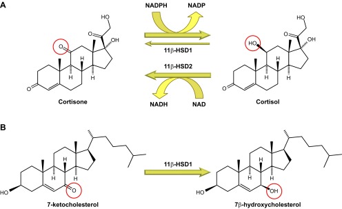
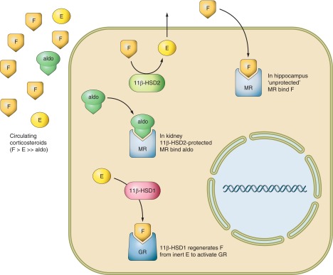
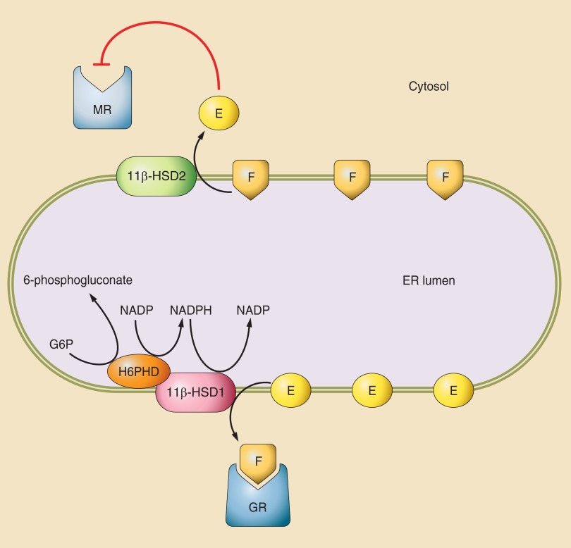
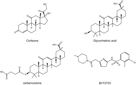
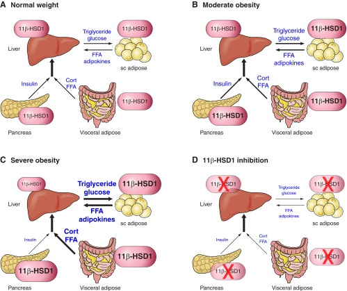
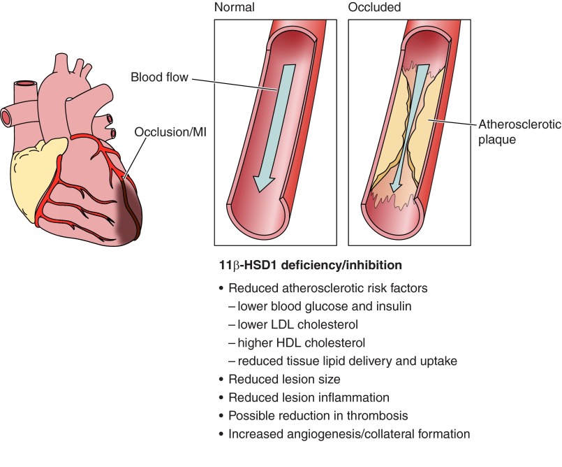
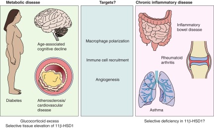
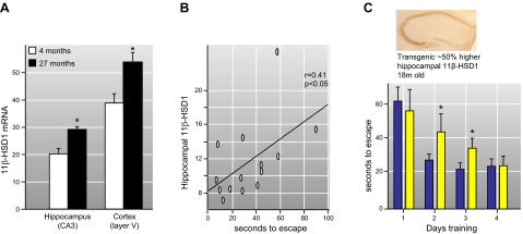
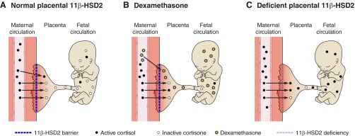
Glucocorticoid action on target tissues is determined by the density of "nuclear" receptors and intracellular metabolism by the two isozymes of 11β-hydroxysteroid dehydrogenase (11β-HSD) which catalyze interconversion of active cortisol and corticosterone with inert cortisone and 11-dehydrocorticosterone. 11β-HSD type 1, a predominant reductase in most intact cells, catalyzes the regeneration of active glucocorticoids, thus amplifying cellular action. 11β-HSD1 is widely expressed in liver, adipose tissue, muscle, pancreatic islets, adult brain, inflammatory cells, and gonads. 11β-HSD1 is selectively elevated in adipose tissue in obesity where it contributes to metabolic complications. Similarly, 11β-HSD1 is elevated in the ageing brain where it exacerbates glucocorticoid-associated cognitive decline. Deficiency or selective inhibition of 11β-HSD1 improves multiple metabolic syndrome parameters in rodent models and human clinical trials and similarly improves cognitive function with ageing. The efficacy of inhibitors in human therapy remains unclear. 11β-HSD2 is a high-affinity dehydrogenase that inactivates glucocorticoids. In the distal nephron, 11β-HSD2 ensures that only aldosterone is an agonist at mineralocorticoid receptors (MR). 11β-HSD2 inhibition or genetic deficiency causes apparent mineralocorticoid excess and hypertension due to inappropriate glucocorticoid activation of renal MR. The placenta and fetus also highly express 11β-HSD2 which, by inactivating glucocorticoids, prevents premature maturation of fetal tissues and consequent developmental "programming." The role of 11β-HSD2 as a marker of programming is being explored. The 11β-HSDs thus illuminate the emerging biology of intracrine control, afford important insights into human pathogenesis, and offer new tissue-restricted therapeutic avenues.


Figures

Figure 1.
Figure 1.
Reactions catalyzed by 11β-hydroxysteroid dehydrogenase (11β-HSD) isozymes. A: interconversion of cortisol and cortisone by 11β-HSD1 and -2. In intact cells and in vivo, 11β-HSD1 is predominantly a reductase, catalyzing NADPH-dependent reduction of cortisone to cortisol, predominantly in the liver. Under some circumstances and in some cells, it may act as an NADP-dependent dehydrogenase, inactivating cortisol. 11β-HSD2, in contrast, catalyzes the NAD+-dependent inactivation of cortisol, converting it to cortisone, predominantly in the kidney. B: conversion of 7-ketocholesterol to 7β-hydroxycholesterol by 11β-HSD1. Other reactions catalyzed by 11β-HSD1, including oxysterol metabolism, are probably of physiological importance. The 11β- and 7α-positions of the steroid nucleus show rotational symmetry, probably explaining the metabolism of 7-ketocholesterol to 7β-hydroxycholesterol by 11β-HSD1 as well as its metabolism of other 7-oxygenated sterols and steroids.
Figure 2.
Figure 2.
Diagrammatic representation of the reactions catalyzed by 11β-HSDs. The adrenal cortex secretes nanomolar concentrations of cortisol (F) and picomolar concentrations of aldosterone (Aldo) into the circulation. While mineralocorticoid receptors (MR) in the kidney only bind Aldo in vivo, identical MR in the hippocampus are occupied by F in vivo and MR bind F and Aldo with similar affinity in vitro. The solution to this conundrum lies in the collocation of renal MR with 11β-HSD2, which catalyzes the rapid inactivation of cortisol to inert cortisone (E) thus only allowing the nonsubstrate Aldo to access MR. 11β-HSD2 is absent from hippocampus so MR bind F. In tissues such as the liver, adipose, and adult brain, 11β-HSD2 is absent, but there is abundant 11β-HSD1. This catalyzes the reverse reaction in intact cells and organs and thus regenerates active F from inert E, amplifying the local glucocorticoid signal particularly at glucocorticoid receptors (GR). GR have 10-fold lower affinity for F than MR and are thus partially unoccupied by F at physiological concentrations allowing a dynamic range for 11β-HSD1 amplification inside cells to impact on signaling. In contrast, MR are largely occupied by physiological F concentrations where 11β-HSD2 is absent so 11β-HSD1 may make less impact on signaling via MR.
Figure 3.
Figure 3.
Cartoon of the possible intracellular relationships of 11β-HSDs. Despite having a lower affinity for F (or corticosterone) than MR, 11β-HSD2 is able to successfully exclude glucocorticoids from MR. While the basis for this is unknown, a possible scenario invokes lipophilic steroids preferentially localizing to the membranes inside cells including those of the endoplasmic reticulum (ER). 11β-HSD2 is located on the cytosolic surface of the ER and is in close association with the MR complex. Therefore, F may perhaps have to pass via 11β-HSD2 before it can gain access to MR. If there is sufficient enzyme and its turnover is suitably fast, it may successfully form the biochemical equivalent of an anatomical moat around MR. In addition, some data suggest that the 11-dehydrocorticosteroid products of 11β-HSD2 (cortisone/E; 11-dehydrocorticosterone) functionally antagonize aldosterone activation of MR. 11β-HSD1 is bidirectional in homogenates and microsomal preparations, but a predominant reductase in intact cells and in vivo. 11β-HSD1 is located inside the inner leaflet of the ER in close association with hectose-6-phosphate dehydrogenase (H6PDH), a powerful generator of NADP(H). NADP(H) drives the 11β-reductase direction of 11β-HSD1 and maintains this in many cell types. Other redox active processes may be important in organs such as brain where H6PDH may be at low levels.
Figure 7.
Figure 7.
Structures of nonselective licorice-based 11β-HSD inhibitors, glycyrrhetinic acid and carbenoxolone, BVT2733, a synthetic selective 11β-HSD1 inhibitor, and the natural cortisone substrate for 11β-HSD1.
Figure 4.
Figure 4.
Structure of the human HSD11B1 gene, associated promoters and relevant transcription factor binding sites. Schematic representation of the HSD11B1 gene (not to scale). Exonic sequences are shown as boxes; white boxes encode the open reading frame with red boxes and a blue box indicating the 5′ leader (dependent on the promoter used) and the 3′ untranslated sequence, respectively. Arrows indicate the positions of the 3 promoters. The gene spans 30 kb with a large intron (∼25 kb) between exons 5 and 6. The position of 2 conserved C/EBP binding sites, located between −112 and −160 in the promoter (244, 775), are shown. These sites are bound by C/EBPα and/or C/EBPβ in hepatocytes and adipocytes and are implicated in HSD11B1 regulation by glucocorticoids, cAMP, ceramide, AMPK, and DHEA. See text for details.
Figure 5.
Figure 5.
Structure of human 11β-hydroxysteroid dehydrogenase in complex with NADP and carbenoxolone. Structure of a dimer of human 11β-HSD1 with NADP cosubstrate (bottom middle) and the inhibitor carbenoxolone (top right) bound. [Image from the RCSB PDB (www.pdb.org) of PDB ID 2BEL (339).]
Figure 6.
Figure 6.
Structure of the human HSD11B2 gene showing positions of relevant transcription factor binding sites. Schematic representation of the human HSD11B2 gene (not to scale) showing positions of relevant transcription factor binding sites. The gene spans 6.4 kb. Exonic sequences are shown as boxes; white boxes encode the open reading frame, with a red box and a blue box indicating the 5′ leader and the 3′ untranslated sequence, respectively. An arrow indicates the position of the promoter, although it should be noted that transcription starts at a number of sites clustered around the site shown (see text for details). Sp1/Sp3 sites are shown as green ovals (these overlap with Egr1 sites), an NF1 site is shown as a pink hexagon, and an NF-κB binding site is shown as a purple diamond. The positions of 2 polymorphisms that reduce transcription factor binding and promoter activity (18) are indicated by asterisks. See text for details.
Figure 8.
Figure 8.
Cartoon of potential role of 11β-HSD1 in metabolic organ interrelationships in obesity. A: in normal weight health, 11β-HSD1 acts largely as a reductase. This performs an important endocrine role in the splanchnic bed by regenerating glucocorticoids, thus contributing ∼40% of daily glucocorticoid production. In addition, it has intracrine actions, amplifying the glucocorticoid signal inside hepatocytes, pancreatic islets (beta and alpha cells), and adipocytes. B: with modest obesity, 11β-HSD1 is elevated in adipocytes and beta cells but not hepatocytes. This increases insulin resistance in adipose tissues, increases release of proinflammatory/antimetabolic adipokines, and portal blood glucocorticoid and fatty acid deliver to the liver, but also increases islet insulin release to glucose, plausibly without changing hepatic insulin sensitivity. This may support storage of calories in adipose tissue in preparation for less bountiful nutritional circumstances. C: with severe obesity, greater rises in adipose and beta cell 11β-HSD1 lead to failure of pancreatic insulin release, worsening peripheral insulin resistance and metabolic disease despite potentially “compensatory” declines in hepatic glucocorticoid regeneration, which might even contribute to HPA axis activation due to loss of bulk glucocorticoid regeneration. D: 11β-HSD1 inhibition maximizes subcutaneous adipose and liver insulin sensitivity and visceral adipose AMPK signaling and reduces its inflammation and portal glucocorticoid and fatty acid release. Despite reduced beta cell insulin release and HPA axis activation (without elevation of glucocorticoid levels), the balance of metabolism favors reduced hepatic gluconeogenesis, increase beta-oxidation of fats, “safe” calorie storage in subcutaneous adipose depots, reduction of visceral adipose mass, and metabolic health.
Figure 9.
Figure 9.
Possible impacts of 11β-HDSD1 inhibition/deficiency on atherosclerosis. Known effects include reductions of multiple risk factors and reduced atherosclerotic lesion size. If a myocardial infarction occurs, the angiogenic effects of 11β-HSD1 deficiency/inhibition promote angiogenesis and recovery of myocardial function for the same initial infarct size.
Figure 10.
Figure 10.
11β-HSD1 in macrophages reflects activation state and shapes the subsequent response towards resolution. Monocytes have negligible levels of 11β-HSD1 (703). After differentiation of human peripheral blood monocytes to macrophages, 11β-HSD1 expression is increased (703). Polarization to M1 with LPS and IFN-γ further increases 11β-HSD1 mRNA levels, whereas polarization to M2 with IL-4 and/or IL-13 has no further effect (326). Treatment of monocytes with IL-4 or IL-13 also increases 11β-HSD1 mRNA and activity levels (703), which are further increased by PPARγ activation (117). Differentiation of monocytes in the presence of glucocorticoid produces a highly phagocytic macrophage phenotype (230). Similarly, glucocorticoid treatment of macrophages promotes a phagocytic phenotype (412). High expression of 11β-HSD1 in M1 macrophages may promote the subsequent transition to a phagocytic phenotype, thus promoting resolution of inflammation. 11β-HSD1 is markedly downregulated following phagocytosis of apoptotic leukocytes (115). Dashed lines indicate speculative connections, whereas solid lines have been demonstrated.
Figure 11.
Figure 11.
11β-HSD1 in the ageing brain. A: 11β-HSD1 is elevated in the ageing (27 mo old) mouse brain, specifically in regions of the hippocampus and cortex underpinning learning and memory. B: with ageing there is considerable interindividual variation in 11β-HSD1 that correlates with cognitive function (seconds to find a hidden platform after training in the watermaze). C: modeling this by transgenic overexpression of 11β-HSD1 in the forebrain from the age of weaning causes cognitive impairments that only emerge with ageing. [Adapted from Holmes et al. (289).]
Figure 12.
Figure 12.
Glucocorticoid programming and placental 11β-HSD2. Placental 11β-HSD2 debulks the much higher levels of active glucocorticoids in the maternal blood. A: normally the enzyme oxidizes cortisol to inert cortisone that cannot be regenerated in the fetus which lacks 11β-HSD1 until near term. Thus the major source of active cortisol in the fetus is its own adrenal glands. B: maternal treatment with dexamethasone, which is a poor substrate for 11β-HSD2 and thus passes the placenta intact, increases glucocorticoid action on the fetus and placenta reducing growth and altering the developmental trajectory of specific tissues. C: similarly inhibition or relative deficiency of placental 11β-HSD2 allows increased passage of active maternal glucocorticoids to the fetus and placental receptors. Lowering of placental 11β-HSD2 occurs with genetic mutations, consumption of licorice, maternal malnutrition, infection, or stress.

References

    1. Abdallah BM, Beck-Nielsen H, Gaster M. Increased expression of 11beta-hydroxysteroid dehydrogenase type 1 in type 2 diabetic myotubes. Eur J Clin Invest 35: 627–634, 2005 - PubMed
    1. Ackermann D, Vogt B, Escher G, Dick B, Reichen J, Frey BM, Frey FJ. Inhibition of 11beta-hydroxysteroid dehydrogenase by bile acids in rats with cirrhosis. Hepatology 30: 623–629, 1999 - PubMed
    1. Agarwal AK, Giacchetti G, Lavery G, Nikkila H, Palermo M, Ricketts M, McTernan C, Bianchi G, Manunta P, Strazzullo P, Mantero F, White PC, Stewart PM. CA-repeat polymorphism in intron 1 of HSD11B2: effects on gene expression and salt sensitivity. Hypertension 36: 187–194, 2000 - PubMed
    1. Agarwal AK, Monder C, Eckstein B, White PC. Cloning and expression of rat cDNA encoding corticosteroid 11 beta-dehydrogenase. J Biol Chem 264: 18939–18943, 1989 - PubMed
    1. Agarwal AK, Mune T, Monder C, White PC. Mutations in putative glycosylation sites of rat 11-beta-hydroxysteroid dehydrogenase affect enzymatic-activity. Biochim Biophys Acta 1248: 70–74, 1995 - PubMed

Publication types

MeSH terms

Substances