Review
. 2013:119:221-82.
doi: 10.1016/B978-0-12-396971-2.00009-9.

The circadian clock in cancer development and therapy

Affiliations
Review

The circadian clock in cancer development and therapy

Loning Fu et al. Prog Mol Biol Transl Sci. 2013.

Abstract

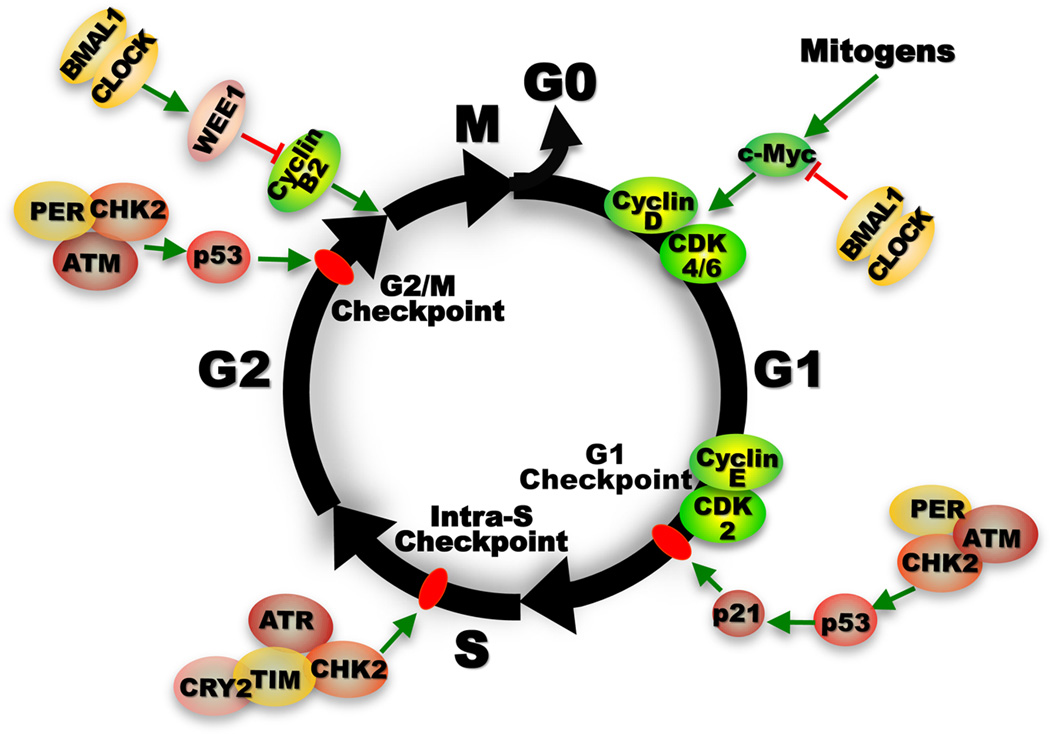
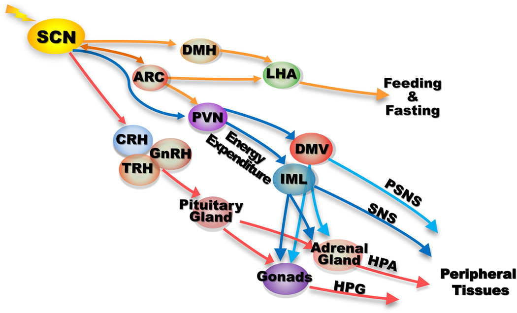
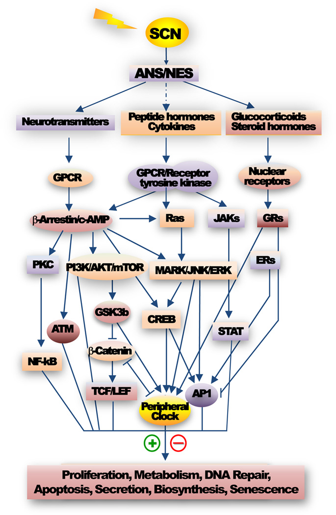
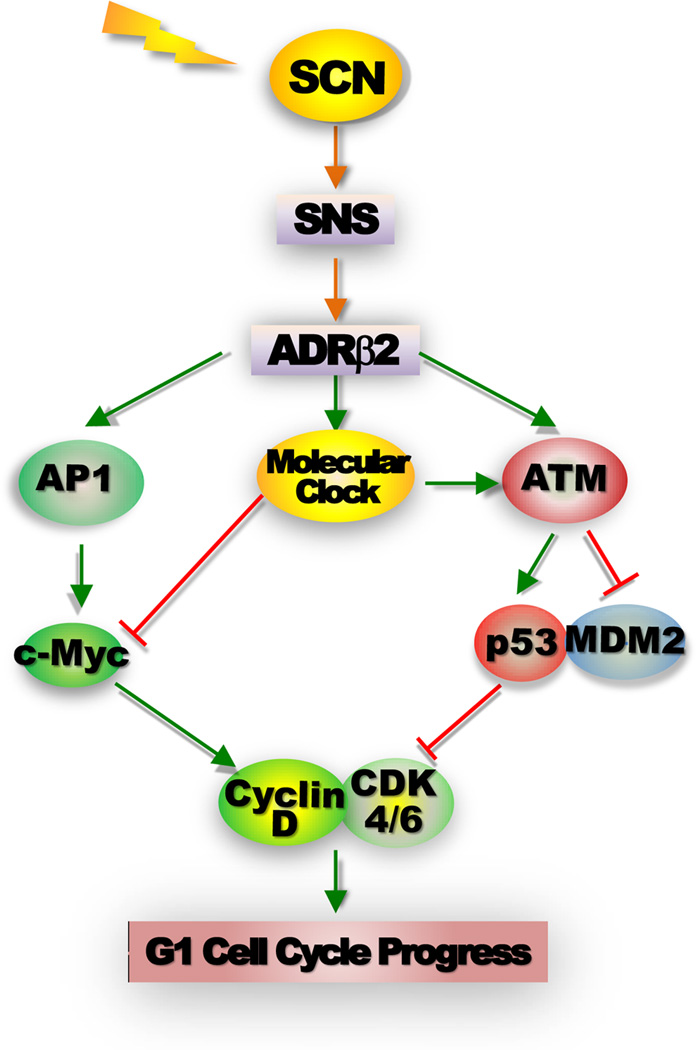
Most aspects of mammalian function display circadian rhythms driven by an endogenous clock. The circadian clock is operated by genes and comprises a central clock in the brain that responds to environmental cues and controls subordinate clocks in peripheral tissues via circadian output pathways. The central and peripheral clocks coordinately generate rhythmic gene expression in a tissue-specific manner in vivo to couple diverse physiological and behavioral processes to periodic changes in the environment. However, with the industrialization of the world, activities that disrupt endogenous homeostasis with external circadian cues have increased. This change in lifestyle has been linked to an increased risk of diseases in all aspects of human health, including cancer. Studies in humans and animal models have revealed that cancer development in vivo is closely associated with the loss of circadian homeostasis in energy balance, immune function, and aging, which are supported by cellular functions important for tumor suppression including cell proliferation, senescence, metabolism, and DNA damage response. The clock controls these cellular functions both locally in cells of peripheral tissues and at the organismal level via extracellular signaling. Thus, the hierarchical mammalian circadian clock provides a unique system to study carcinogenesis as a deregulated physiological process in vivo. The asynchrony between host and malignant tissues in cell proliferation and metabolism also provides new and exciting options for novel anticancer therapies.

Keywords: Aging; Cancer immune surveillance; Cellular senescence; Chronotherapy; Circadian clock; Energy homeostasis; G1 cell cycle progression; Phase shift; Signal transduction; Tumor suppression.


Figures

Figure 1
Figure 1. Control of cell proliferation by the molecular clock
Unlike the molecular clock, the cell cycle does not free run before passing the G1/S phase transition. The initiation of cell cycle progression is strictly controlled by extracellular mitogenic signals that transiently activate immediate early genes such as c-Myc, which then induces Cyclin D leading to activation of CyclinD/CDK4/6 complex that in turn activates E2F-dependent Cyclin E expression by suppressing tumor suppressor RB (not shown). The interaction of Cyclin E with CDK2 allows G1 to S phase transition. G1 is the longest phase in the cell cycle during which most biosynthesis essential for supporting cell cycle progression occurs. c-MYC or E2F oncogenic activation induced elevation of G1 Cyclin expression or genomic DNA damage leads to activation of G1 checkpoint mediated by p16Ink4A and p21WAF1/CIP1, controlled by RB and p53 respectively. P16Ink4A disrupts Cyclin D/CDK4/6 complex (not shown), whereas p21WAF1/CIP1 disrupts Cyclin E/CDK2 interaction. The activation G1 checkpoint leads cells to pause before entering S-phase to repair damaged DNA or exit cell cycle to enter G0 phase (non-dividing status). Under certain conditions, excessive DNA damage or uncontrolled oncogenic signaling can both activate RB and/or p53 tumor suppression pathways leading to cellular senescence. DNA damage induced by UV radiation leads to activation of ATR/CHK1-mediated intra-S checkpoint that couples DNA damage repair with replication. Whereas double-stranded DNA damage induced by γ-radiation activates ATM/CHK2-mediated G1/S and G2/M checkpoints to prevent damaged cells to enter S or mitotic (M) phase. Prolonged G2/M checkpoint arrest is also often associated with p53-mediated apoptosis. G2/M transition is also regulated by WEE1, a kinase that phosphorylates and inactivates the Cyclin B1/Cell Division Cycle 2 (CDC2) complex essential for G2/M transition. Upon the completion of mitosis, cells either enter the next cell cycle stimulated by extracellular mitogen, or withdraw from cell cycle to enter the G0 phase in the absence of mitogenic signals,. The molecular clock functions in all phases of the cell cycle to prevent neoplastic growth. At the early G1 phase, the BMAL1/CLOCK heterodimer down regulates Myc transcription to prevent its overexpression,,. PER1 directly interacts with ATM and CHK2 to control G1 checkpoint in response to double-strand DNA damage. In the S phase, CRY2/TIM complex directly interacts with ATR/CHK1 to control intra-S checkpoint. In the G2 phase, PER-mediated ATM/CHK2/p53 signaling in response to DNA double-strand breaks and BMAL1/CLOCK activated Wee1 expression both lead to activation of G2/M checkpoint to prevent inappropriate M phase entry,.
Figure 2
Figure 2. Peripheral control by the SCN pacemaker
The SCN clock targets a variety of brain centers within the hypothalamus to control homeostasis of endogenous physiology. It controls nutrient intake and energy expenditure by targeting the brain energy homeostasis center arcuate nucleus (ARC) and catabolic center paraventricular nucleus (PVN) directly, and feeding and satiety center LHA indirectly via ARC and dorsomedial hypothalamus (DMH). It also controls the neuroendocrine system (NES) by directly targeting the corticotropin-releasing hormone (CRH), thyrotropin-releasing hormone (TRH) and gonadotropin-releasing hormone (GnRH) neurons that control the adrenal and gonadal glands via pituitary glands. The SCN pacemaker directly targets the autonomic paraventricular (aPVN) neurons that project to the preganglionic parasympathatic and sympathetic neurons in the dorsal motor nucleus of the vagus (DMV) and intermediolateral cell columns (IML) of the spinal cord to control parasympathatic and sympathetic nervous systems (PSNS and SNS). Both PSNS and SNS also crosstalk with the HPA and HPG axes by directly innervating the adrenal and gonadal glands. The NES and ANS innervate all peripheral tissues in vivo to generate circadian rhythm of internal physiology by controlling extracellular signaling and peripheral clock activity,–,,.
Figure 3
Figure 3. Circadian control of intracellular signaling
The central pacemaker controlled autonomic nervous and neuroendocrine systems (ANS and NES) rhythmically signal to all of their target tissues. The resulting circadian rhythm in peripheral tissue function also generates local and/or circulating signaling molecules that rhythmically act on their targets. Together, these extracellular signals including neurotransmitters, steroid hormones, peptide hormones, chemokines, growth factors and cytokines activate intracellular signaling mediated by G-protein coupled receptors (GPCRs), tyrosine kinase receptors, integrins (not shown), and nuclear receptors in a tissue and cell-type specific manner. These same intracellular signaling pathways also activate the peripheral clock. The coordinated activities of the central and peripheral clocks orchestrate the complicated extracellular and intracellular signaling to maintain tissue homeostasis by controlling a network of gene expression. Disruption of the central clock-controlled extracellular signaling or mutations in core circadian genes both abolish peripheral clock activity leading to loss of circadian homeostasis in peripheral tissues. The representative intracellular signaling pathways directly or indirectly controlled by the central clock shown in the figure include the c-AMP/PKA/CREB/AP1, Ras/MARK/JNK/ERK and PI3K/AKT/β-Catenin/TCF/LEF pathways essential for c-Myc activation and cell cycle progression, the PI3K/AKT/mTOR signaling controlling biosynthesis and drug resistance,, the GPCR/ATM signaling for p53 activation, the GPCR/PKC/NF-κB pathway that regulates stress and immune response, the JAK/STAT pathway controlling apoptotic response, and the GR and ERα signaling pathways cross-talking with the AP1 signaling,. These signaling pathways also control the expression and function of circadian genes leading to a coupled activation of the molecular clock with tissue-specific function in vivo including cell proliferation, metabolism, apoptosis, DNA repair, biosynthesis, secretion and senescence.
Figure 4
Figure 4. Control of G1 cell cycle progression by the peripheral clock and the SNS
The activation of the β-adrenergic receptor 2 (ADRβ2) by SNS signaling leads a coupled induction of Ap1 and Period genes via CREB-mediated transcriptional regulation, which in turn activates AP1-controlled Myc induction and Myc-dependent G1 cell cycle progression as well as peripheral clock that prevents Myc overexpression via BMAL1/CLOCK-mediated transcriptional regulation. The activation of ADRβ2 intracellular signaling and the peripheral clock also synergistically activate ATM, which induces p53 by blocking p53-MDM2 interaction to provide an additional mechanism for preventing MYC oncogenic activation. Disruption of the central clock-SNS-peripheral clock axis in mice by chronic jet-lag or ablation of Per genes suppresses peripheral clock activation in response to ADRβ2 activation and abolishes ATM-mediated p53 induction but had no effect on Ap1-Myc signaling. Together these events lead to uncontrolled G1 cell cycle progression and neoplastic growth of osteoblasts both in vitro and in vivo,.

References

    1. Young MW, Kay SA. Time zones: a comparative genetics of circadian clocks. Nat Rev Genet. 2001 Sep;2(9):702–715. - PubMed
    1. Reppert SM, Weaver DR. Coordination of circadian timing in mammals. Nature. 2002 Aug 29;418(6901):935–941. - PubMed
    1. Buijs RM, Scheer FA, Kreier F, et al. Organization of circadian functions: interaction with the body. Prog Brain Res. 2006;153:341–360. - PubMed
    1. Guo H, Brewer JM, Lehman MN, Bittman EL. Suprachiasmatic regulation of circadian rhythms of gene expression in hamster peripheral organs: effects of transplanting the pacemaker. J Neurosci. 2006 Jun 14;26(24):6406–6412. - PMC - PubMed
    1. Takahashi JS, Hong HK, Ko CH, McDearmon EL. The genetics of mammalian circadian order and disorder: implications for physiology and disease. Nat Rev Genet. 2008 Oct;9(10):764–775. - PMC - PubMed

Publication types