. 2013 Sep 11;54(9):6118-32.
doi: 10.1167/iovs.13-11673.

Subretinal gene therapy of mice with Bardet-Biedl syndrome type 1

Affiliations

Subretinal gene therapy of mice with Bardet-Biedl syndrome type 1

Seongjin Seo et al. Invest Ophthalmol Vis Sci. .

Abstract

Purpose: To study safety and efficacy of subretinal adeno-associated virus (AAV) vector AAV-Bbs1 injection for treatment of a mouse model of Bardet-Biedl syndrome type 1 (BBS1).

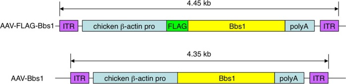
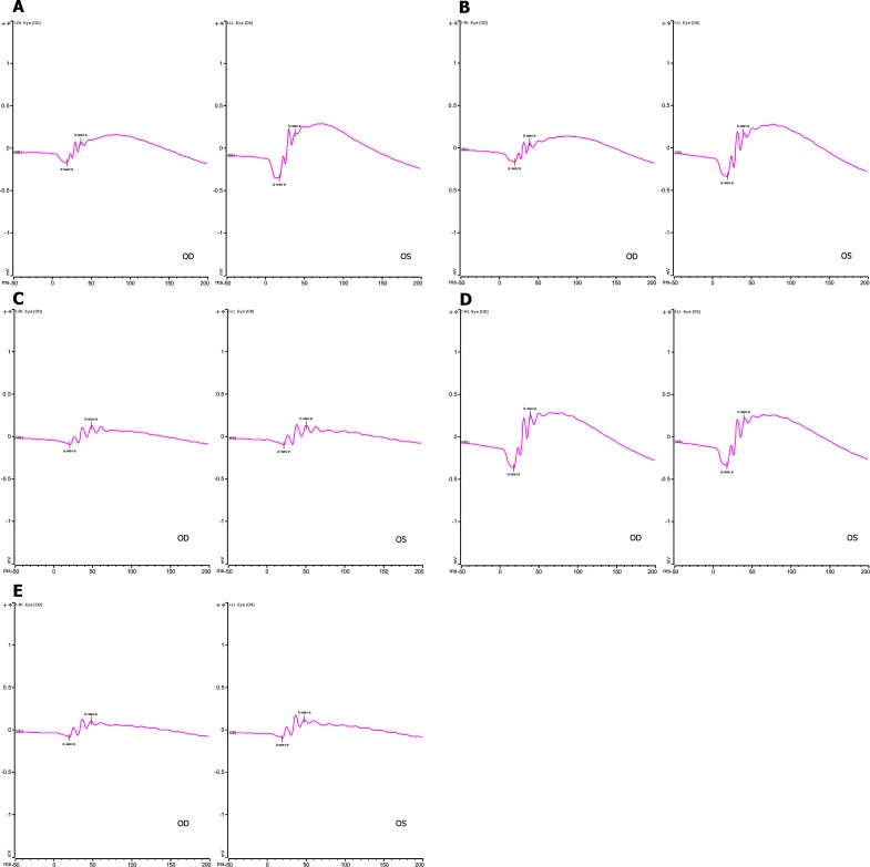
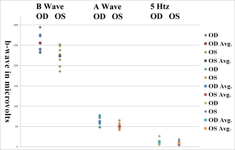
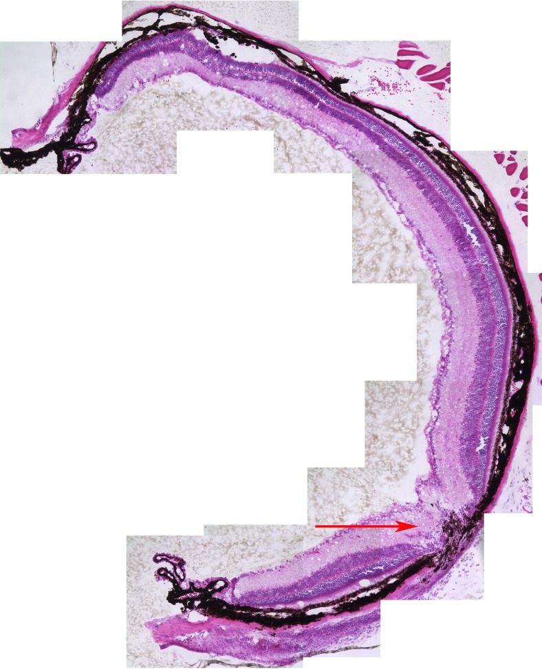
Methods: Constructs containing a wild-type (WT) Bbs1 gene with and without a FLAG tag in AAV2/5 vectors were generated. Viral genomes were delivered by subretinal injection to right eyes and sham injections to left eyes at postnatal day 30 (P30) to P60. Transgene expression and BBSome reconstitution were evaluated by immunohistochemistry and Western blotting following sucrose gradient ultracentrifugation. Retinal function was analyzed by electroretinogram (ERG) and structure by optical coherence tomography (OCT). Histology and immunohistochemistry were performed on selected eyes.

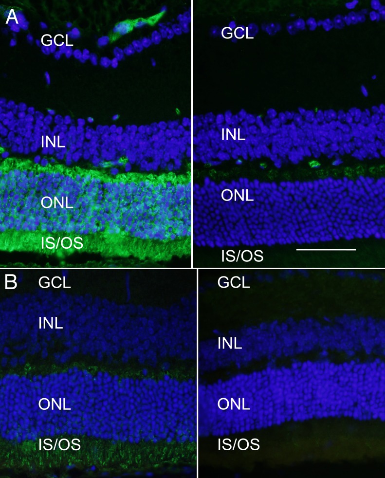
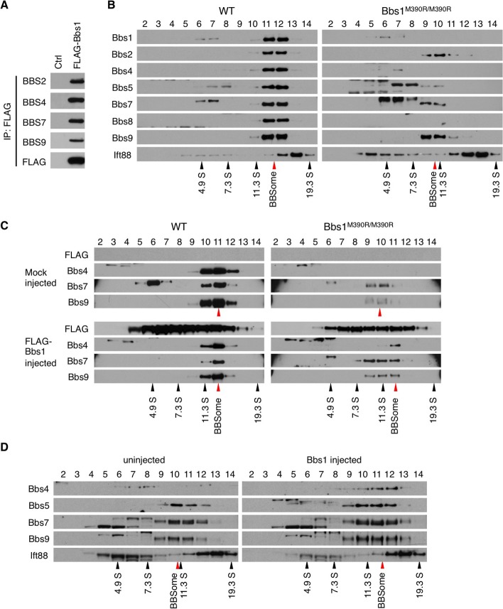
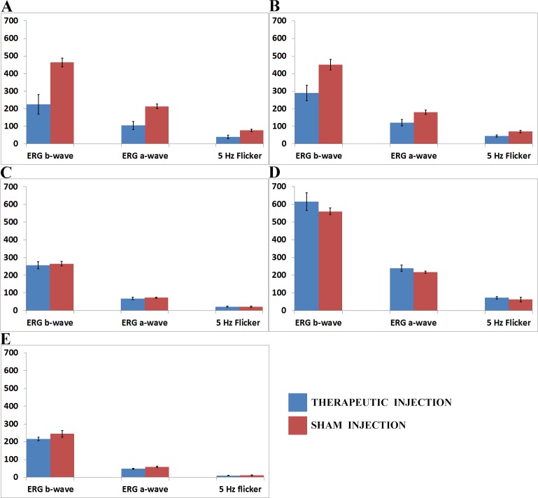
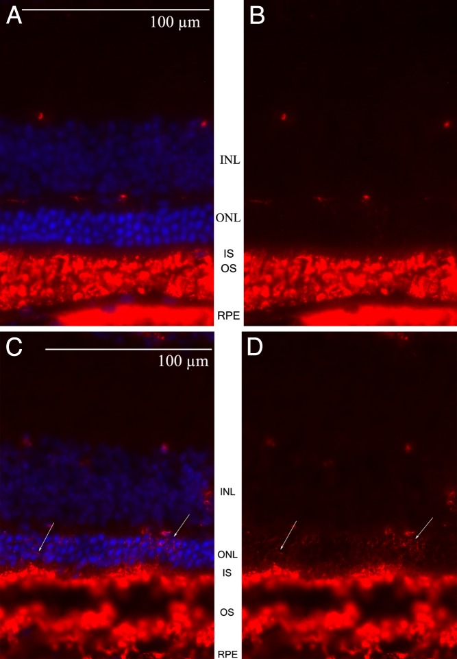
Results: Expression of FLAG-tagged Bbs1 was demonstrated in photoreceptor cells using antibody directed against the FLAG tag. Coinjection of AAV-GFP demonstrated transduction of 24% to 32% of the retina. Western blotting demonstrated BBS1 protein expression and reconstitution of the BBSome. ERG dark-adapted bright flash b-wave amplitudes were higher in AAV-Bbs1-injected eyes than in sham-injected fellow eyes in more than 50% of 19 animals. Anti-rhodopsin staining demonstrated improved localization of rhodopsin in AAV-Bbs1-treated eyes. WT retinas injected with AAV-Bbs1 with or without a FLAG tag showed outer retinal degeneration on ERG, OCT, and histology.

Conclusions: In a knock-in model of BBS1, subretinal delivery of AAV-Bbs1 rescues BBSome formation and rhodopsin localization, and shows a trend toward improved ERG. BBS is challenging to treat with gene therapy due to the stoichiometry of the BBSome protein complex and overexpression toxicity.

Keywords: AAV vector; Bardet Biedl syndrome; gene therapy; mouse model; retinal degeneration.


Figures

Figure 1.
Figure 1.
Structure of the FLAG-tagged and non-FLAG–tagged Bbs1 inserts. The 4.45-kb insert, composed of the chicken β-actin promoter, 3× FLAG sequences in reading frame with the mouse Bbs1 gene, and a poly A tail sequence, is flanked by two inverted terminal repeat sequences (ITR). In the second construct, the insert is 4.35 kb and lacks the FLAG tag.
Figure 2.
Figure 2.
Whole mount of a retina that received 1/10 AAV-GFP mixed with the AAV-FLAG-Bbs1 as a subretinal injection. Subretinal injection was performed in a 1-month-old WT animal. The animal was killed and the whole mount prepared 2 weeks after injection. Note the substantial degree of retinal transduction.
Figure 3.
Figure 3.
Expression of FLAG-BBS1 in photoreceptor cells. (A) Wild-type retina was transduced with the AAV-FLAG-Bbs1 (without AAV-GFP) and labeled with anti-FLAG antibody. Animals received subretinal injection of AAV-FLAG-Bbs1 vector at 1 month of age and were killed 2 weeks after injection. Left: Anti-FLAG immunofluorescence is seen in the retina which was over the injection site. Note the extensive labeling of the FLAG fusion protein in the outer plexiform layer, the inner segments, and to a lesser extent the outer nuclear layer. Right: A field from the same retina remote from the injection site. Scale bar: 50 μm. GCL, ganglion cell layer; INL, inner nuclear layer; ONL, outer nuclear layer; IS/OS, inner and outer segments. (B) Localization of native BBS1 protein. Left: An area of retina from a 6-week-old WT mouse that had received a subretinal injection of AAV-FLAG-Bbs1 2 weeks before. This section was stained with anti-BBS1 antibody (not anti-FLAG antibody). Right: A different section of the same retina remote from the injection site (i.e., not exposed to the vector) labeled with anti-BBS1 antibody. The anti-BBS1 antibody is labeled with Alexa Fluor 488. The amount of immunoreactive BBS1 protein in the injected area is much greater than in the uninjected area. BBS1 protein can be detected without the anti-FLAG stain; however, this antibody does not bind as avidly as the anti-FLAG antibody.
Figure 4.
Figure 4.
AAV-Bbs1 and AAV-FLAG-Bbs1 partly restore the BBSome assembly. (A) FLAG-BBS1 forms the BBSome. HEK293T cells were transduced with AAV-FLAG-Bbs1, and cell lysate was subjected to immunoprecipitation (IP) using anti-FLAG antibody and Western blotting against indicated BBS proteins. Normal HEK293T cells were used as a control. (B) Disruption of the BBSome in Bbs1M390R/M390R mutant eyes. Protein extracts from WT and Bbs1M390R/M390R mutant eyes were fractionated on a 10% to 40% sucrose linear gradient. Collected fractions were subjected to SDS-PAGE and Western blotting using the appropriate antibodies. Fraction numbers are shown at the top. Black and red arrowheads indicate the migration of sedimentation coefficient standards and the BBSome, respectively. In the WT gel, BBS1, -2, -4, -5, -7, and -9 migrate together, indicating a complex. In the Bbs1 gel, only BBBS2, -7, and -9 migrate together at a slightly lower fraction, indicating that the abnormal Bbs1M390R/M390R proteinis not incorporated into the BBSome and disrupts the complex. (C) Virally delivered FLAG-Bbs1 partly rescues the BBSome assembly. Protein extracts from treated animals were analyzed by sucrose gradient ultracentrifugation as in (B). (D) Virally delivered WT Bbs1 (without the FLAG) partly rescued the BBSome assembly just as the FLAG-tagged version did. Note the reappearance of BBS4 in fractions 11 and 12 of the treated eye.
Figure 5.
Figure 5.
Electroretinogram amplitudes of wild-type (WT) and Bbs1M390R/M390R mice injected with therapeutic and control AAV vectors. The x-axes show right and left eye b-wave and a-wave amplitudes for the dark-adapted bright flash standard combined response (SCR) ERG and the light-adapted 5-Hz flicker ERG. The y-axis is the amplitude of the ERG responses in microvolts. (A) Comparison of ERG amplitudes in 11 WT mice whose right eyes (OD) were injected with 1 × 10¹⁰ viral genomes (VG) AAV-FLAG-Bbs1 in 2 μL versus left eyes (OS) injected with 1 × 10¹⁰ VG AAV-GFP in 2 μL. Note that the WT right eyes injected with therapeutic vector had significantly worse b-wave amplitudes than control left eyes. (B) Comparison of ERG amplitudes in seven WT mice whose right eyes (OD) were injected with 1 × 10¹⁰ VG AAV-Bbs1 without a FLAG tag in 2 μL versus left eyes (OS) injected with 1 × 10¹⁰ VG AAV-GFP in 2 μL. Note that the right eyes injected with therapeutic vector had significantly worse b-wave amplitudes than control left eyes, just as seen in the FLAG-tagged vector–injected eyes. (C) Comparison of ERG amplitudes in five Bbs1M390R/M390R mice whose right eyes were injected with 1 × 10¹⁰ VG AAV-FLAG-Bbs1 in a volume of 2 μL versus left eyes (OS) injected with sterile saline solution 2 μL. The right eyes injected with therapeutic vector did not have improved ERG compared to control, but neither did they have a worse ERG as in the WT eyes. (D) Comparison of ERG amplitudes in five WT mice whose eyes were injected with a 1/1000 dilution of 1 × 10¹⁰ VG AAV-FLAG-Bbs1 in 2 μL, right eyes (OD), versus left eyes (OS) injected with 1/1000 dilution of empty AAV viral capsids in 2 μL. Diluting the therapeutic vector mitigated its toxicity in WT eyes. (E) Comparison of ERG amplitudes in six Bbs1M390R/M390R mice whose right eyes were injected with a 1/1000 dilution of 1 × 10¹⁰ VG AAV-FLAG-Bbs1 in a volume of 2 μL, right eyes (OD), versus left eyes (OS) injected with 1/1000 dilution of empty AAV capsids in 2 μL. Diluting the therapeutic vector showed a trend toward less effect in the Bbs1 eyes. Data shown are mean ± standard error of the mean.
Figure 6.
Figure 6.
Representative ERG SCR waveforms in the experimental groups shown graphically in Figure 5. The y-axis is SCR ERG b-wave amplitude in microvolts. The x-axis is in milliseconds. OD, right eye; OS, left eye. In all experiments, animals received subretinal injection at 4 to 6 weeks of age with therapeutic AAV vector in right eyes and control AAV-GFP in the left eyes. 1 × 10¹⁰ VG per 2 μL was the stock vector solution. (A) WT mouse; right eye 2 μL AAV-FLAG-Bbs1, left eye 2 μL AAV-GFP. (B) WT mouse; right eye 2 μL AAV-Bbs1 without FLAG, left eye 2 μL AAV-GFP. (C) Bbs1M390R/M390R mouse; right eye 2 μL AAV-FLAG-Bbs1, left eye 2 μL AAV-GFP. (D) WT mouse; right eye 2 μL 1/1000 dilution of AAV-FLAG-Bbs1, left eye 2 μL 1/1000 dilution of AAV-GFP. (E) Bbs1M390R/M390R mouse; right eye 2 μL 1/1000 dilution of AAV-FLAG-Bbs1, left eye 2 μL 1/1000 dilution AAV-GFP.
Figure 7.
Figure 7.
SCR ERG b-wave amplitudes in Bbs1M390R/M390R mice treated with 1-μL injections. N = 15 eyes. Right eyes were treated with 1-μL injections containing 1 × 10¹⁰ AAV-FLAG-Bbs1 versus left eyes treated with the same volumes of AAV-GFP. Eyes were injected when mice were 5 weeks old, and ERG was performed 3 weeks later. The b-wave amplitudes are statistically significantly different (P = 0.02, Wilcoxon).
Figure 8.
Figure 8.
Rate of decline of SCR ERG b-wave amplitude over time in AAV-FLAG-Bbs1–treated versus AAV-GFP–treated Bbs1M390R/M390R mice. The therapeutically treated right eyes (OD) showed better ERG amplitudes at all time points than the sham-treated left eyes (OS); however, the rate of decline remained the same. N = 15 eyes.
Figure 9.
Figure 9.
Needle entry site demonstrated in the retinal periphery. Retinal section of a 3-month-old mouse retina 1 month after subretinal injection on the left; on the right, OCT of a living mouse at the same age after the same procedure. Note the normal retina on either side of the needle entry site. In most cases, subretinal injection causes minimal or no damage to the retina detectable on OCT or histology.
Figure 10.
Figure 10.
OCT demonstration of retinal toxicity due to subretinal injection of AAV-FLAG-Bbs1 in WT mice retinas versus Bbs1 retinas. (A, B) The outer retinal layers to the right of the optic nerve in the photos of the right eyes (OD) from two different WT mice demonstrate disruption of the ELM and IS/OS junction and ellipsoid region in the area where the subretinal injection was delivered (A). In corresponding areas of the left eyes (OS) of the same two WT mice (B), which received subretinal AAV-GFP, the retinal lamination is retained and ONL appears normal. The right and left eye pairs in (A) and (B) are from two different animals at the same age after the same treatment. (C) In Bbs1M390R/M390R eyes, the AAV-FLAG-Bbs1–injected right eye (OD) looks similar to the AAV-GFP–injected left eye (OS), shown in (D). All OCTs were performed 1 month after injections given at 4 to 8 weeks old.
Figure 11.
Figure 11.
Histology of WT mouse retinas following subretinal injection of AAV-FLAG-Bbs1 (A) or AAV-GFP (B). The retina to the left of the optic nerves was under the injection bleb. ONL measured 26.8 μm in the right eye in the area of the therapeutic bleb, versus 49.6 μm in the left eye in the area of the control bleb. Histology was performed 1 month after injection of 4- to 8-week-old mice.
Figure 12.
Figure 12.
Histology of Bbs1 mice retinas following subretinal injection. (A) Histology of Bbs1M390R/M390R right eye, after injection of AAV-FLAG-Bbs1, compared to (B) left eye injected with AAV-GFP. There is no sign of disruption of outer retina, but neither is there a dramatic improvement in the AAV-Bbs1–injected eye, with the ONL thickness measuring 14.2 μm in the right eye and 14.5 μm in the left eye. The retina to the left of the optic nerve was under the injection bleb. Histology was performed 1 month after injection of 4- to 8-week-old mice.
Figure 13.
Figure 13.
Montage of eye cup of Bbs1M390R/M390R eyes treated with AAV-FLAG-Bbs1. Histology was done at P90; injection was performed at age 5 weeks. Note the thicker retina around the needle site (arrow) compared to the contralateral side of the eye cup. The area around the needle entry site is the site of the retinal bleb, under which transduction occurs. Two of three eyes examined in this way demonstrated the difference.
Figure 14.
Figure 14.
Rhodopsin mislocalization is improved in Bbs1 mice treated with subretinal AAV-FLAG-Bbs1. (A) Anti-rhodopsin stain (red) and DAPI (blue) of Bbs1M390R/M390R right eye treated with subretinal AAV-FLAG-Bbs1. (B) Only the rhodopsin stain is shown. Note that there is very little rhodopsin mislocalized to the outer nuclear layer in this treated eye. (C) 4′,6-diamidino-2-phenylindole (DAPI) (blue) and anti-rhodopsin (red) stains of retina of the left untreated eye of the same Bbs1M390R/M390R mouse. There are multiple cells in the outer nuclear layer with mislocalized rhodopsin (arrows). (D) Only the anti-rhodopsin stain in this untreated Bbs1M390R/M390R eye is shown.

References

    1. Drack AV, Mullins RF, Seo S. RP syndromes: Bardet Biedl. In: Hartnett ME. Pediatric Retina. 2nd ed. Philadelphia: Lippincott Williams & Wilkins; 2012.
    1. Estrada-Cuzcano A, Roepman R, Cremers FP, den Hollander AI, Mans DA. Non-syndromic retinal ciliopathies: translating gene discovery into therapy. Hum Mol Genet. 2012; 21(R1): R111–R124 - PubMed
    1. Nachury MV, Loktev AV, Zhang Q, et al. A core complex of BBS proteins cooperates with the GTPase Rab8 to promote ciliary membrane biogenesis. Cell. 2007; 129: 1201–1213 - PubMed
    1. Seo S, Baye LM, Schultz NP, et al. BBS6, BBS10, and BBS12 form a complex with CCT/TRiC family chaperonins and mediate BBSome assembly. Proc Natl Acad Sci U S A. 2010; 107: 1488–1493 - PMC - PubMed
    1. Coppieters F, Lefever S, Leroy BP, De Baere E. CEP290, a gene with many faces: mutation overview and presentation of CEP290base. Hum Mutat. 2010; 31: 1097–1108 - PubMed

Publication types

MeSH terms

Substances

Supplementary concepts