. 2013 Jun;72(6 Suppl 2):58-62.

Expression of recombinant antigenic proteins from Angiostrongylus cantonensis: a brief report

Affiliations

Expression of recombinant antigenic proteins from Angiostrongylus cantonensis: a brief report

Alessandra L Morassutti et al. Hawaii J Med Public Health. 2013 Jun.

Abstract

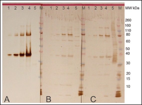
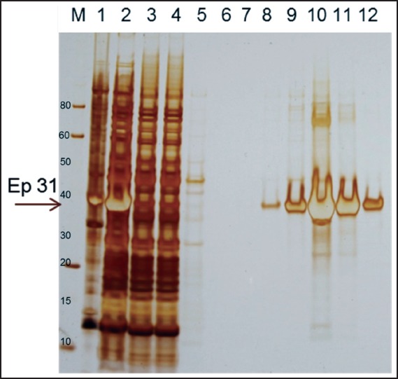
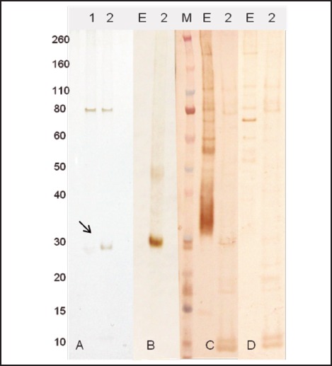
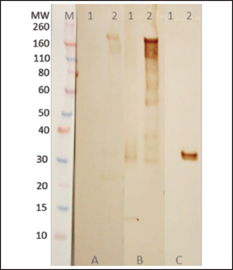
Cerebral angiostrongyliasis is an acute inflammation caused by the infection of the nematode Angiostrongylus cantonensis that results in eosinophilic meningitis. The current immunological assay of choice is an immunoblot that detects antibodies to a 31 kDa protein present in crude extracts of the female worm. Recently we have identified diagnostic targets from excretion and secretion products and determined the composition of the 31 kDa antigen after 2-D gel electrophoresis and mass spectrometry. Here we cloned and expressed five proteins in prokaryotic and eukaryotic systems. Recombinant proteins were purified and analysed by Western blot assays and among them 14-3-3, Lec5 and ES7 were recognized by Angiostrongylus-specific serum, although the signal was weak.

Keywords: 31 kDa antigen; Angiostrongylus; Eosinophilic meningitis; Recombinant protein.


Figures

Figure 1
Figure 1
Expression of Ep31. Putative epsilon subunit of coatomer protein complex isoform 2 (Ep31) of Angiostrongylus cantonens expressed in E. coli. Western blot analyses were performed using monoclonal anti-His tag (A), normal human (B) and Angiostrongylus-specific (C) serum. Lane 1 - Ep31 0.5 µg /lane; lane 2 - Ep31 0.3 µg/lane; lane 3 - Ep31 0.2 µg/lane; lane 4 - Ep31 0.1 µg /lane; lane 5 - crude extract of A. cantonensis 0.8 µg/lane. M - molecular weight in kDa.
Figure 2
Figure 2
Purification of Ep31. The recombinant protein Ep31 purified by affinity chromatography to nickel. Lane 1 - Ep31 cell lysed insoluble proteins; lane 2 - Ep31 cell lysed soluble proteins; lane 3 - column unbound proteins; lanes 4 and 5 - column washes; lane 6 - Ep31 elution 1; lane 7 - Ep31 elution 2; lane 8 - Ep31 elution 3; lane 9 - Ep31 elution 4; lane 10 - Ep31 elution 5; lane 11 - Ep31 elution 6; lane 12 - Ep31 elution 7; M - molecular weight in kDa. The arrow indicates the Ep31.
Figure 3
Figure 3
Expression and purification of ES7 protein. A - ES7 expressed in E. coli cells and purified by affinity chromatography to nickel: 1 - elution 1; 2 - elution 2. Western blot analyses were performed using monoclonal anti-His tag (B), Angiostrongylus-specific serum (C), and normal human serum (D). E - crude extract of A. cantonensis 0.8 µg. M - molecular weight in kDa. The arrow indicates the ES7 recombinant protein.
Figure 4
Figure 4
Expression and immunoblotting analyses of 14-3-3 and Lec5 recombinant proteins in E. coli cells. Western blot analyses were performed using monoclonal anti-His tag (A), Angiostrongylus-specific serum (B), and normal human serum (C). Lane 1 - 14-3-3 5 µg/lane; lane 2 - Lec5 0.8 µg/lane; lane 3 - 14-3-3 1 µg/lane; lane 4 - 14-3-3 3 µg/lane; lane 5 - 14-3-3 5 µg/lane; lane 6 - Lec5 0.1 µg/lane; lane 7 - Lec5 0.5µg/lane; lane 8 - Lec5 0.8 µg/lane M - molecular weight in kDa. The arrows indicate 14-3-3 (lane 1) and Lec5 (lane 2) recombinant proteins.
Figure 5
Figure 5
Expression of 14-3-3 recombinant protein in insect cells. Western blot analyses were performed using normal human serum (A), Angiostrongylus-specific serum (B), and monoclonal anti-His tag (C). Lane 1 - crude extract of A. cantonensis 0.5 µg/lane; lane 2 - 14-3-3 0.5 µg/lane. M - molecular weight in kDa.

References

    1. Graeff-Teixeira C, da Silva AC, Yoshimura K. Update on eosinophilic meningoencephalitis and its clinical relevance. Clin Microbiol Rev. 2009;22(2):322–348. - PMC - PubMed
    1. Eamsobhana P, Yong HS. Immunological diagnosis of human angiostrongyliasis due to Angiostrongylus cantonensis (Nematoda: Angiostrongylidae) Int J Infect Dis. 2009;13(4):425–431. - PubMed
    1. Eamsobhana P, Yoolek A, Suvouttho S, Suvuttho S. Purification of a specific immunodiagnostic Parastrongylus cantonensis antigen by electroelution from SDS-polyacrylamide gels. Southeast Asian J Trop Med Public Health. 2001;32:308–313. - PubMed
    1. Eamsobhana P, Yoolek A, Punthuprapasa P. Dot-blot ELISA for the immunological detection of specific antibody to Parastrongylus cantonensis. Trop Biomed. 2003;20:1–6.
    1. Morassutti AL, Levert K, Perelygin A, da Silva AJ, Wilkins P, Graeff-Teixeira C. The 31 kDa antigen of Angiostrongylus cantonensis comprises distinct antigenic glycoproteins. Vector-Borne Zoonot Dis. 2012;12(11):961–968. - PMC - PubMed

MeSH terms

Substances

Supplementary concepts