Review
. 2014 Aug;81(8):679-711.
doi: 10.1002/mrd.22223. Epub 2014 Jul 22.

The biology of the germ line in echinoderms

Affiliations
Review

The biology of the germ line in echinoderms

Gary M Wessel et al. Mol Reprod Dev. 2014 Aug.

Abstract

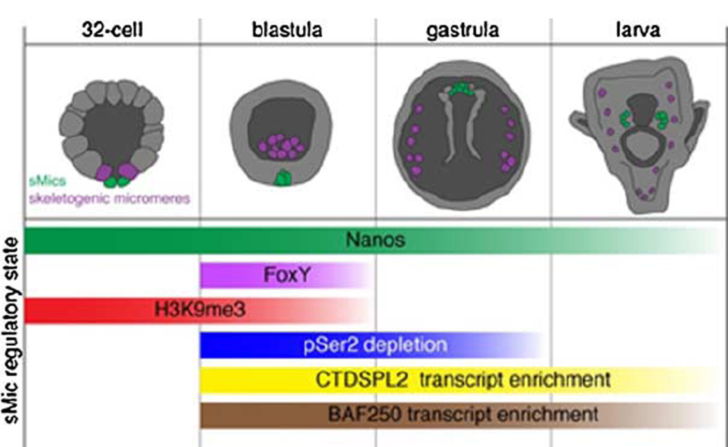
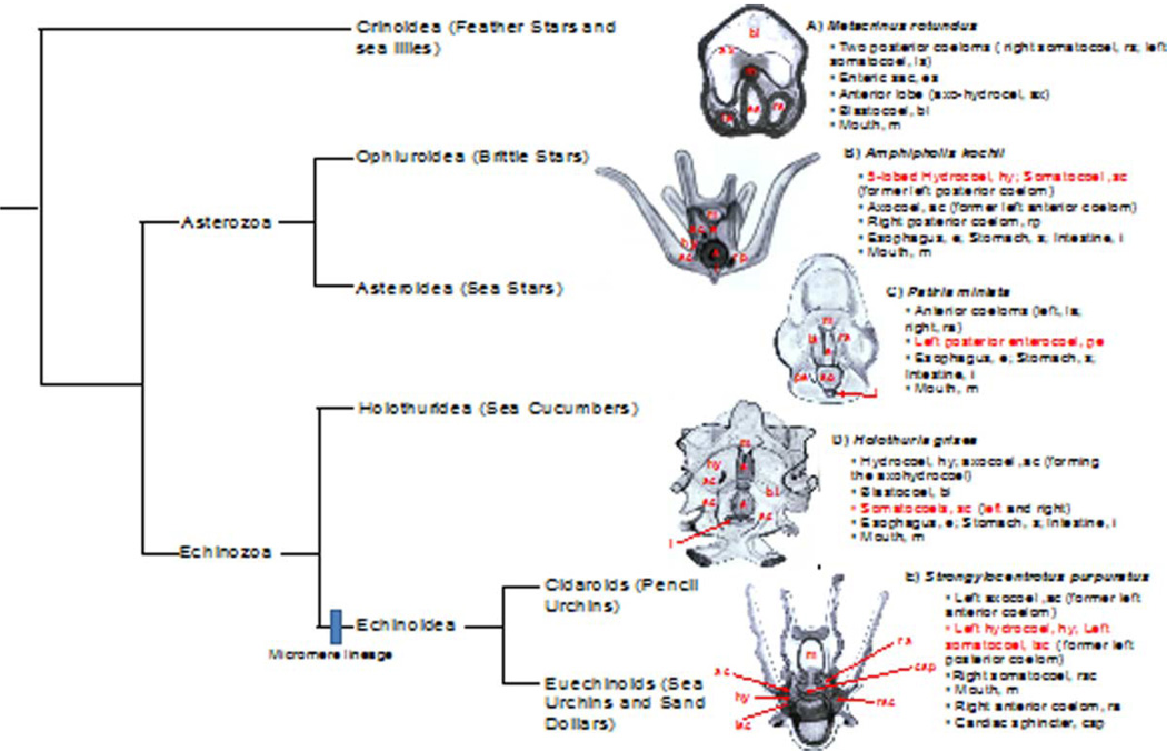
The formation of the germ line in an embryo marks a fresh round of reproductive potential. The developmental stage and location within the embryo where the primordial germ cells (PGCs) form, however, differs markedly among species. In many animals, the germ line is formed by an inherited mechanism, in which molecules made and selectively partitioned within the oocyte drive the early development of cells that acquire this material to a germ-line fate. In contrast, the germ line of other animals is fated by an inductive mechanism that involves signaling between cells that directs this specialized fate. In this review, we explore the mechanisms of germ-line determination in echinoderms, an early-branching sister group to the chordates. One member of the phylum, sea urchins, appears to use an inherited mechanism of germ-line formation, whereas their relatives, the sea stars, appear to use an inductive mechanism. We first integrate the experimental results currently available for germ-line determination in the sea urchin, for which considerable new information is available, and then broaden the investigation to the lesser-known mechanisms in sea stars and other echinoderms. Even with this limited insight, it appears that sea stars, and perhaps the majority of the echinoderm taxon, rely on inductive mechanisms for germ-line fate determination. This enables a strongly contrasted picture for germ-line determination in this phylum, but one for which transitions between different modes of germ-line determination might now be experimentally addressed.


Figures

Figure 1
Figure 1
Diagram of the development of a sea urchin. Early development yields Vasa-positive cells (shown in red, beginning with a uniform Vasa positive egg and early embryo). At the 32-cell stage, the sMics are uniquely Vasa-positive. These cells move into the coelom during gastrulation, segregate into the left and right coelomic pouches, and expand to contribute to the germ cells of the adult, and likely also to some somatic cells of the rudiment.
Figure 2
Figure 2
The morphology of the sMics. The early embryo of Lytechinus variegates was imaged in a variety of ways to expose the various cells. A: DAPI-labeled 32-cell stage viewed from the vegetal pole. B–D: lateral view of 32-cell stage embryos viewed with differential interference contrast (B), scanning electron microscopy (SEM) (C), and pseudocoloring (D) to identify the various tiers of cells. E–H: DAPI-stained progressions, viewed from the vegetal pole, of the formation of the four sMics (centered) followed by cell division of the large micromeres adjacent to them. The cell cycle of the sMics is shown relative to their neighboring and sibling cells. K: Spli 32-cell embryo fixed for SEM showing the various tiers of cells and the LMics and sMics at the bottom. M: A stereo view of a 32-cell stage embryo viewed form the vegetal pole. Stereo view glasses (B/W) or slightly crossed eyes are needed to view this pair. For more detailed information on the other cell types, see, for example, Horstadiuis (1976), McClay (2011), Gilbert (2013). Figure courtesy of John Morrill, personal communication.
Figure 3
Figure 3
Sea urchin sMics exhibit slow cell-cycling, as revealed by bromo-deoxyuridine (BrdU) pulse-chase studies. The sMics divide much slower relative to the somatic cells. Late gastrula (A, side view; B, top view) showing sMics selectively labeled with bromo-deoxyuridine (BrdU). BrdU was pulsed in the embryo following fertilization, washed, and the embryos were incubated until various time points. The sMics retain the BrdU relative to other cells of the embryo because they divide much slower than other cells (soma) and thereby do not dilute the BrdU in their genome (Tanaka and Dan, 1990; similar results were first shown by Pehrson and Cohen, 1986). Scale bar in A. 10 µm; in B 20 µm.
Figure 4
Figure 4
The sMics are dispersible for development. Diagram at top pointing out the sMic lineage (red). A: Method of sMic removal with a glass pipette. The resulting embryos develop into a larva (B–E), form an adult rudiment (D, E, arrow in E), and metamorphos into an adult (F) that does not produce gametes. Scale bars, __ µm.
Figure 5
Figure 5
Summary of the Vasa-compensatory mechanism. Vasa mRNA is abundant in S. purpuratus 16-cell stage embryos, but not in L. variegates embryos of the same stage. When micromeres are removed from embryos of each species, Vasa protein is upregulated in the blastulae in proportionl to the amount of vasa mRNA present. Adults are obtained from each species, at a frequency proportional to the amount of Vasa protein upregulated. When sMics are removed from either species at the 32-cell stage, no Vasa up-regulation is detectable and no germ cell production is seen.
Figure 6
Figure 6
Vasa associates with the mitotic spindle in early cleavage divisions. Vasa is present throughout the cytoplasm in early embryos, but becomes enriched in the spindle. during mitosis. Scale bars, __ µm. Reprinted with permission from Yajima and Wessel (2011).
Figure 7
Figure 7
Vasa is asymmetrically partitioned into the micromeres. During anaphase of the 4th cell division in the vegetal tier of blastomeres, Vasa becomes enriched in the future micromere region of the spindle and concentrates in the vegetal-most region of the embryo. This phenomenon is repeated again to form the sMics, which retain the highest concentration of Vasa Whole embryos are shown in A and B and are each ~90 microns in diameter, whereas the micromeres shown in C, D, and E are each ~10 microns in diameter. in the embryo. Scale bars, __ µm. Reprinted with permission from Yajima and Wessel (2011).
Figure 8
Figure 8
The endoplasmic reticulum is enriched at the vegetal pole in early development. Scale bar, __ µm. scale bar is 50 microns Reprinted with permission from Yazaki et al. (2004).
Figure 9
Figure 9
Calcium fluxes (red circle in photos, and red line in graph) are unique in the sMics. Scale bar, __ µm. Scale bar - 50 microns Reprinted with permission from Yazaki (2001).
Figure 10
Figure 10
Post-translational Vasa regulation during sea urchin embryogenesis. A: Whole-mount in situ RNA hybridization of endogenous vasa transcript at the indicated developmental stages. B: Immunofluorescence localization of endogenous Vasa protein at the indicated embryonic stages. C: Two synthetic mRNAs were co-injected into fertilized eggs, then imaged at the blastula stage. One mRNA contained the Vasa open reading frame with a C-terminal GFP tag flanked by β-globin 5′- and 3′-UTRs. The control mRNA contained only the mCherry open reading frame flanked by β-globin 5′- and 3′-UTRs. D: Immunofluorescence localization of endogenous Vasa protein in embryos cultured in artificial seawater (ASW) alone, ASW containing 0.5% dimethyl sulfoxide (DMSO), or ASW containing 25 or 50 µM MG132. Scale bars, _ µm. "control blastulae are 100 microns in diameter."
Figure 11
Figure 11
Temporal dynamics of sMic transcriptional activity. Sea urchin embryonic stages are schematized with the sMics indicated in green. Colored bars below indicate timing of events in the sMics
Figure 12
Figure 12
sMic retention of an exogenous RNA containing the mCherry open reading frame surrounded by the Xenopus β-globin UTRs. A: mCherry fluorescence at the gastrula stage after RNA injection. B: In situ RNA hybridization at the gastrula stage after injection of the RNA.
Figure 13
Figure 13
Summary of the RNA constructs tested for sMic RNA retention. The open reading frame is surrounded by Xenopus β-globin UTRs. The black circle represents the m7GTP cap, and the polyadenylation tail is symbolized by A30. In the second construct, the "x" indicates the mutation of the polyadenylation signal in the 3′-UTR from β-globin.
Figure 14
Figure 14
Sp GNARLE is necessary for selective RNA retention and protein accumulation in the sMics. A: Schematic representation of nanos2 RNA, indicating the location of GNARLE in the 3′-UTR. B: RNAs containing the Sp nanos2 5′-UTR followed by a GFP open reading frame and by either GNARLE or ΔGNARLE 3′-UTR were injected in S. purpuratus fertilized eggs. At blastula stage, RNA retention was tested by in situ hybridization and protein accumulation was followed by GFP fluorescence.
Figure 15
Figure 15
Differential accumulation of CNOT6 in somatic and germ-line blastomeres. A: Cnot6 transcript (green) is detectable in all cells of an 18-hr blastula by fluorescence in situ hybridization, except in the sMics, which are labeled by Vasa immunofluorescence (red). B,C: Schematization of differential RNA retention in somatic cells versus sMics. B: Cnot6 RNA is present in all somatic cells, and is translated and likely incorporated into a CCR4-NOT related complex, where it affects the degradation of germ-line RNAs. C: Nanos/Pumilio targets the Cnot6 transcript for degradation, leading to a reduction in de-adenylase activity in the sMics. This reduction creates a generally stable environment for both endogenous germ-line RNAs and microinjected reporters.
Figure 16
Figure 16
ABC transporters are involved in PGC success. A: In Drosophila, the ABC transporter mdr49 is required for germ-cell migration to the mesoderm. An mdr49D3.16 embryo (bottom) shows lost germ cells. Reprinted with permission from Ricardo and Lehmann (2009). B: The fluorescent indicator calcein indicates ABC activity the absence of fluorescence indicates active efflux, whereas the presence indicates a lack of ABC efflux activity. Remarkably, the sMics have low ABC-effluxing activity and, as a result, retain calcein in the cytoplasm. 10 microns Reprinted with permission from Campanale and Hamdoun (2012)
Figure 17
Figure 17
Members of the phylum Echinodermata. There are five extant classes of echinoderms: Crinoidea, Ophiuroidea, Asteroidea, Holothuridea, and Echinoidea. Representatives of these classes: sea lily Metacrinus rotundus; brittle star Amphipholis kochii; sea star P. miniata; sea cucumber Holothuria grisea; sea urchin S. purpuratus. Schematic drawings of larval stages indicating different tissues for each organism. A: A 3.5-day, non-feeding auricularia larva. (B) A 6-day, eight-armed ophiopluteus. (C) A 5-day bipinnaria larva. (D) A 4-day aspidochirotid auricularia. (E) A 4-armed pluteus larva. Dorsal views of the larvae are shown. Members of the class Echinoidea, which have a micromere lineage, are split into the subclasses Euechinoids (sea urchins and sand dollars) and Cidaroids (pencil urchins).
Figure 18
Figure 18
Late gastrula stage from P. miniata This dynamic sequence shows mesenchyme migrating from the apical plate (ap) and coming in contact with the anterior vesicle (av). 1: The archenteron bears the usual two enterocele pouches and a single, left posterior one. 2: Anterior end of bipinnaria showing the completion of the mesenchyme formation at the apical pole and its migration to the esophageal region. The left hydrocele is provided with a pore canal and hydropore. lp, left posterior vesicle. 3: Section of gastrula showing the apical plate with segns of mesenchyme formation. 4: Bipinnaria showing mesenchyme migrating past the anterior vesicle (av) to the esophagus. The left hydrocele has severed its connection with the archenteron. lp, left posterior vesicle in process of formation. 5: Surface overview of gastrula immediately before mesenchyme development from the apical plate. Reprinted with permission from Heath (1917).

References

    1. Amit S, Hatzubai A, Birman Y, Andersen JS, Ben-Shushan E, Mann M, Ben-Neriah Y, Alkalay I. Axin-mediated CKI phosphorylation of beta-catenin at Ser 45: A molecular switch for the Wnt pathway. Genes Dev. 2002;16:1066–1076. - PMC - PubMed
    1. Ancelin K, Lange UC, Hajkova P, Schneider R, Bannister AJ, Kouzarides T, Surani MA. Blimp1 associates with Prmt5 and directs histone arginine methylation in mouse germ cells. Nat Cell Biol. 2006;8:623–630. - PubMed
    1. Anderson P, Kedersha N. RNA granules. J Cell Biol. 2006;172:803–808. - PMC - PubMed
    1. Andrews S, Snowflack DR, Clark IE, Gavis ER. Multiple mechanisms collaborate to repress nanos translation in the Drosophila ovary and embryo. RNA. 2011;17:967–977. - PMC - PubMed
    1. Angerer LM, Angerer RC. Animal–vegetal axis patterning mechanisms in the early sea urchin embryo. Dev Biol. 2000;218:1–12. - PubMed

Publication types

MeSH terms

LinkOut - more resources