. 2014 Dec;24(12):3301-9.
doi: 10.1093/cercor/bht188. Epub 2013 Jul 30.

Impaired prefrontal sleep spindle regulation of hippocampal-dependent learning in older adults

Affiliations

Impaired prefrontal sleep spindle regulation of hippocampal-dependent learning in older adults

Bryce A Mander et al. Cereb Cortex. 2014 Dec.

Abstract

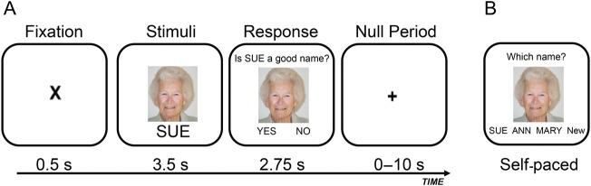
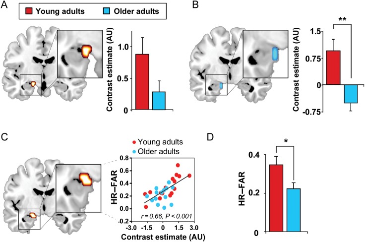
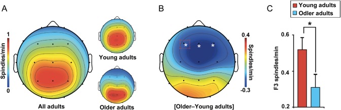
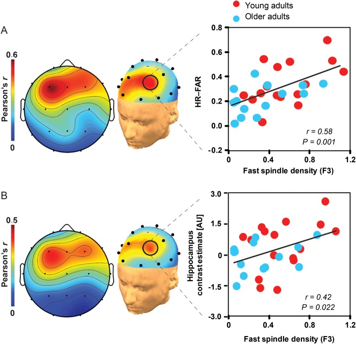
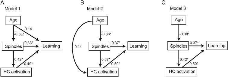
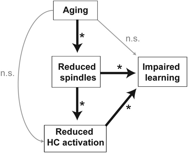
A hallmark feature of cognitive aging is a decline in the ability to form new memories. Parallel to these cognitive impairments are marked disruptions in sleep physiology. Despite recent evidence in young adults establishing a role for sleep spindles in restoring hippocampal-dependent memory formation, the possibility that disrupted sleep physiology contributes to age-related decline in hippocampal-dependent learning remains unknown. Here, we demonstrate that reduced prefrontal sleep spindles by over 40% in older adults statistically mediates the effects of old age on next day episodic learning, such that the degree of impaired episodic learning is explained by the extent of impoverished prefrontal sleep spindles. In addition, prefrontal spindles significantly predicted the magnitude of impaired next day hippocampal activation, thereby determining the influence of spindles on post-sleep learning capacity. These data support the hypothesis that disrupted sleep physiology contributes to age-related cognitive decline in later life, the consequence of which has significant treatment intervention potential.

Keywords: aging; fMRI; hippocampus; learning; sleep.


Figures

Figure 1.
Figure 1.
(A) Example face-name encoding fMRI trial (read left to right). During each trial, a fixation was first presented for 0.5 s, followed by a face-name pair for 3.5 s. This was followed by a screen for 2.75 s during which participants had to determine whether the name “fits” the face while still presenting the face-name pair, allowing for a confirmation that participants attended to each face-name pair, and promoted deeper encoding of each face-name pair. Sixty null events, consisting of a fixation display and varying in duration from 1.5 to 10 s, were interspersed to jitter trial onsets. (B) Example face-name recognition trial, presenting a studied face-name pair. During each self-paced recognition trial, a face was first presented on the screen with 4 options presented below that participants chose among allowing for a determination of associative memory recognition: (1) the original name previously paired with that face (correct “Hit” response), (2) a name previously seen before at encoding, but with a different face (incorrect “Lure” response), (3) a new name never shown during encoding (incorrect response), or (4) an option “new” rejecting the trial as a foil trial. Face-name recognition memory was calculated by subtracting the proportion of new faces endorsed as “studied” faces and incorrectly paired with a name (false alarm rate) from the proportion of studied faces correctly named (hit rate).
Figure 2.
Figure 2.
Effects of age on hippocampal functioning. (A) Hippocampal activation greater during successful associative episodic encoding than unsuccessful associative encoding (Hits–Lures; 8-mm sphere ROI: x = −21, y = −6, z = −15) (Miller et al. 2008) and (B) corresponding age effects (Hits–Lures; older < young adults) in the same ROI. (C) Regression between episodic learning and encoding-related hippocampal activation (Hits–Lures) in the above-mentioned ROI. (D) Episodic learning in young and older adults, both performing significantly greater than chance (HR-FAR: hit rate to originally studied faces–false alarm rate to new, unstudied faces). Activations are displayed and considered significant at the voxel level of P < 0.05 FWE, corrected for multiple comparisons within the a priori hippocampal ROIs (Miller et al. 2008). Hot colors represent the extent of activation in both young and older adults, and cold colors represent the extent of decreased activation in older, relative to younger, adults. While the 8-mm sphere ROI used to correct for multiple comparisons did extend outside the hippocampus, no effects were detected or presented outside the hippocampus in the current report. Bilateral effects were detected in the hippocampus, when using an anatomical hippocampal mask, albeit at a lower statistical threshold (P < 0.005 uncorrected; Supplementary Fig. 2). *P < 0.05, **P < 0.001.
Figure 3.
Figure 3.
Age effects on fast sleep spindle density. (A) electroencephalography (EEG) topographic plots of fast spindle density in all adults with young (top) and older (bottom) adult plots to the right, and (B) the fast sleep spindle density difference between young and older adults, significant over prefrontal EEG derivations. Red box indicates the stage 2 NREM sleep fast spindle density at the F3 derivation, plotted as a bar graph (C) in young (red) and older (blue) adults. * denotes significance at P < 0.05.
Figure 4.
Figure 4.
Associations between sleep spindles, hippocampal activation, and episodic learning. (A) EEG topographic plots of the association between fast sleep spindle density and episodic learning, with the corresponding regression for young (red) and older (blue) adults plotted to the right, focusing on the left prefrontal EEG derivation (F3) where the spindle-learning association is significant false discovery rate corrected (Benjamini and Hochberg 1995). (B) EEG topographic plots of the association between fast sleep spindle density and encoding-related hippocampal activation (described in Fig. 2C), with the corresponding regression for young (red) and older (blue) adults plotted to the right, focusing on the left prefrontal EEG derivation (F3). Associations were specific to fast sleep spindles, as subjective measures of sleepiness and alertness (all P > 0.12), objective alertness response times collected during encoding and recognition testing (all P > 0.20), absolute and relative slow-wave activity (0.8–4.6 Hz calculated as described previously, Mander et al. 2011, all P > 0.10), circadian preference (all P > 0.72), and scanning time relative to wake time (all P > 0.16) did not correlate with: (1) Episodic learning, (2) hippocampal activation, or (3) prefrontal fast sleep spindles, in either young or older adults. Associations were also specific to the hippocampus, as activation within an encoding-relevant region of the left prefrontal cortex (Spaniol et al. 2009) did not differ between age groups (P = 0.244) and was not associated with either frontal fast sleep spindles (r2 < 0.01, P = 0.969) or episodic learning ability across participants (r2 < 0.1, P = 0.156; see Supplementary Results).
Figure 5.
Figure 5.
Path analysis models examining the relative contributions of age, frontal fast sleep spindles (spindles), and hippocampal (HC) activation to learning ability (learning) in 3 hypothesized models (AC). Values represent standardized regression weights. Models are estimated and model fit for model 1 (A, AICc = 40.191; BIC = 31.191; RMR = 0.019; GFI = 0.990), model 2 (B, AICc = 40.658; BIC = 31.658; RMR = 0.003; GFI = 0.983), and model 3 (C, AICc = 36.837; BIC = 28.837; RMR = 0.019; GFI = 0.973) are compared against a saturated model (AICc = 44.012; BIC = 34.012; RMR = 0.000; GFI = 1.000) and an independence model (AICc = 52.320; BIC = 48.320; RMR = 0.070; GFI = 0.602). * denotes path significance at P < 0.05.
Figure 6.
Figure 6.
Model schematic of path and mediation analyses findings. Aging is associated with reduced frontal fast sleep spindles, which mediates the degree of reduced next day, encoding-related hippocampal (HC) activation and impaired episodic learning ability in older adults. Furthermore, age did not significantly impact learning or hippocampal activation independently of its impact on frontal fast sleep spindles in the present study. *Denotes paths that are significant at P < 0.05.

References

    1. Andrade KC, Spoormaker VI, Dresler M, Wehrle R, Holsboer F, Samann PG, Czisch M. Sleep spindles and hippocampal functional connectivity in human NREM sleep. J Neurosci. 2011;31:10331–10339. - PMC - PubMed
    1. Ashburner J. A fast diffeomorphic image registration algorithm. Neuroimage. 2007;38:95–113. - PubMed
    1. Ashburner J, Friston KJ. Voxel-based morphometry—the methods. Neuroimage. 2000;11:805–821. - PubMed
    1. Benjamini Y, Hochberg Y. Controlling the false discovery rate—a practical and powerful approach to multiple testing. J R Stat Soc Ser B Method. 1995;57:289–300.
    1. Bollinger J, Rubens MT, Masangkay E, Kalkstein J, Gazzaley A. An expectation-based memory deficit in aging. Neuropsychologia. 2011;49:1466–1475. - PMC - PubMed

Publication types

MeSH terms