. 2013 Aug 13;110(33):13504-9.
doi: 10.1073/pnas.1309618110. Epub 2013 Jul 30.

Structural basis for the stabilization of the complement alternative pathway C3 convertase by properdin

Affiliations

Structural basis for the stabilization of the complement alternative pathway C3 convertase by properdin

Martín Alcorlo et al. Proc Natl Acad Sci U S A. .

Abstract

Complement is an essential component of innate immunity. Its activation results in the assembly of unstable protease complexes, denominated C3/C5 convertases, leading to inflammation and lysis. Regulatory proteins inactivate C3/C5 convertases on host surfaces to avoid collateral tissue damage. On pathogen surfaces, properdin stabilizes C3/C5 convertases to efficiently fight infection. How properdin performs this function is, however, unclear. Using electron microscopy we show that the N- and C-terminal ends of adjacent monomers in properdin oligomers conform a curly vertex that holds together the AP convertase, interacting with both the C345C and vWA domains of C3b and Bb, respectively. Properdin also promotes a large displacement of the TED (thioester-containing domain) and CUB (complement protein subcomponents C1r/C1s, urchin embryonic growth factor and bone morphogenetic protein 1) domains of C3b, which likely impairs C3-convertase inactivation by regulatory proteins. The combined effect of molecular cross-linking and structural reorganization increases stability of the C3 convertase and facilitates recruitment of fluid-phase C3 convertase to the cell surfaces. Our model explains how properdin mediates the assembly of stabilized C3/C5-convertase clusters, which helps to localize complement amplification to pathogen surfaces.


Conflict of interest statement

The authors declare no conflict of interest.

Figures
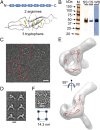
Fig. 1.
Fig. 1.
Structure of properdin oligomers by electron microscopy. (A) Schematic cartoon of the arrangement of TSR domains in a properdin monomer (Upper) and a view of the atomic structure of one homolog TSR domain from thrombospondin (PDB 3R6B, Lower) (10). Side chains of the proposed key arginine and tryptophan residues are shown in blue and yellow, respectively. Disulfide bonds are shown in pink. (B) Final preparation of purified properdin analyzed by SDS/PAGE. SS, silver staining; CS, Coomassie staining; WB, Western blotting using polyclonal antibodies against properdin. (C) Typical micrograph of properdin. Selected single molecule images for several properdin oligomers are highlighted within a red square. (Scale bar, 26 nm.) (D) Reference-free 2D averages of properdin vertexes extracted from the micrographs reveal several views of the structure connecting two monomers. (Scale bar, 7 nm.) (E) 3D structure of the properdin vertex and pseudoatomic model obtained by fitting a crystal structure of a TSR domain from thrombospondin (PDB 3R6B) (10) into the EM density. (Scale bar, 1.2 nm.) (F) Carton representation of a properdin tetramer (Lower) and a raw image for a properdin tetramer (Upper). Vertexes are represented as a blue circle and the region whose distance was measured is indicated.
Fig. 2.
Fig. 2.
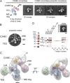
Purification and electron microscopy of the properdin–C3bBb convertase complex. (A) Chromatograms (Upper) and silver-stained SDS/PAGE (Lower) for the fractions of size-exclusion chromatography experiments performed in a Superdex 200 gel-filtration column (GE Healthcare) using properdin, C3b, FD, and either wild-type FB or the FB-D279G mutant. Chromatograms show profiles for properdin injected alone (P, blue line), the incubation of C3b, FB, and FD to assemble a C3bBb convertase (C3bBb, magenta discontinuous line), and the incubation of properdin, C3b, FB-D279G, and FD to assemble a properdin–C3bBb convertase complex (PC3bBb, green line). Lower shows SDS/PAGE of selected fractions from the chromatographies above. (Left) Assembly of a C3bBb convertase (C3bBb, magenta discontinuous line). (Right) Properdin–C3bBb convertase complex (PC3bBb, green line). (Center) SDS/PAGE of a chromatography analyzing the interaction of properdin with the C3bB proconvertase. The input to the gel-filtration column is indicated as IN, and C3b, FB, and properdin are loaded as controls. Chains of C3b detected in the SDS/PAGE are indicated. The formation of the properdin–C3bBb convertase complex is revealed by the advanced elution of C3bBb (factions 10–15) in the presence of properdin compared with the elution of C3bBb convertase alone (C3bBb, fractions 17–21), as well as the appearance of a new band corresponding to the FB fragment Bb (labeled Bb) resulting from the proteolysis of the input FB (labeled FB). The fraction selected for EM analysis is labeled. (B) Representative micrograph corresponding to properdin–C3bBb convertase complexes collected at two experimental conditions generating partial (Left) or high occupancy (Right) of properdin by C3bBb convertase. Selected C3bBb convertase molecules bound to properdin have been labeled with an open arrow. Black arrows stand for unbound C3bBb convertase molecules. (Scale bar, 14 nm.) (C) Gallery of raw images of properdin–C3bBb convertase complexes selected from the micrographs and panelled according to the oligomeric state of properdin, and showing, from left to right, increased occupancy of properdin vertexes with C3bBb convertase molecules. (Scale bar, 14 nm.)
Fig. 3.
Fig. 3.
Structure of the properdin–C3bBb convertase complex. (A) Representative reference free 2D averages of C3bBb convertase molecules bound to properdin vertexes (Right), compared with a view of the crystallographic and EM structures of C3bBb convertase (Left) (PDB 2WIN) (17). Each domain has been colored differently and labeled. (Scale bar, 5 nm.) (B) Selected average of the properdin–C3bBb convertase complex. Different domains and regions are labeled. (Scale bar, 5 nm.) (C) Two views of the structure of the properdin–C3 convertase complex at 29.3-Å resolution. A pseudoatomic model of the properdin–C3 convertase complex was obtained by fitting the atomic structure of C3bBb convertase (PDB 2WIN) (17) into the EM structure. The MG ring is displayed in blue. C345, CUB, and TED domains are colored in orange, red, and green, respectively. vWA and SP domains from the Bb fragment are colored in pink. Densities corresponding to properdin vertex are labeled with asterisks. (Scale bar, 2 nm.) (D) Fractions from a size-exclusion chromatography loaded with the incubation of CVF, FB-D279 mutant, properdin, and FD were analyzed by SDS/PAGE. Properdin does not interact with CVF-FB in the conditions tested, as revealed by the absence of comigration of the CVFB complex with properdin. Inset, Upper Right corner shows an average of images obtained for the purified CVFB complex using electron microscopy. (Scale bar, 5 nm.)
Fig. 4.
Fig. 4.
Positioning of the TED domain in the properdin–C3bBb convertase complex. (A) Representative 2D averages of the minor conformation of the properdin–C3bBb convertase complex. These show that the TED domain is not in the proximities of the MG3 domain, but in the location found in the crystal structure of C3b. (Scale bar, 5 nm.) (B) One view of the structure of the minor conformation of the properdin–C3bBb convertase complex at 33.0-Å resolution. Densities corresponding to properdin vertex are labeled with asterisks. (Scale bar, 2 nm.) (C) Processing and classification of images of C3b revealed that most molecules show the TED domain in the classical conformation, whereas a small percentage of molecules display the TED domain in alternative conformations. An arrow points to the TED domain placed close to the MG3 domain, found in 3.5% of the images analyzed. A view from the crystal structure of C3b (PDB 2I07) is shown to help comparison with the EM images. Each domain has been colored differently and labeled. (Scale bar, 5 nm.)
Fig. 5.
Fig. 5.
C3bBb convertase decay in the presence of MCP, FI, and properdin. (A) C3bBb convertase was formed by incubating C3b, FB, and FD in the absence (P) (−) or the presence of two amounts of properdin (P) (0.9 µg + and 1.8 µg ++) and incubated with MCP and FI. SDS/PAGE shows the result of this reaction after incubating for 15 min. Each experiment was performed in duplicate. (B) The amount of C3b remaining after incubation was estimated by quantifying the ratio between α′ chain/β chain of C3b. Experiments labeled as 1–4 correspond to the matching experiment in A (0.9 µg of P, gray and 1.8 µg of P, black). Error bars indicate the mean ± SD of two independent experiments.
Fig. 6.
Fig. 6.
Model for complement activation by properdin. (A) Idealized images of properdin and properdin–C3bBb convertase complexes generated by combining the averages and the dimensions of experimental single molecule images. (B) Cartoon representing the three alternative models for the arrangement of subunits at the vertex of properdin oligomers (see Discussion). Alternating oligomers are shown in black and gray. The TSR domains are numbered from 0 to 6.

Similar articles

Cited by

References

    1. Ricklin D, Hajishengallis G, Yang K, Lambris JD. Complement: A key system for immune surveillance and homeostasis. Nat Immunol. 2010;11(9):785–797. - PMC - PubMed
    1. Rodríguez de Córdoba S, Harris CL, Morgan BP, Llorca O. Lessons from functional and structural analyses of disease-associated genetic variants in the complement alternative pathway. Biochim Biophys Acta. 2011;1812(1):12–22. - PubMed
    1. Gros P, Milder FJ, Janssen BJ. Complement driven by conformational changes. Nat Rev Immunol. 2008;8(1):48–58. - PubMed
    1. Lea SM, Johnson S. Putting the structure into complement. Immunobiology. 2012;217(11):1117–1121. - PMC - PubMed
    1. Hourcade DE. The role of properdin in the assembly of the alternative pathway C3 convertases of complement. J Biol Chem. 2006;281(4):2128–2132. - PubMed

Publication types