. 2013 Jul 30;6(286):ra63.
doi: 10.1126/scisignal.2004177.

miR-29 acts as a decoy in sarcomas to protect the tumor suppressor A20 mRNA from degradation by HuR

Affiliations

miR-29 acts as a decoy in sarcomas to protect the tumor suppressor A20 mRNA from degradation by HuR

M Y Balkhi et al. Sci Signal. .

Erratum in

  • Sci Signal. 2013 Sep 10;6(292):er6. Balkhi, Mumtaz Y [corrected to Balkhi, M Y]

Abstract

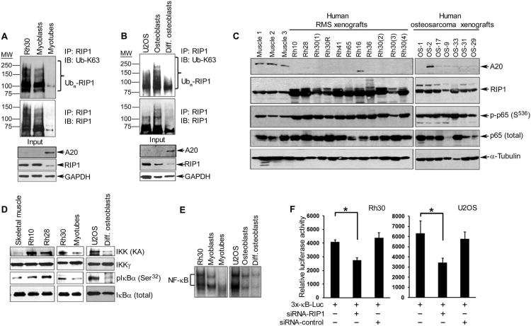
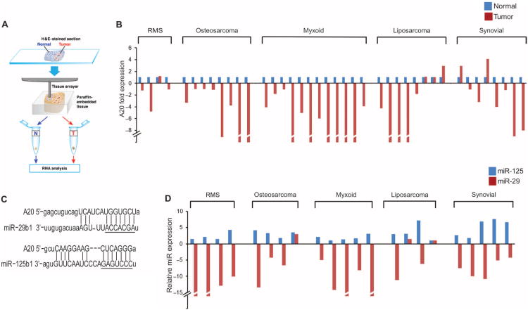
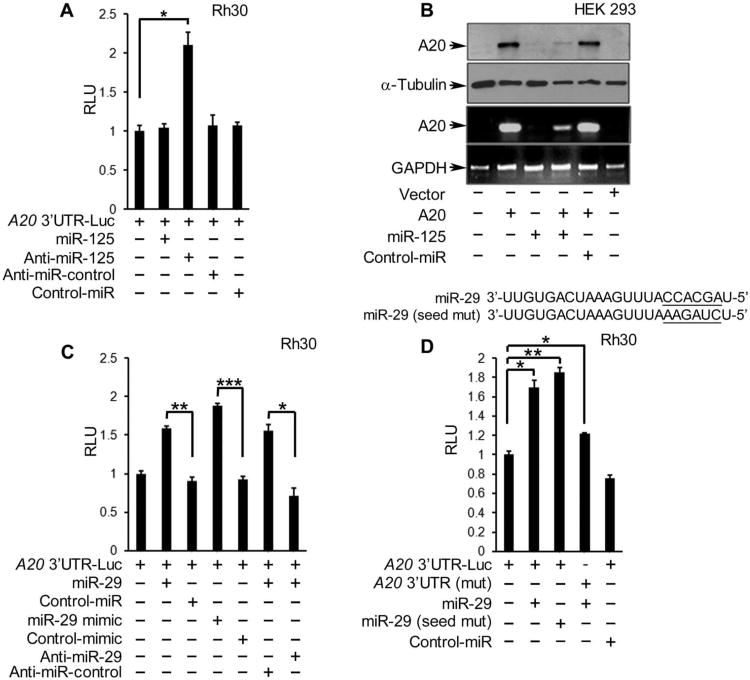
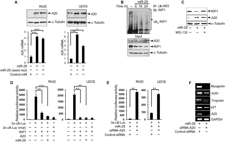
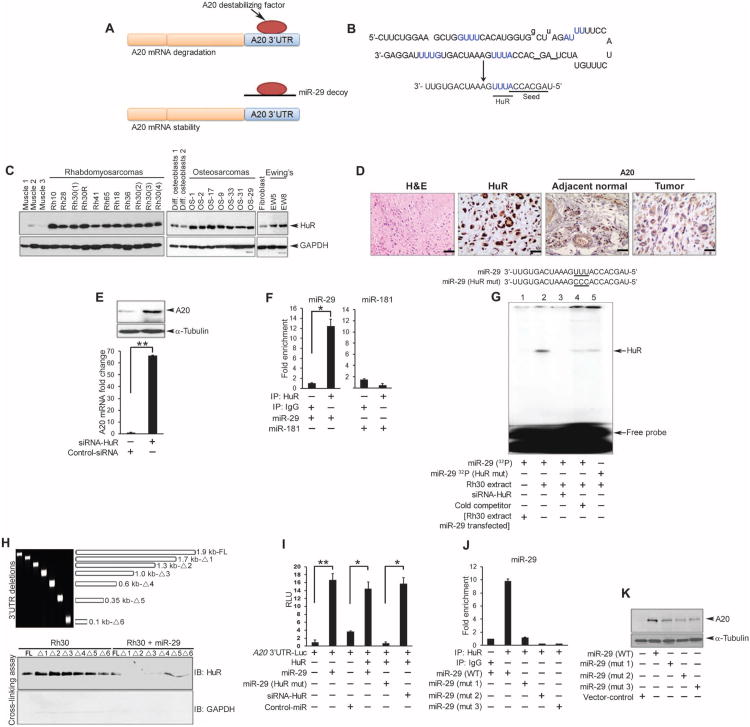
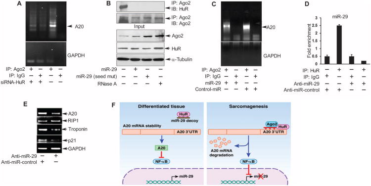
In sarcoma, the activity of NF-κB (nuclear factor κB) reduces the abundance of the microRNA (miRNA) miR-29. The tumor suppressor A20 [also known as TNFAIP3 (tumor necrosis factor-α-induced protein 3)] inhibits an upstream activator of NF-κB and is often mutated in lymphomas. In a panel of human sarcoma cell lines, we found that the activation of NF-κB was increased and, although the abundance of A20 protein and mRNA was decreased, the gene encoding A20 was rarely mutated. The 3' untranslated region (UTR) of A20 mRNA has conserved binding sites for both of the miRNAs miR-29 and miR-125. Whereas the expression of miR-125 was increased in human sarcoma tissue, that of miR-29 was decreased in most samples. Overexpression of miR-125 decreased the abundance of A20 mRNA, whereas reconstituting miR-29 in sarcoma cell lines increased the abundance of A20 mRNA and protein. By interacting directly with the RNA binding protein HuR (human antigen R; also known as ELAVL1), miR-29 prevented HuR from binding to the A20 3'UTR and recruiting the RNA degradation complex RISC (RNA-induced silencing complex), suggesting that miR-29 can act as a decoy for HuR, thus protecting A20 transcripts. Decreased miR-29 and A20 abundance in sarcomas correlated with increased activity of NF-κB and decreased expression of genes associated with differentiation. Together, the findings reveal a unique role of miR-29 and suggest that its absence may contribute to sarcoma tumorigenesis.


Conflict of interest statement

Competing interests: C.K. received honoraria for presentations from Novartis, GlaxoSmithKline, and Takeda.

Figures

Fig. 1
Fig. 1. RIP1 activates NF-κB in RMS and osteosarcoma in association with loss of A20
(A) Immunoprecipitation (IP) for RIP1, followed by Western blotting (IB) for K63-linked ubiquitin (Ub-K63), RIP1, and A20 in lysates from human Rh30 cells and nontransformed mouse C2C12 muscle cells (undifferentiated myoblasts and differentiated myotubes). Blots were stripped and reprobed for glyceraldehyde-3-phosphate dehydrogenase (GAPDH) as a loading control. (B) Same as in (A) except that reactions were performed with U2OS cell lines and human primary undifferentiated and differentiated osteoblasts. (C) Western blotting for A20, RIP1, phosphorylated p65 (Ser536), and total p65 (against α-tubulin as a loading control) in lysates from human RMS and osteosarcoma xenograft tumors compared with normal human skeletal muscle tissue. (D) Kinase assays followed by Western blotting to assess IKK activity in lysates from xenograft tumors or sarcoma cell lines, differentiated myotubes or osteoblasts, and murine skeletal muscle tissue used as control. (E) Electrophoretic mobility shift assay to measure the DNA binding activity of NF-κB in Rh30 and U2OS cell lines compared with their respective nontransformed control cell lines. (F) NF-κB reporter assays in Rh30 and U2OS sarcoma cell lines in the presence of either RIP1 small interfering RNA (siRNA) or a control siRNA. Data are means ± SD of relative luciferase activity, normalized to β-galactosidase to control for transfection efficiency, from a representative of three independent experiments; *P < 0.05, two-tailed Student′s t test. All blots (A to E) are representative of three independent experiments.
Fig. 2
Fig. 2. Abundance of A20 and miR-29 is decreased in human sarcomas and is inversely correlated with that of miR-125
(A) Schematic of the extraction of tumor and adjacent normal tissue from paraffin-embedded blocks. (B) qRT-PCR for A20 (normalized to GAPDH) in adjacent normal and tumor tissue sections of sarcoma biopsies. Data are representative of three technical repeats per sample; P = 6.055 × 10⁻⁵, two-tailed parametric t test. (C) TargetScan/MicroRNA prediction analysis for complementary sequences to the 3′UTR of A20. The seed regions of miR-29 and miR-125 sequences are underlined. (D) qRT-PCR for miR-29 and miR-125 in patient tumor material as in (B).
Fig. 3
Fig. 3. A20 abundance and reporter activity is regulated by miR-29 and miR-125
(A) A20 3′UTR luciferase reporter activity in Rh30 cells cotransfected with the indicated miRNAs, normalized to that in nontransfected cells and those transfected with the reporter alone. RLU, relative light units. (B) Western blotting and qRT-PCR in HEK 293 cells cotransfected with an A20 expression plasmid and miR-125 or a control (control-miR). Blots are representative of three experiments. (C and D)A20 3′UTR luciferase reporter assays in Rh30 cells cotransfected with (C) a wild-type (WT) UTR reporter construct and the indicated miRNAs or (D) a reporter containing WT or mutated miR-29 binding site [A20 3′UTR (mut)] and either WT or seed sequence-mutated miR-29. Graphs in (A), (C), and (D) are means ± SD from a representative of at least three independent experiments. *P < 0.05, **P < 0.01, and ***P< 0.005, two-tailed Student's t test.
Fig. 4
Fig. 4. MiR-29 rescues A20 abundance and promotes a differentiated phenotype in RMS tumor cells
(A) Western blotting and qRT-PCR for A20 protein and mRNA, respectively, in Rh30 and U2OS cells 24 hours after transfection with miR-29, miR-29 (seed mut), or control-miR. (B) Immunoprecipitation and Western blotting for K36-linked ubiquitinated RIP1 in Rh30 cells performed at the indicated times after transfection with miR-29. (C) Western blotting for A20 and RIP1 in Rh30 cells transfected with miR-29 treated with or without MG-132 for 6 hours. (D) NF-κB promoter reporter luciferase assays in Rh30 and U2OS cells cotransfected with different combinations of RIP1, A20, and miR-29. (E) NF-κB reporter assays in Rh30 cells cotransfected with miR-29 and A20 or control siRNA. (F) RT-PCR for skeletal muscle differentiation markers in Rh30 cells cotransfected with miR-29 and A20 or control siRNA. Blots are representative of a minimum of two repeats, and data are means ± SEM from three independent experiments; *P < 0.05, **P < 0.01, and ***P < 0.005.
Fig. 5
Fig. 5. MiR-29 functions as a decoy for HuR
(A) Model of miR-29 as a decoy against an RNA-destabilizing factor that contributes to A20 mRNA turnover. (B) Sequences of precursor and mature miR-29. Predicted HuR binding sites are in blue. The seed sequence and HuR binding site are underlined in mature miR-29. (C) Western blot for HuR in human xenograft sarcoma tumors compared with control murine muscle tissue, human differentiated osteoblasts, or murine fibroblasts. (D) H&E staining and immunohistochemistry for A20 and HuR in formalin-fixed paraffin-embedded osteosarcoma. Images are representative of four samples, stained in duplicate; scale bars, 4 μm. (E) Western blotting and qRT-PCR for A20 in lysates from Rh30 cells transfected with control or HuR siRNA. (F) Immunoprecipitation for HuR or IgG followed by qRT-PCR for miR-29 or miR-181 in Rh30 cell lysates. (G) RMSA with a 32P-labeled miR-29 probe and extracts from Rh30 cells transfected with HuR (lanes 2 to 5), miR-29 (lane 1), HuR siRNA (lane 3), unlabeled miR-29 (lane 4, cold competitor), or miR-29 containing a mutated HuR binding site (lane 5, HuR mut). (H) Top: PCR fragments were amplified from the full-length (FL) A20 3′UTR or deletion constructs (Δ1 to Δ6) (table S1). Bottom: Western blotting for HuR in ultraviolet–cross-linked lysates from Rh30 cells transfected with or without miR-29 and the indicated fragment. Data are representative of two experiments. (I) A20 3′UTR luciferase reporter activity in Rh30 cells cotransfected as indicated. (J) Immunoprecipitation for HuR and qRT-PCR for miR-29 in lysates from U2OS cells transfected with plasmids expressing WT or mutated (mut 1 to mut 3, fig. S9A) precursor miR-29. (K) Western blotting for A20 in U2OS lysates transfected with WT or mutated m i R - 2 9. Blots are representative of at least two independent repeats, and data are means ± SEM from three independent experiments; *P < 0.05, **P < 0.01.
Fig. 6
Fig. 6. MiR-29 protects A20 by preventing a HuR-Ago2 complex
(A to C) Immuno-precipitation for Ago2 in lysates from Rh30 cells transfected with (A) HuR siRNA, (B) WT or seed-mutant miR-29, or (C) miR-29 or control-miR, followed by RT-PCR for endogenous A20 mRNA (A and C) or Western blotting for HuR and Ago2. (D) Immunoprecipitation for HuR followed by qRT-PCR for miR-29 in lysates from C2C12 myoblasts transfected with anti–miR-29 or the indicated control and differentiated for 48 hours. (E) RT-PCR for A20, RIP1, p21, and troponin T2 fast against GAPDH in lysates of C2C12 myoblasts transfected as in (D) and 48 hours in differentiation medium. Data from gel and bar graphs in (A) to (E) are from a minimum of two and three independent experiments, respectively. *P < 0.05. (F) A model for how miR-29 protects A20 transcript stability and inhibits NF-κB activity in differentiated tissue compared with its absence in sarcoma tumors. Loss of A20 abundance perpetuates a feed-forward loop whereby increased NF-κB activity further silences miR-29, leading to a less differentiated state.

References

    1. Didonato JA, Mercurio F, Karin M. NF-κB and the link between inflammation and cancer. Immunol Rev. 2012;246:379–400. - PubMed
    1. Maniatis T. Catalysis by a multiprotein IκB kinase complex. Science. 1997;278:818–819. - PubMed
    1. Huang TT, Kudo N, Yoshida M, Miyamoto S. A nuclear export signal in the N-terminal regulatory domain of lκBα controls cytoplasmic localization of inactive NF-κB/IκBα complexes. Proc Natl Acad Sci U S A. 2000;97:1014–1019. - PMC - PubMed
    1. Harhaj EW, Dixit VM. Regulation of NF-κB by deubiquitinases. Immunol Rev. 2012;246:107–124. - PMC - PubMed
    1. Lee EG, Boone DL, Chai S, Libby SL, Chien M, Lodolce JP, Ma A. Failure to regulate TNF-induced NF-κB and cell death responses in A20-deficient mice. Science. 2000;289:2350–2354. - PMC - PubMed

Publication types

MeSH terms