. 2013 Jul 25:19:1667-76.
Print 2013.

The neuroprotective effect of resveratrol on retinal ganglion cells after optic nerve transection

Affiliations

The neuroprotective effect of resveratrol on retinal ganglion cells after optic nerve transection

Seok Hwan Kim et al. Mol Vis. .

Abstract

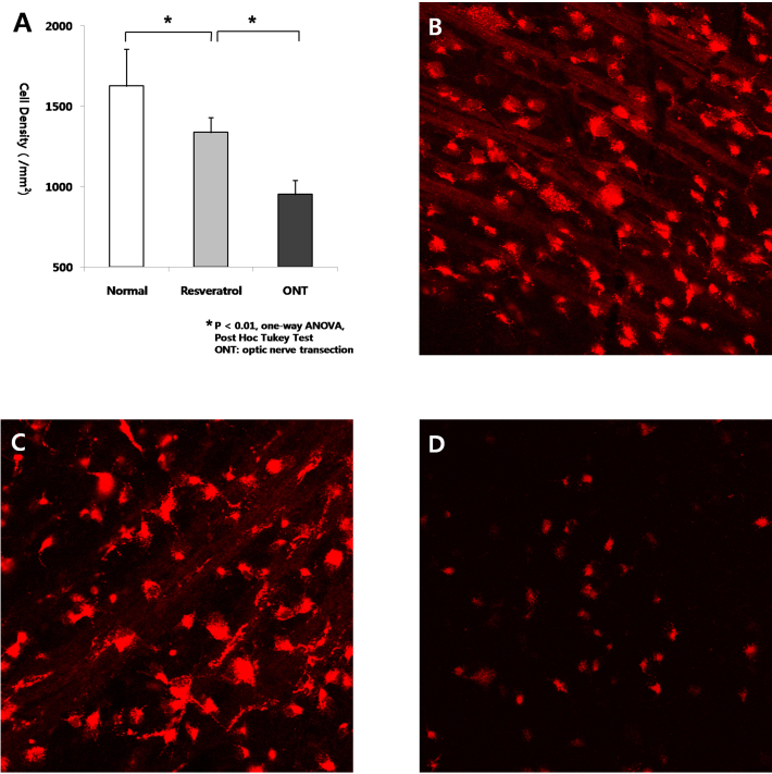
Purpose: This study aimed to investigate the neuroprotective effect of resveratrol in an optic nerve transection (ONT) model and to identify the neuroprotective mechanism of resveratrol in retinal ganglion cells (RGCs).

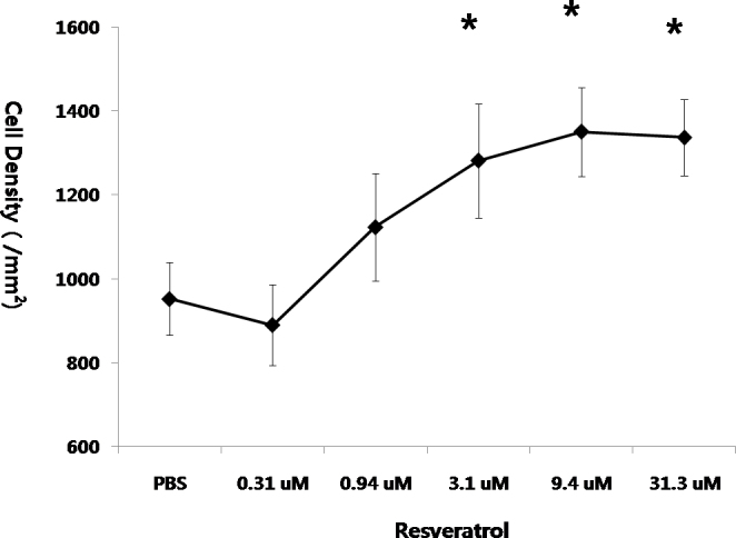
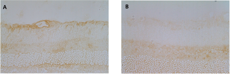
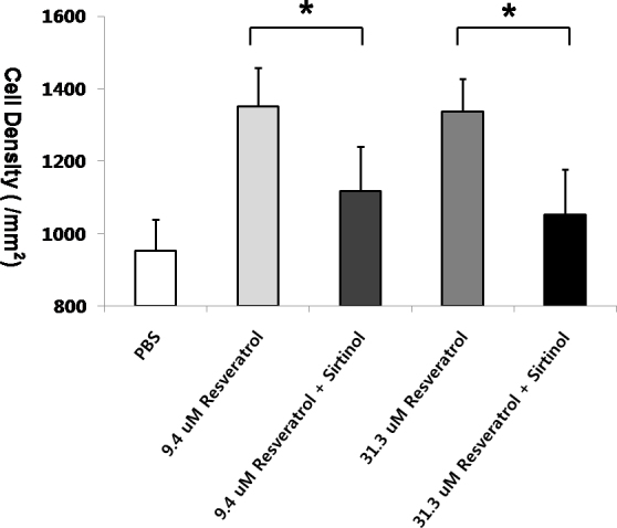
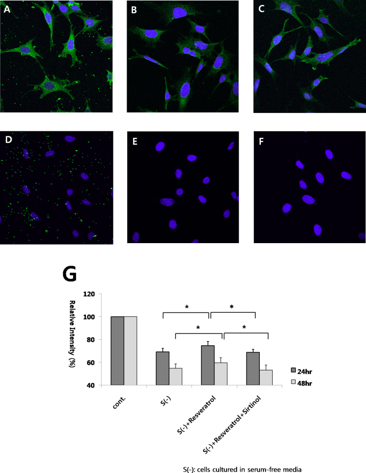
Methods: ONT and retrograde labeling were performed in Sprague-Dawley rats. Various concentrations of resveratrol were injected intravitreally immediately after ONT. The number of labeled RGCs was determined at 1 and 2 weeks after ONT. The effect of resveratrol and sirtinol (a sirtuin 1 inhibitor) co-injection was investigated. RGC-5 cells were cultured and treated with staurosporine to induce differentiation. 3-(4,5-Dimethylthiazol-2-yl)-2,5-diphenyltetrazolium bromide (MTT) assay was performed to evaluate the effect of resveratrol on RGC-5 cell survival under serum-free conditions. RGC-5 cells were cultured with sirtinol to investigate the neuroprotective mechanism of resveratrol.

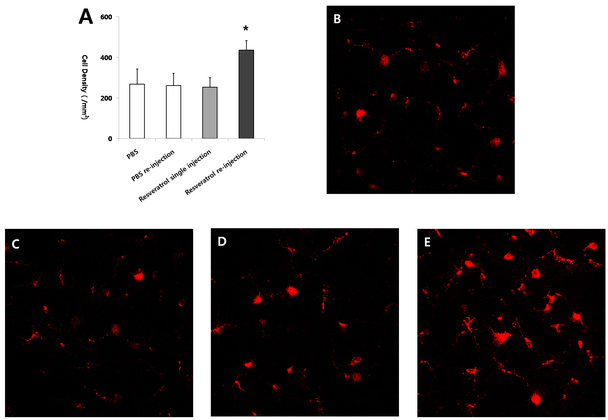
Results: A dose-response relationship was observed between resveratrol and RGC survival. A single intravitreal injection of resveratrol was neuroprotective in RGCs at 1 week after ONT (p<0.01). Repeated intravitreal injection of resveratrol showed a neuroprotective effect at 2 weeks after ONT (p<0.01). However, co-injection of resveratrol and sirtinol diminished the neuroprotective effect of resveratrol (p<0.05). The neuroprotective effect of resveratrol was observed in RGC-5 cells under serum-free conditions, and sirtinol diminished this neuroprotective effect.

Conclusions: Resveratrol exerts its neuroprotective effect on RGCs via activation of the sirtuin 1 pathway in an ONT model. This finding demonstrates the therapeutic potential of resveratrol in treating optic nerve diseases.


Figures

Figure 1
Figure 1
Analysis of neuroprotective effect of resveratrol at 1 week after optic nerve transection. A: The numbers of retinal ganglion cells (RGCs) of eyes treated with intravitreal injection of resveratrol (31.3 uM) in the transected rats (C) were significantly larger than those with injection of PBS in the transected rats (D), but smaller than those with injection of PBS in the nontransected rats (B; n=5 for each group, *p<0.01, one-way analysis of variance [ANOVA], post hoc Tukey test, error bars represent standard deviation).
Figure 2
Figure 2
Comparison of the neuroprotective effect of resveratrol according to the different concentrations. In optic nerve transected (ONT) eyes, resveratrol in the concentration of 3.1 uM or more showed statistically significant neuroprotective effect for the retinal ganglion cells (RGCs) compared to the control group (n≥4 for each group, *p<0.01, one-way analysis of variance [ANOVA], post hoc Tukey test, error bars represent standard deviation).
Figure 3
Figure 3
Analysis of neuroprotective effect of resveratrol at 2 week after injection. A: The numbers of retinal ganglion cells (RGCs) of eyes treated with resveratrol (31.3 uM) in the transected rats (D) were not significantly different from those with injection of PBS in the transected rats (B, C). However, the numbers of the RGCs of eyes with intravitreal reinjection of resveratrol (E) were significantly larger than those with PBS in transected rats (B, C; n≥5 for each group, *p<0.01, one-way analysis of variance [ANOVA], post hoc Tukey test, error bars represent standard deviation).
Figure 4
Figure 4
Immunolocalization of sirtuin 1 in the normal rat retina. Immunohistochemical staining showed that sirtuin 1 (SIRT1) was strongly expressed in the retinal ganglion cell (RGC) layer (GCL), inner nuclear layer (INL), outer nuclear layer, and photoreceptor layer (PRL; A). SIRT1 was also expressed in the optic nerve axon (B). The negative controls showed no detectable staining in retina (C, D).
Figure 5
Figure 5
Expression of sirtuin 1 in retinal ganglion cells three days after optic nerve transection in eyes treated with resveratrol injection (A) and in eyes treated with co-injections of sirtinol and resveratrol (B). The expression of sirtuin 1 (SIRT1) in retinal ganglion cells (RGCs) decreased in eyes treated with co-injections of sirtinol and resveratrol.
Figure 6
Figure 6
Neuroprotection of resveratrol blocked by sirtinol, a sirtuin 1 inhibitor. The numbers of retinal ganglion cells (RGCs) in eyes co-injected with resveratrol and sirtinol were smaller than injected with resveratrol at 1 week after treatment with concentrations of 9.4 uM and 31.3 uM of resveratrol (n≥5 for each group, *p<0.05, Mann-Whitney test, error bars represent standard deviation).
Figure 7
Figure 7
Effect of resveratrol and sirtinol on the survival of RGC-5 cells in serum-free media were shown. The control group had cells cultured in 10% fetal bovine serum; the label S(-) indicated a group with cells cultured in serum-free media. The group with resveratrol addition showed more cell viability than the group with PBS addition in cells cultured in serum-free media. Coaddition of resveratrol and sirtinol resulted in lower cell viabilities than the group with resveratrol only at 24 h and 48 h after addition. (G; n ≥ 3 for each group, *p < 0.05, one-way analysis of variance [ANOVA], post hoc Tukey test, error bars represent standard deviation) Immunocytochemistry showed the expression of Brn-3 (A), neurofilament (B) and Thy-1 (C) in RGC-5 cells. The negative controls of Brn-3 (D), neurofilament (E) and Thy-1 (F) were shown, which were stained with only secondary antibody.

References

    1. Mey J, Thanos S. Intravitreal injections of neurotrophic factors support the survival of axotomized retinal ganglion cells in adult rats in vivo. Brain Res. 1993;602:304–17. - PubMed
    1. Mansour-Robaey S, Clarke DB, Wang YC, Bray GM, Aguayo AJ. Effects of ocular injury and administration of brain-derived neurotrophic factor on survival and regrowth of axotomized retinal ganglion cells. Proc Natl Acad Sci USA. 1994;91:1632–6. - PMC - PubMed
    1. Weishaupt JH, Bähr M. Degeneration of axotomized retinal ganglion cells as a model for neuronal apoptosis in the central nervous system - molecular death and survival pathways. Restor Neurol Neurosci. 2001;19:19–27. - PubMed
    1. Kermer P, Ankerhold R, Klocker N, Krajewski S, Reed JC, Bahr M. Caspase-9: involvement in secondary death of axotomized rat retinal ganglion cells in vivo. Brain Res Mol Brain Res. 2000;85:144–50. - PubMed
    1. Kermer P, Klocker N, Labes M, Thomsen S, Srinivasan A, Bahr M. Activation of caspase-3 in axotomized rat retinal ganglion cells in vivo. FEBS Lett. 1999;453:361–4. - PubMed

Publication types

LinkOut - more resources