Comparison of Placido disc and Scheimpflug image-derived topography-guided excimer laser surface normalization combined with higher fluence CXL: the Athens Protocol, in progressive keratoconus
- PMID: 23901251
- PMCID: PMC3720663
- DOI: 10.2147/OPTH.S44745
Comparison of Placido disc and Scheimpflug image-derived topography-guided excimer laser surface normalization combined with higher fluence CXL: the Athens Protocol, in progressive keratoconus
Abstract
Background: The purpose of this study was to compare the safety and efficacy of two alternative corneal topography data sources used in topography-guided excimer laser normalization, combined with corneal collagen cross-linking in the management of keratoconus using the Athens protocol, ie, a Placido disc imaging device and a Scheimpflug imaging device.
Methods: A total of 181 consecutive patients with keratoconus who underwent the Athens protocol between 2008 and 2011 were studied preoperatively and at months 1, 3, 6, and 12 postoperatively for visual acuity, keratometry, and anterior surface corneal irregularity indices. Two groups were formed, depending on the primary source used for topoguided photoablation, ie, group A (Placido disc) and group B (Scheimpflug rotating camera). One-year changes in visual acuity, keratometry, and seven anterior surface corneal irregularity indices were studied in each group.
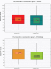
Results: Changes in visual acuity, expressed as the difference between postoperative and preoperative corrected distance visual acuity were +0.12 ± 0.20 (range +0.60 to -0.45) for group A and +0.19 ± 0.20 (range +0.75 to -0.30) for group B. In group A, K1 (flat keratometry) changed from 45.202 ± 3.782 D to 43.022 ± 3.819 D, indicating a flattening of -2.18 D, and K2 (steep keratometry) changed from 48.670 ± 4.066 D to 45.865 ± 4.794 D, indicating a flattening of -2.805 D. In group B, K1 (flat keratometry) changed from 46.213 ± 4.082 D to 43.190 ± 4.398 D, indicating a flattening of -3.023 D, and K2 (steep keratometry) changed from 50.774 ± 5.210 D to 46.380 ± 5.006 D, indicating a flattening of -4.394 D. For group A, the index of surface variance decreased to -5.07% and the index of height decentration to -26.81%. In group B, the index of surface variance decreased to -18.35% and the index of height decentration to -39.03%. These reductions indicate that the corneal surface became less irregular (index of surface variance) and the "cone" flatter and more central (index of height decentration) postoperatively.
Conclusion: Of the two sources of primary corneal data, the Scheimpflug rotating camera (Oculyzer™) for topography-guided normalization treatment with the WaveLight excimer laser platform appeared to provide more statistically significant improvement than the Placido disc topographer (Topolyzer™). Overall, the Athens protocol, aiming both to halt progression of keratoconic ectasia and to improve corneal topometry and visual performance, produced safe and satisfactory refractive, keratometric, and topometric results. The observed changes in visual acuity, along with keratometric flattening and topometric improvement, are suggestive of overall postoperative improvement.
Keywords: Athens protocol; EX500 excimer laser; WaveLight/Alcon excimer laser; anterior Pentacam indices; cross-linking; higher fluence collagen cross-linking; keratoconus.
Figures


Similar articles
-
Keratoconus management: long-term stability of topography-guided normalization combined with high-fluence CXL stabilization (the Athens Protocol).J Refract Surg. 2014 Feb;30(2):88-93. doi: 10.3928/1081597X-20140120-03. J Refract Surg. 2014. PMID: 24763473
-
Long-Term Stability With the Athens Protocol (Topography-Guided Partial PRK Combined With Cross-Linking) in Pediatric Patients With Keratoconus.Cornea. 2019 Aug;38(8):1049-1057. doi: 10.1097/ICO.0000000000001996. Cornea. 2019. PMID: 31169612
-
Corneal refractive power and symmetry changes following normalization of ectasias treated with partial topography-guided PTK combined with higher-fluence CXL (the Athens Protocol).J Refract Surg. 2014 May;30(5):342-6. doi: 10.3928/1081597X-20140416-03. J Refract Surg. 2014. PMID: 24893359
-
Outcomes of Simultaneous and Sequential Cross-linking With Excimer Laser Surface Ablation in Keratoconus.J Refract Surg. 2018 Oct 1;34(10):690-696. doi: 10.3928/1081597X-20180824-01. J Refract Surg. 2018. PMID: 30296330
-
Scheimpflug-Derived Keratometric, Pachymetric and Pachymetric Progression Indices in the Diagnosis of Keratoconus: A Systematic Review and Meta-Analysis.Clin Ophthalmol. 2023 Dec 20;17:3941-3964. doi: 10.2147/OPTH.S436492. eCollection 2023. Clin Ophthalmol. 2023. PMID: 38143558 Free PMC article. Review.
Cited by
-
Scheimpflug vs Scanning-Slit Corneal Tomography: Comparison of Corneal and Anterior Chamber Tomography Indices for Repeatability and Agreement in Healthy Eyes.Clin Ophthalmol. 2020 Sep 4;14:2583-2592. doi: 10.2147/OPTH.S251998. eCollection 2020. Clin Ophthalmol. 2020. PMID: 32943840 Free PMC article.
-
Forme Fruste Keratoconus Imaging and Validation via Novel Multi-Spot Reflection Topography.Case Rep Ophthalmol. 2013 Oct 25;4(3):199-209. doi: 10.1159/000356123. eCollection 2013. Case Rep Ophthalmol. 2013. PMID: 24348403 Free PMC article.
-
Short- and long-term safety and efficacy of corneal collagen cross-linking in progressive keratoconus: A systematic review and meta-analysis of randomized controlled trials.Taiwan J Ophthalmol. 2022 Nov 28;13(2):191-202. doi: 10.4103/2211-5056.361974. eCollection 2023 Apr-Jun. Taiwan J Ophthalmol. 2022. PMID: 37484615 Free PMC article. Review.
-
Pentacam topographic changes after collagen cross-linking in patients with keratoconus.Adv Biomed Res. 2015 Feb 23;4:62. doi: 10.4103/2277-9175.151886. eCollection 2015. Adv Biomed Res. 2015. PMID: 25802831 Free PMC article.
-
Higher incidence of steroid-induced ocular hypertension in keratoconus.Eye Vis (Lond). 2016 Feb 23;3:4. doi: 10.1186/s40662-016-0035-9. eCollection 2016. Eye Vis (Lond). 2016. PMID: 26909354 Free PMC article.
References
-
- Krachmer JH, Feder RS, Belin MW. Keratoconus and related noninflammatory corneal thinning disorders. Surv Ophthalmol. 1984;28:293–322. - PubMed
-
- Belin MW, Asota IM, Ambrosio R, Jr, Khachikian SS. What’s in a name: keratoconus, pellucid marginal degeneration, and related thinning disorders. Am J Ophthalmol. 2011;152:157–162. - PubMed
-
- Ambrósio R, Caldas DL, da Silva RS, Pimentel LN, de Freitas VB. Impact of the wavefront analysis in refraction of keratoconus patients. Rev Bras Oftalmol. 2010;69:294–300. Portuguese.
-
- Kosaki R, Maeda N, Bessho K, et al. Magnitude and orientation of Zernike terms in patients with keratoconus. Invest Ophthalmol Vis Sci. 2007;48:3062–3068. - PubMed
-
- Zadnik K, Steger-May K, Fink BA, et al. Collaborative longitudinal evaluation of keratoconus. Between-eye asymmetry in keratoconus. Cornea. 2002;21:671–679. - PubMed
LinkOut - more resources
Full Text Sources
Other Literature Sources

