

Invasive micropapillary carcinoma of the breast: MR imaging findings
- PMID: 23901311
- PMCID: PMC3725348
- DOI: 10.3348/kjr.2013.14.4.551
Invasive micropapillary carcinoma of the breast: MR imaging findings
Abstract
Objective: To analyze the magnetic resonance (MR) imaging findings of invasive micropapillary carcinoma of the breast.
Materials and methods: MR images were retrospectively evaluated in 14 patients (age range: 37-67, mean age: 49 years) with pathologically confirmed invasive micropapillary carcinoma of the breast. The enhancement type (mass/non-mass), shape, margin, contrast enhancement, and time-intensity curve pattern on the dynamic study were correlated with the histopathologic features. Associated findings, such as edema, nipple change, skin change and enlarged axillary lymph nodes were also studied.
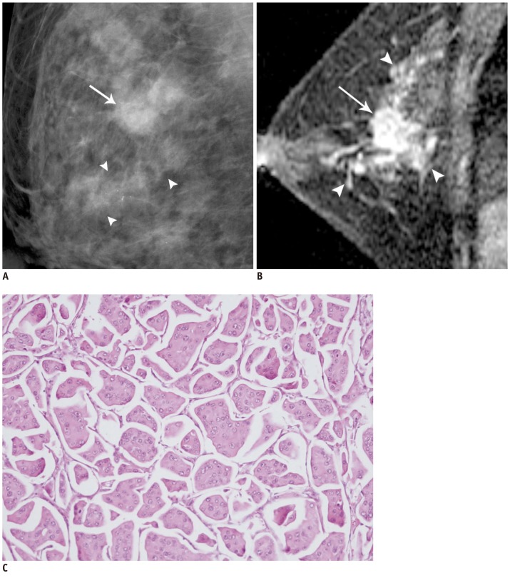
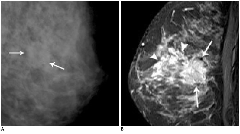
Results: The most common features of the masses were irregular shape (12 of 14 patients, 85.8%) and irregular or spiculated margin (11 of 14 patients, 78.7%). The contrast enhancement was heterogeneous in 11 patients (78.7%), rim enhancement in 2 cases (14.2%), and homogeneous in one patient (7.1%). The predominant kinetic pattern was rapid increase (14 of 14, 100%) in the initial phase and washout (11 of 14, 78.7%) in the delayed phase. Associated non-mass like enhancement was shown in 4 patients, representing ductal carcinoma in situ. MR imaging helped detect additional sites of cancer other than the index lesion in 3 patients (21.4%). Enlarged axillary lymphadenopathy was identified in 7 of the 14 patients (50%).
Conclusion: Invasive micropapillary carcinoma appears as a mass with an irregular shape, irregular or spiculated margin and heterogeneous enhancement on MR imaging. Though these findings are not specific and are also observed with other breast malignancies, invasive micropapillary carcinoma frequently showed multiple lesions, accompanying non-mass enhancement and axillary lymph node enlargement.
Keywords: Breast neoplasms; Diagnosis; MR; Micropapillary carcinoma.
Figures
References
-
- Luna-Moré S, Gonzalez B, Acedo C, Rodrigo I, Luna C. Invasive micropapillary carcinoma of the breast. A new special type of invasive mammary carcinoma. Pathol Res Pract. 1994;190:668–674. - PubMed
-
- Paterakos M, Watkin WG, Edgerton SM, Moore DH, 2nd, Thor AD. Invasive micropapillary carcinoma of the breast: a prognostic study. Hum Pathol. 1999;30:1459–1463. - PubMed
-
- Kuroda H, Sakamoto G, Ohnisi K, Itoyama S. Clinical and pathologic features of invasive micropapillary carcinoma. Breast Cancer. 2004;11:169–174. - PubMed
-
- Pettinato G, Manivel CJ, Panico L, Sparano L, Petrella G. Invasive micropapillary carcinoma of the breast: clinicopathologic study of 62 cases of a poorly recognized variant with highly aggressive behavior. Am J Clin Pathol. 2004;121:857–866. - PubMed
-
- Yu JI, Choi DH, Park W, Huh SJ, Cho EY, Lim YH, et al. Differences in prognostic factors and patterns of failure between invasive micropapillary carcinoma and invasive ductal carcinoma of the breast: matched case-control study. Breast. 2010;19:231–237. - PubMed
MeSH terms
LinkOut - more resources
Full Text Sources
Other Literature Sources
Medical
