. 2013;9(7):e1003591.
doi: 10.1371/journal.pgen.1003591. Epub 2013 Jul 18.

Combinatorial regulation of meiotic holliday junction resolution in C. elegans by HIM-6 (BLM) helicase, SLX-4, and the SLX-1, MUS-81 and XPF-1 nucleases

Affiliations

Combinatorial regulation of meiotic holliday junction resolution in C. elegans by HIM-6 (BLM) helicase, SLX-4, and the SLX-1, MUS-81 and XPF-1 nucleases

Ana Agostinho et al. PLoS Genet. 2013.

Erratum in

  • PLoS Genet. 2013 Aug;9(8). doi:10.1371/annotation/d8c73205-151d-4e22-89c6-3aa574037d10

Abstract

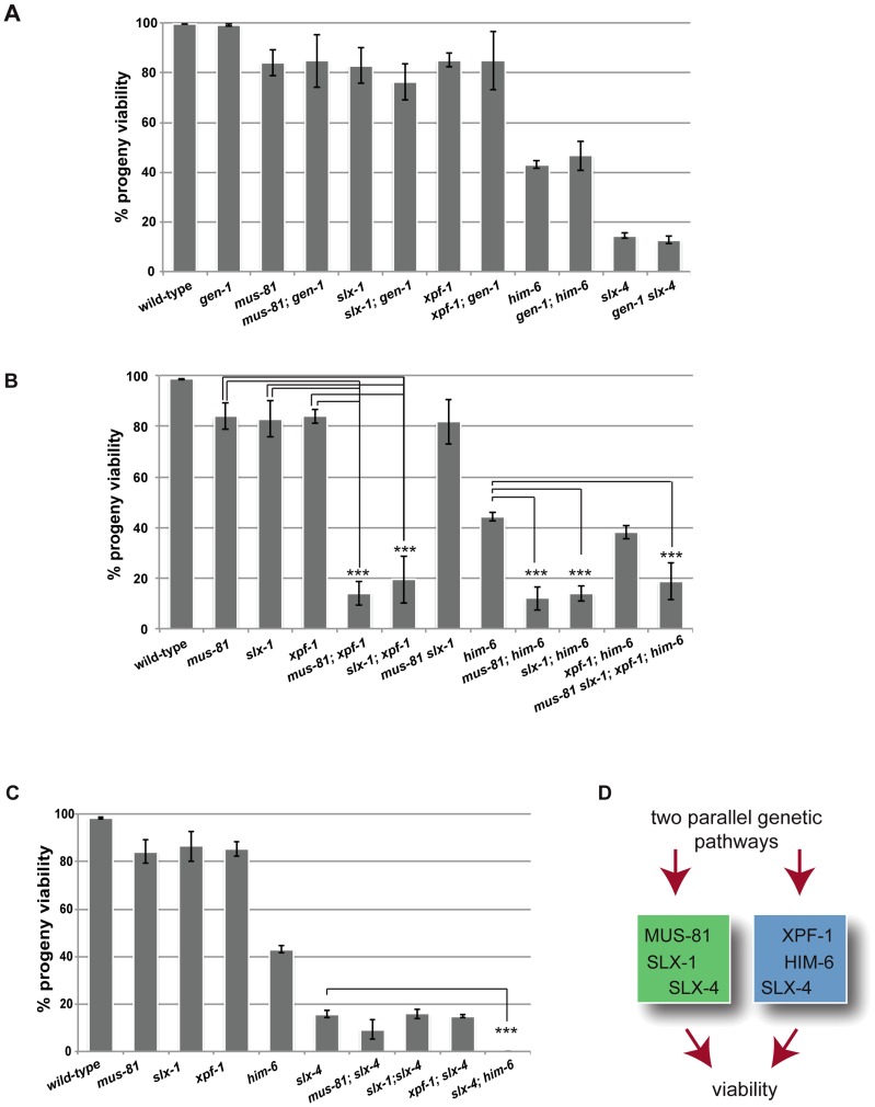
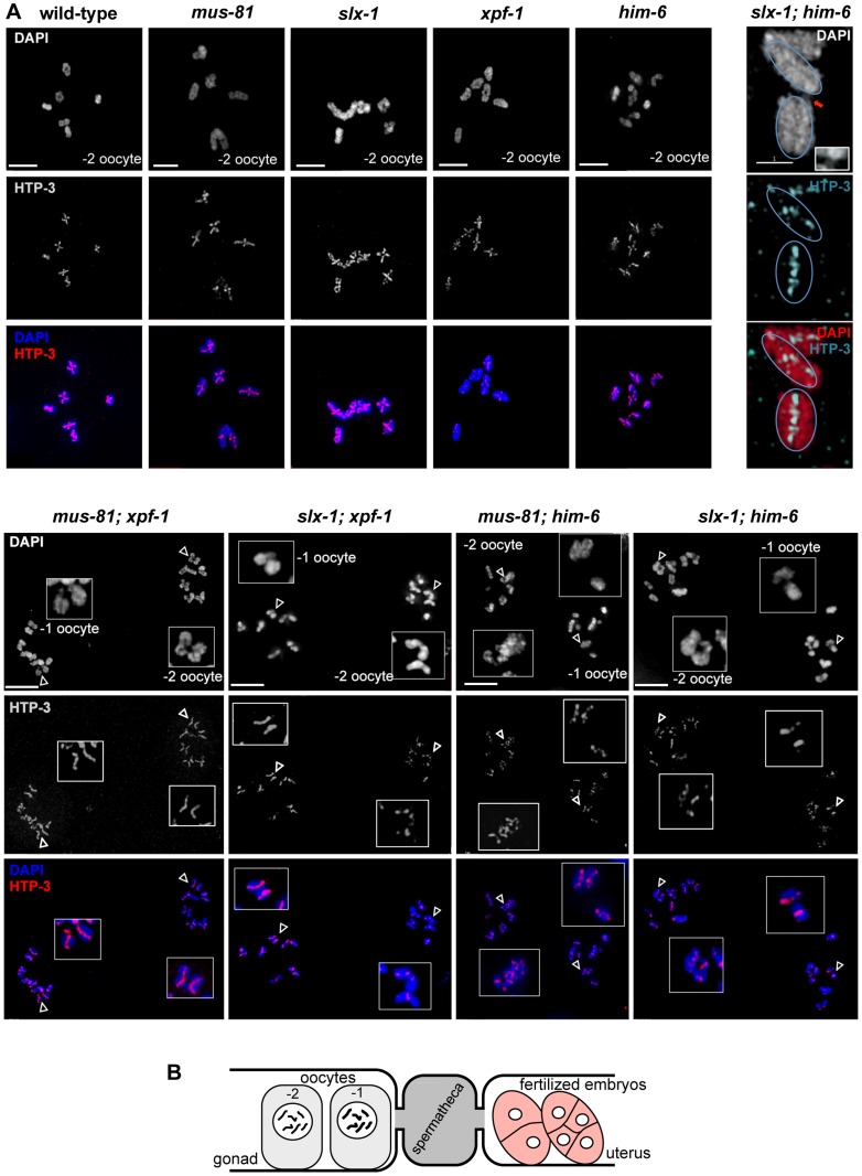
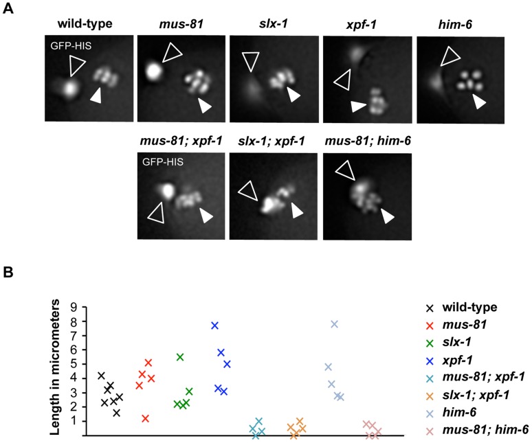
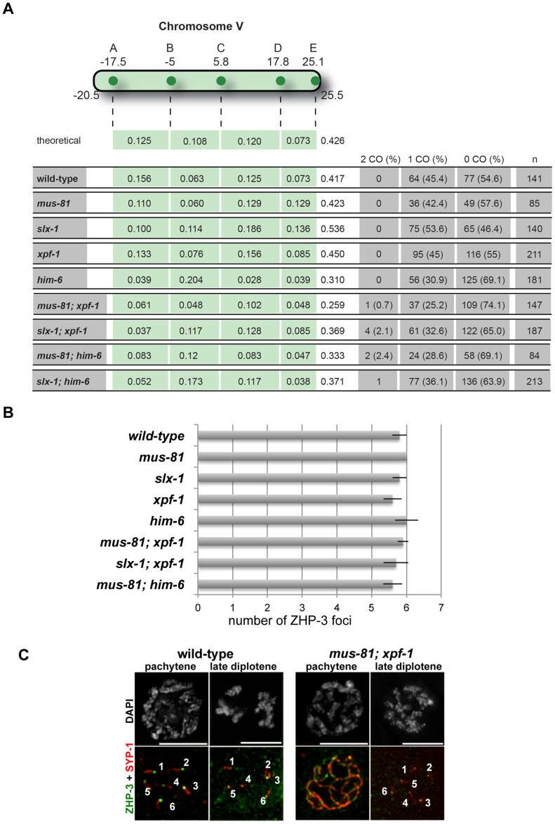
Holliday junctions (HJs) are cruciform DNA structures that are created during recombination events. It is a matter of considerable importance to determine the resolvase(s) that promote resolution of these structures. We previously reported that C. elegans GEN-1 is a symmetrically cleaving HJ resolving enzyme required for recombinational repair, but we could not find an overt role in meiotic recombination. Here we identify C. elegans proteins involved in resolving meiotic HJs. We found no evidence for a redundant meiotic function of GEN-1. In contrast, we discovered two redundant HJ resolution pathways likely coordinated by the SLX-4 scaffold protein and also involving the HIM-6/BLM helicase. SLX-4 associates with the SLX-1, MUS-81 and XPF-1 nucleases and has been implicated in meiotic recombination in C. elegans. We found that C. elegans [mus-81; xpf-1], [slx-1; xpf-1], [mus-81; him-6] and [slx-1; him-6] double mutants showed a similar reduction in survival rates as slx-4. Analysis of meiotic diakinesis chromosomes revealed a distinct phenotype in these double mutants. Instead of wild-type bivalent chromosomes, pairs of "univalents" linked by chromatin bridges occur. These linkages depend on the conserved meiosis-specific transesterase SPO-11 and can be restored by ionizing radiation, suggesting that they represent unresolved meiotic HJs. This suggests the existence of two major resolvase activities, one provided by XPF-1 and HIM-6, the other by SLX-1 and MUS-81. In all double mutants crossover (CO) recombination is reduced but not abolished, indicative of further redundancy in meiotic HJ resolution. Real time imaging revealed extensive chromatin bridges during the first meiotic division that appear to be eventually resolved in meiosis II, suggesting back-up resolution activities acting at or after anaphase I. We also show that in HJ resolution mutants, the restructuring of chromosome arms distal and proximal to the CO still occurs, suggesting that CO initiation but not resolution is likely to be required for this process.


Conflict of interest statement

The authors have declared that no competing interests exist.

Figures

Figure 1
Figure 1. Dissection of genetic interactions between gen-1, mus-81, slx-1, xpf-1, him-6 and slx-4.
(A) gen-1 does not alter viability, alone or in conjunction with nucleases, slx-4 or him-6. Progeny viability in % was determined as described in Material and Methods. (B) Evidence for MUS-81/SLX-1 and XPF-1/HIM-6 acting in two genetic pathways. Scoring was done as in A. For double mutants with reduced viability the F1 progeny of double homozygous mothers derived from hT2-balanced lines was scored (for strain list see Materials and Methods). In contrast slx-1; him-6 double mutants derived from a strain were him-6 is balanced by nT1 behave as synthetic lethal (data not shown) . We think that this lethality is due the nT1 balancer, which has been described as prone to brake down, as preferably segregating to the male germ line and as conferring a reduced brood size . (C) Reduced progeny viability of slx-4 is not enhanced by nuclease mutations. (D) Genetic model. Asterisks indicate statistical significance between different genotypes as determined by two-tailed Mann-Whitney test (*** indicates P<0.01).
Figure 2
Figure 2. Normal chromosome morphology and HTP-3 and SYP-1 loading in pachytene stage nuclei in [mus-81; xpf-1], [slx-1; xpf-1], [mus-81; him-6] and [slx-1; him-6] double mutants.
(A) Projections of representative nuclei from whole-mount gonads stained with α-HTP-3 antibody (white) and DAPI (white), or red and blue, respectively, in merged images. (B) Representative pachytene nuclei stained with α-SYP-1 antibody and DAPI (SPY-1, green in merged images). Scale bars are shown in white (1 µm). (C) FISH images were taken with 100× and 60× magnification.
Figure 3
Figure 3. Evidence for DNA bridges linking homologous chromosomes in in [mus-81; xpf-1], [slx-1; xpf-1], [mus-81; him-6] and [slx-1; him-6] double mutants.
(A) DAPI stained diakinesis chromosomes. Images represent projected Z-stacks obtained by deconvolution microscopy. Red arrows indicate thin chromatin bridges, blue arrows indicate univalents. The asterisk points to a sperm nucleus. Scale bars are shown in white (5 µm). (B) FISH analysis reveals that chromatin linkages occur between homologous chromosomes. Scale bars shown in white (2 µm). (C) Quantification of bivalents, ‘dissociated bivalents’ and univalents. 100% would reflect all oocytes scored containing six bivalents. A pair of ‘dissociated bivalents’ as well as two (unlinked) univalents are scored as one, in the respective categories. A minimum of 15 oocytes was scored for each genotype. Chromosomes often overlap in cytological preparations. In these cases chromosomes were scored as “n/d”. In addition, in mus-81, slx-1 and [xpf-1; him-6] in a very limited number of cases, fusions, which appear to occur between different chromosomes could be observed (Figure S1, data not shown, also see co-submitted paper by Saito et al. [48]). (D) Images of representative chromosomes taken by super-resolution SIM microscopy. DNA bridges are indicated by red arrows. Scale bars are shown in white (1 µm). (E) Close up of DNA bridges obtained by SIM microscopy.
Figure 4
Figure 4. Absence of chiasmata on ‘dissociated bivalents’ revealed by HTP-3 staining.
(A) Projections of representative nuclei of diakinesis oocytes stained with α-HTP-3 antibody and DAPI. Open arrowheads indicate the linked chromosomes shown in close-ups. The upper right panels show SIM images of a representative ‘dissociated bivalents’ of an [slx-1; him-6] oocyte. Linked univalent are encircled in blue. Scale bars are shown in white (4 µm). (B) Schematic representation of the proximal germ line, spermatheca and early embryogenesis in the C. elegans germ line.
Figure 5
Figure 5. Chromosome linkages depend on SPO-11-induced meiotic double-strand breaks.
(A) Images of DAPI-stained -1 and -2 oocytes (position distal to spermatheca) of the indicated genotypes. Scale bars are 4 µm unless indicated otherwise. (B) Irradiation induced double-strand breaks bypass the SPO-11 requirement. Red arrows indicate chromosome linkages. Blue arrows indicate DNA fragments, which we observed in [slx-1; him-6 spo-11] but not in [slx-1; him-6]. Scale bars are shown in white (5 µm).
Figure 6
Figure 6. Impaired chromosome segregation during meiosis.
(A) Representative images taken from time-lapse recordings of GFP-Histone H2B expressing embryos. Images were taken during metaphase of meiosis II. Open arrowheads indicate the first polar body; filled arrowheads indicate chromosomes aligned on the metaphase plate. Note five chromosomes in the him-6 panel indicating chromosome missegregation. (B) Graph depicting the distance between the first polar body and the metaphase plate one minute prior to the onset of anaphase II. A minimum of five embryos were analysed for each genotype.
Figure 7
Figure 7. Despite showing reduced crossover recombination frequencies, wild-type numbers of ZHP-3 foci are detected in [mus-81; xpf-1] and [slx-1; xpf-1] double mutants.
(A) Measurements of CO recombination frequency and distribution between five snip-SNPs, highlighted as A to E, was scored on Chromosome V. The genetic map position of the SNP used is indicated. n is the number of cross-progeny scored. The frequency of 2 COs, 1 CO or 0 CO per chromosome is indicated in absolute numbers and as percentage (in brackets). (B) Quantification of α-ZHP-3 foci in pachytene/diplotene nuclei (a minimum of 8 nuclei was scored per genotype). (C) Representative images of SYP-1 (red), ZHP-3 (green) stainings.
Figure 8
Figure 8. CO initiation but not CO completion is required for the reciprocal localization of HTP-1/2 and SYP-1 in diakinesis.
(A) SYP-1 staining of representative diplotene nuclei. Scale bars are 2 µm. (B) HTP-1/2 (red) and SYP-1 (green) staining of wild-type diakinesis chromosomes. (C) The same staining of [mus-81; xpf-1], [slx-1; xpf-1], [mus-81; him-6] and [slx-1; him-6] double mutants nuclei. Filled arrowheads indicate DNA bridges. Open arrowheads indicate the chromosomes shown in the close ups. Scale bars are shown in red.
Figure 9
Figure 9. Model of redundant resolution pathways.

Comment in

  • An elegans Solution for Crossover Formation.
    Bellendir SP, Sekelsky J. Bellendir SP, et al. PLoS Genet. 2013;9(7):e1003658. doi: 10.1371/journal.pgen.1003658. Epub 2013 Jul 18. PLoS Genet. 2013. PMID: 23874241 Free PMC article. No abstract available.

References

    1. Sun H, Treco D, Schultes NP, Szostak JW (1989) Double-strand breaks at an initiation site for meiotic gene conversion. Nature 338: 87–90. - PubMed
    1. Keeney S, Giroux CN, Kleckner N (1997) Meiosis-specific DNA double-strand breaks are catalyzed by Spo11, a member of a widely conserved protein family. Cell 88: 375–384. - PubMed
    1. Lui DY, Colaiacovo MP (2013) Meiotic Development in Caenorhabditis elegans . Adv Exp Med Biol 757: 133–170. - PMC - PubMed
    1. Buhler C, Borde V, Lichten M (2007) Mapping meiotic single-strand DNA reveals a new landscape of DNA double-strand breaks in Saccharomyces cerevisiae . PLoS Biol 5: e324. - PMC - PubMed
    1. Moens PB, Kolas NK, Tarsounas M, Marcon E, Cohen PE, et al. (2002) The time course and chromosomal localization of recombination-related proteins at meiosis in the mouse are compatible with models that can resolve the early DNA-DNA interactions without reciprocal recombination. J Cell Sci 115: 1611–1622. - PubMed

Publication types