. 2014 Nov 10;21(14):1960-73.
doi: 10.1089/ars.2013.5416. Epub 2013 Sep 20.

Mitophagy is required for acute cardioprotection by simvastatin

Affiliations

Mitophagy is required for acute cardioprotection by simvastatin

Allen M Andres et al. Antioxid Redox Signal. .

Abstract

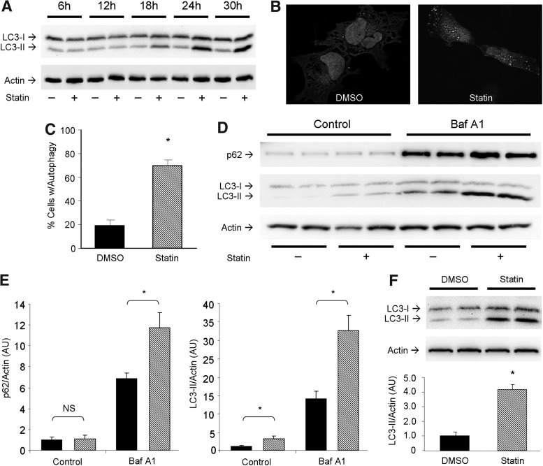
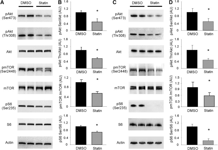
Aims: We have shown that autophagy and mitophagy are required for preconditioning. While statin's cardioprotective effects are well known, the role of autophagy/mitophagy in statin-mediated cardioprotection is not. In this study, we used HL-1 cardiomyocytes and mice subjected to ischemia/reperfusion to elucidate the mechanism of statin-mediated cardioprotection.

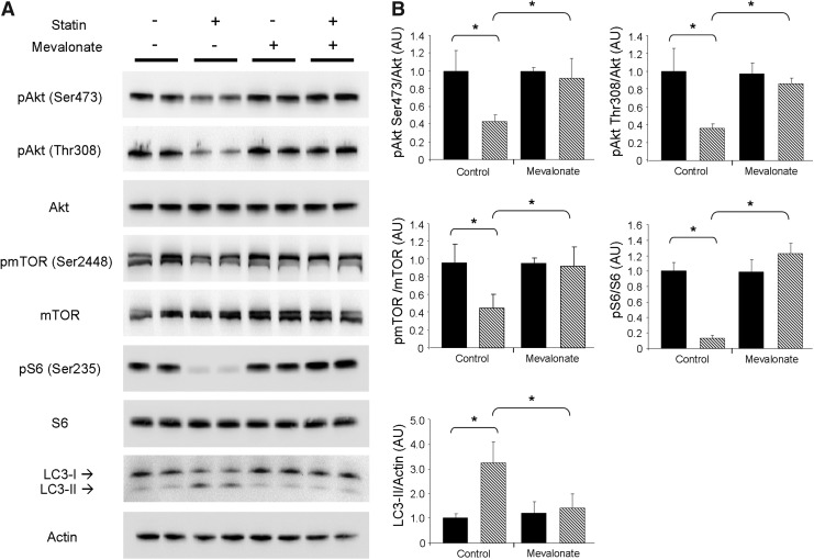
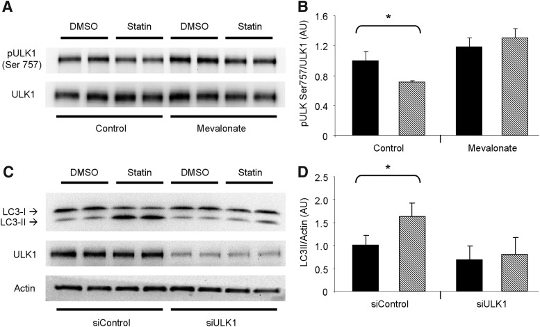
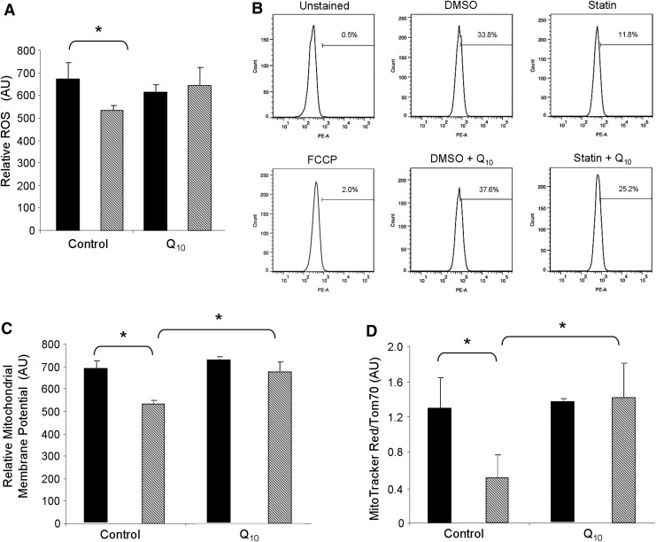
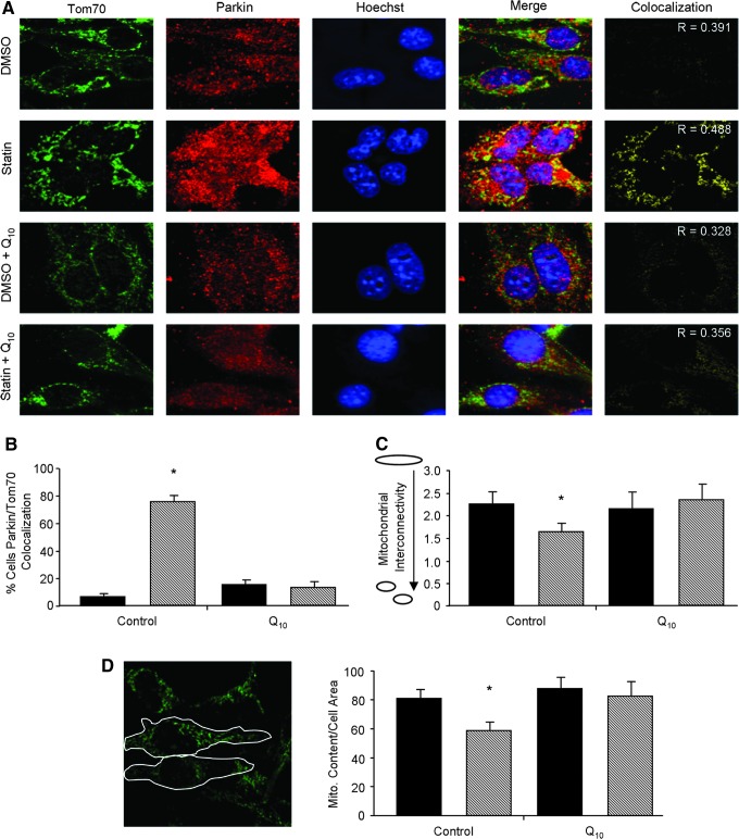
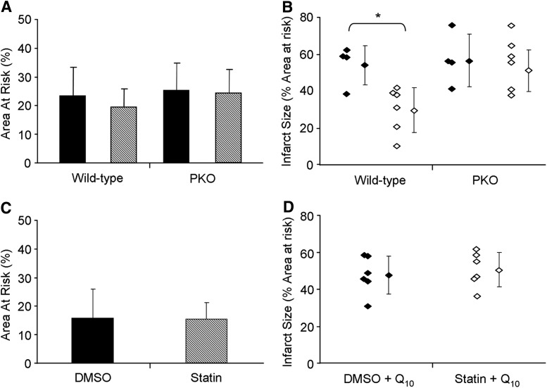
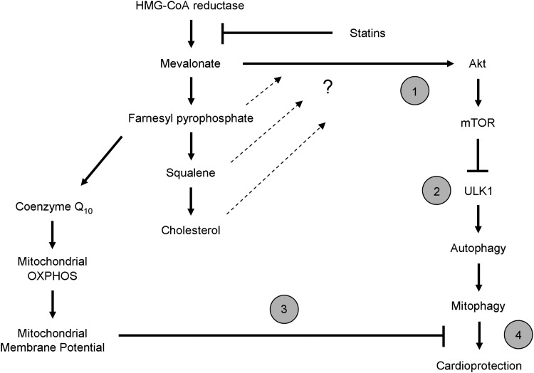
Results: HL-1 cardiomyocytes exposed to simvastatin for 24 h exhibited diminished protein kinase B (Akt)/mammalian target of rapamycin (mTOR) signaling, increased activation of unc-51-like kinase 1, and upregulation of autophagy and mitophagy. Similar findings were obtained in hearts of mice given simvastatin. Mevalonate abolished simvastatin's effects on Akt/mTOR signaling and autophagy induction in HL-1 cells, indicating that the effects are mediated through inhibition of 3-hydroxy-3-methylglutaryl coenzyme A (HMG-CoA) reductase. Simvastatin-treated HL-1 cells exhibited mitochondrial translocation of Parkin and p62/SQSTM1, fission, and mitophagy. Because Parkin is required for mitophagy and is expressed in heart, we investigated the effect of simvastatin on infarct size in Parkin knockout mice. Simvastatin reduced infarct size in wild-type mice but showed no benefit in Parkin knockout mice. Inhibition of HMG-CoA reductase limits mevalonate availability for both cholesterol and coenzyme Q10 (CoQ) biosynthesis. CoQ supplementation had no effect on statin-induced Akt/mTOR dephosphorylation or macroautophagy in HL-1 cells, but it potently blocked mitophagy. Importantly, CoQ supplementation abolished statin-mediated cardioprotection in vivo.

Innovation and conclusion: Acute simvastatin treatment suppresses mTOR signaling and triggers Parkin-dependent mitophagy, the latter which is required for cardioprotection. Coadministration of CoQ with simvastatin impairs mitophagy and cardioprotection. These results raise the concern that CoQ may interfere with anti-ischemic benefits of statins mediated through stimulation of mitophagy.


Figures

<b>FIG. 1.</b>
FIG. 1.
Statins induce cardiac autophagy. (A) Time course ranging from 6 to 30 h examining statin-mediated autophagy induction via LC3-I to LC3-II conversion by immunoblot. (B) HL-1 cells infected with adenovirus for LC3-GFP and treated with vehicle or statin, showing autophagic puncta formation by fluorescence microscopy. (C) Quantitation of autophagy induction by statin treatment. Cells with >20 puncta were scored positive for autophagy. n=3 plates per group; 50 cells were scored for each n. (D) Autophagic flux was examined via use of 100 nM bafilomycin A1 for 2 h immediately before 24 h statin (or vehicle DMSO) treatment of cells. p62/SQSTM1 and LC3 were monitored. (E) Quantitation of results shown in (D) (DMSO, solid bars; and simvastatin, hashed bars). n=4 per group; experiments were performed at least two times. *p-value<0.05. (F) Representative western blot for LC3 from hearts of mice given simvastatin (20 mg/kg) i.p. and sacrificed 4 h after treatment. n=3 mice per group; experiments were performed twice with similar results. *p-value<0.05. DMSO, dimethyl sulfoxide; GFP, green fluorescent protein; i.p., intraperitoneal; LC3, light chain 3.
<b>FIG. 2.</b>
FIG. 2.
Statins attenuate Akt/mTOR signaling. (A) Representative western blots of Akt/mTOR signaling pathway proteins from HL-1 cells treated with vehicle (DMSO, solid bars) or simvastatin (statin, hashed bars) as indicated. (B) Quantitation of results shown in (A). n=3 per group; experiments were performed at least three times. *p-value<0.05. (C) Representative western blots of Akt/mTOR signaling pathway proteins in DMSO or statin-treated mouse hearts. n=4 mice per condition. (D) Quantitation of results shown in (C). Akt, protein kinase B; mTOR, mammalian target of rapamycin.
<b>FIG. 3.</b>
FIG. 3.
Mevalonate inhibits statin-induced attenuation of Akt/mTOR signaling and autophagy. (A) Representative western blots of Akt/mTOR signaling proteins and LC3 from HL-1 cells treated with statin in the absence (control) or presence of 100 μM mevalonic acid (mevalonate). Treatment groups identified as: vehicle (DMSO, solid bars) and simvastatin (statin, hashed bars). (B) Quantitation of results shown in (A). n=4 per group; experiments were performed at least three times with similar results. *p-value<0.05.
<b>FIG. 4.</b>
FIG. 4.
ULK1 is required for statin-mediated autophagy. (A) Representative western blots examining the phosphorylation of ULK1 at Ser757 in HL-1 cells treated with statin in the absence or presence of 100 μM mevalonic acid as indicated. Treatment groups identified as: vehicle (DMSO) and simvastatin. (B) Quantitation of results shown in (A) (DMSO, solid bars; simvastatin, hashed bars). n=4 per group; experiments were performed twice. *p-value<0.05. (C) HL-1 cells were silenced for ULK1 before treatment with simvastatin. Cells were probed for LC3 lipidation as an indicator of autophagy, and ULK1 to confirm siRNA efficacy. (D) Quantitation of results shown in (C). n=4 per group; experiments were performed twice with similar results. *p-value<0.05. ULK1, unc-51-like kinase 1.
<b>FIG. 5.</b>
FIG. 5.
Statin treatment lowers ROS production and mitochondrial membrane potential. (A) HL-1 cells were given 1 μM simvastatin with or without the concurrent supplementation with 5 μM coenzyme Q10 for 24 h. Cells were then loaded with 10 μM H2DCFDA for 30 min at 37°C and subsequently analyzed via FACS for fluorescence intensity per cell as a measure of ROS. (B) Representative FACS histograms of HL-1 cells loaded with membrane potential-sensitive MitoTracker Red CMX Rosamine for 30 min at the end of statin and/or coenzyme Q10 treatments, which were then fixed with 4% paraformaldehyde before analysis. Unstained cells, and cells given 10 μM FCCP for 1 h served as controls. (C) Mean average fluorescence intensity (of the MitoTracker Red CMX Rosamine) was quantified as a measure of relative mitochondrial membrane potential changes. n=3 plates per group; 10,000 cells were examined per plate. *p-value<0.05. (D) Cells were treated as in (A), then immunolabeled with anti-Tom70 and nuclear stain Hoechst 33432. n=3 plates per group; 50 cells per plate were analyzed for MitoTracker Red CMX Rosamine normalized to Tom70 fluorescence. *p-value<0.05. FACS, fluorescence-activated cell sorting; FCCP, carbonyl cyanide 4-(trifluoromethoxy) phenylhydrazone; ROS, reactive oxygen species; Tom70, translocase outer membrane 70 kDa subunit.
<b>FIG. 6.</b>
FIG. 6.
Statins promote mitophagy. (A) Representative images of Parkin translocation to the mitochondria of HL-1 cells given DMSO or 1 μM simvastatin with or without concomitant supplementation with 5 μM coenzyme Q10 for 24 h. Parkin (red) and Tom70 (green) were immunolabeled, and nuclei were stained with Hoeschst33432. To emphasize mitochondrial translocation of Parkin, processed images were thresholded for yellow color representing colocalized points (Parkin and Tom70). The Pearson's correlation coefficient (R) was used to assess colocalization indicated in representative images. (B) HL-1 cells treated as in (A) were scored for Parkin translocation to the mitochondria. Cells exhibiting more than 40% colocalization of Parkin to mitochondria were counted as positive. n=4 plates per group. *p-value<0.05. (C) Mitochondrial interconnectivity was examined to assess mitochondrial fragmentation versus network morphology. The oval signifies fused mitochondria (high connectivity ratio) and the two small spheres signify mitochondrial fragmentation (low connectivity ratio). (D) Determination of mitophagy defined by the loss of Tom70 fluorescence per unit cell area. Perimeters of individual cells were outlined as in the sample image and quantified for fluorescence intensity of Tom70. n=4 per group; at least 100 cells were analyzed per n. *p-value<0.05. Treatment groups identified as: vehicle (DMSO, solid bars) and simvastatin (statin, hashed bars).
<b>FIG. 7.</b>
FIG. 7.
Parkin is required for statin-mediated cardioprotection which is abolished by coenzyme Q10. (A) Area-at-risk of wild-type versus Parkin knockout mice (PKO) treated either with either vehicle (DMSO, solid bars), or simvastatin (statin, hashed bars). (B) Quantitation of infarct size as a percentage of risk area. Treatment groups identified as: vehicle (DMSO, solid diamonds) and simvastatin (statin, open diamonds). Individual diamonds signify the result for each mouse heart. n=4–6 mice per group. *p-value<0.01. (C) Area-at-risk of wild-type mice treated either with either vehicle control (DMSO, solid bars), or simvastatin (statin, hashed bars), followed 1 h later with 10 mg/kg coenzyme Q10 i.p. Coronary artery ligation was performed 4 h after statin administration and infarct size was determined 22 h later. (D) Quantitation of infarct size as a percentage of risk area from (C). Treatment groups identified as: vehicle (DMSO, solid diamonds) and simvastatin (statin, open diamonds). Individual diamonds signify the result for each mouse. n=4–6 mice per group. *p-value<0.05.
<b>FIG. 8.</b>
FIG. 8.
Proposed mechanism for statin-mediated cardioprotection. Statins are able to afford cardioprotection through an unknown mechanism beyond their ability to lower circulating levels of plasma cholesterol. Our findings elucidate this pathway and provide evidence for the first time that statin-mediated cardioprotection involves triggering mitophagy in the heart. Here we propose a mechanism by which statins promote cardiac mitophagy, which is essential for its protective effect: (1) Depletion of mevalonate (via inhibition of HMG-CoA reductase) attenuates Akt/mTOR signaling. (2) Diminished Akt/mTOR signaling relieves the inhibition of ULK1 resulting in increased macroautophagy. (3) Depletion of mevalonate results in loss of coenzyme Q10, thereby impairing the ability of mitochondria to maintain their membrane potential. Autophagy targeting machinery is recruited to depolarized mitochondria. This mitophagy machinery includes (but is not limited to) Parkin and p62/SQSTM1. These elements of statin treatment set the stage for promoting mitophagy in the heart. (4) Increased mitophagy confers cardioprotection against I/R injury. HMG-CoA, 3-hydroxy-3-methylglutaryl coenzyme A; I/R, ischemia/reperfusion.

References

    1. Araki M, Maeda M, and Motojima K.Hydrophobic statins induce autophagy and cell death in human rhabdomyosarcoma cells by depleting geranylgeranyl diphosphate. Eur J Pharmacol 674: 95–103, 2012 - PubMed
    1. Avis HJ, et al. Rosuvastatin lowers coenzyme Q10 levels, but not mitochondrial adenosine triphosphate synthesis, in children with familial hypercholesterolemia. J Pediatr 158: 458–462, 2011 - PubMed
    1. Bentinger M, Dallner G, Chojnacki T, and Swiezewska E.Distribution and breakdown of labeled coenzyme Q10 in rat. Free Radic Biol Med 34: 563–575, 2003 - PubMed
    1. Cafforio P, Dammacco F, Gernone A, and Silvestris F.Statins activate the mitochondrial pathway of apoptosis in human lymphoblasts and myeloma cells. Carcinogenesis 26: 883–891, 2005 - PubMed
    1. Cao P, et al. Statin-induced muscle damage and atrogin-1 induction is the result of a geranylgeranylation defect. FASEB J 23: 2844–2854, 2009 - PMC - PubMed

Publication types

MeSH terms

Substances

LinkOut - more resources