. 2013 Dec;15(12):2093-108.
doi: 10.1111/cmi.12175. Epub 2013 Aug 22.

Mannan core branching of lipo(arabino)mannan is required for mycobacterial virulence in the context of innate immunity

Affiliations
Free PMC article

Mannan core branching of lipo(arabino)mannan is required for mycobacterial virulence in the context of innate immunity

Esther J M Stoop et al. Cell Microbiol. 2013 Dec.
Free PMC article

Abstract

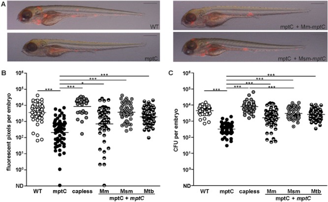
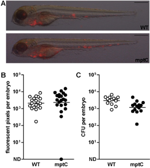
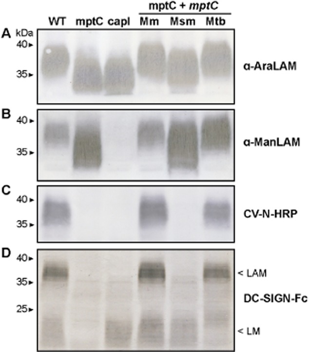
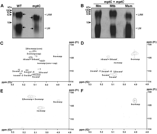
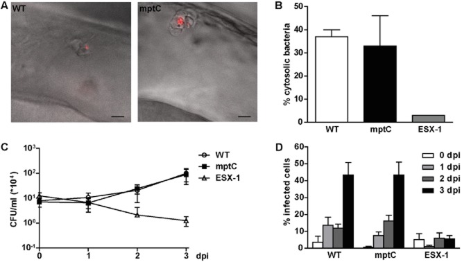
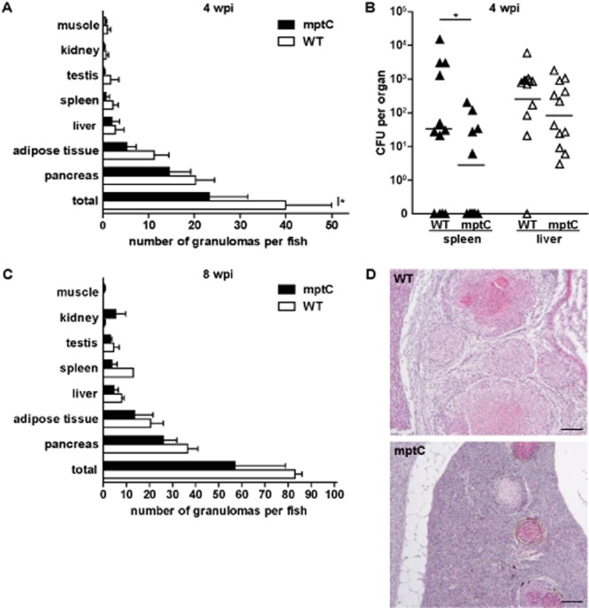
The causative agent of tuberculosis (TB), Mycobacterium tuberculosis, remains an important worldwide health threat. Although TB is one of the oldest infectious diseases of man, a detailed understanding of the mycobacterial mechanisms underlying pathogenesis remains elusive. Here, we studied the role of the α(1→2) mannosyltransferase MptC in mycobacterial virulence, using the Mycobacterium marinum zebrafish infection model. Like its M. tuberculosis orthologue, disruption of M. marinum mptC (mmar_3225) results in defective elongation of mannose caps of lipoarabinomannan (LAM) and absence of α(1→2)mannose branches on the lipomannan (LM) and LAM mannan core, as determined by biochemical analysis (NMR and GC-MS) and immunoblotting. We found that the M. marinum mptC mutant is strongly attenuated in embryonic zebrafish, which rely solely on innate immunity, whereas minor virulence defects were observed in adult zebrafish. Strikingly, complementation with the Mycobacterium smegmatis mptC orthologue, which restored mannan core branching but not cap elongation, was sufficient to fully complement the virulence defect of the mptC mutant in embryos. Altogether our data demonstrate that not LAM capping, but mannan core branching of LM/LAM plays an important role in mycobacterial pathogenesis in the context of innate immunity.


Figures

Figure 1
Figure 1
Transposon mutant in mmar_3225 attenuated for granuloma formation in zebrafish embryo model.A. Representative overlays of brightfield and fluorescent images of embryos at 5 dpi with wildtype M. marinum (WT), mptC mutant (mptC), mptC mutant complemented with M. marinummptC (mptC + mptC Mm) and mptC mutant complemented with M. smegmatismptC (mptC + mptC Msm). Inocula were 117, 158, 138, and 198 cfu respectively. Scale bars represent 500 μm.B and C. Quantification of embryo infection at 5 dpi with wildtype M. marinum, the mptC mutant, the capless mutant (capless), the mptC mutant complemented with M. marinummptC, the mptC mutant complemented with M. smegmatismptC and the mptC mutant complemented with M. tuberculosismptC (mptC + mptC Mtb). (B) Number of red pixels in fluorescent images of infected embryos as determined with custom-designed software and (C) number of cfu of single infected embryos as determined by whole-embryo plating. Both graphs show data of three independent experiments and the bars represent mean after log transformation. Mean ± standard deviation of inocula were 99 ± 6, 149 ± 9, 99 ± 6, 134 ± 18, 144 ± 21 and 104 ± 13 cfu respectively. *P < 0.05, ***P < 0.0001, one-way anova, Bonferroni's multiple comparison test on log transformed data. ND, not detected.
Figure 2
Figure 2
High mptC mutant inoculum restores attenuated virulence in zebrafish embryos.A. Representative overlays of bright field and fluorescent images of embryos at 5 dpi with wildtype M. marinum (WT) or mptC mutant (mptC). Scale bars represent 500 μm.B. Number of red pixels in fluorescent images of infected embryos as determined with custom-designed software and (C) number of cfu of single infected embryos as determined by whole-embryo plating. Both graphs show data of two independent experiments and the bars represent mean after log transformation. Mean ± standard deviation of inocula were 175 ± 81 and 419 ± 108 cfu respectively. ND, not detected.
Figure 3
Figure 3
Mannose cap elongation and mannan core branching disrupted in mptC mutant. Immunoblots of lysates of wildtype M. marinum (WT), the mptC mutant (mptC), the capless mutant (capl), the mptC mutant complemented with M. marinum, M. smegmatis or M. tuberculosismptC, (mptC + mptC Mm, Msm and Mtb respectively). Samples were separated by SDS-PAGE and probed with (A) Mab F30-5 (α-AraLAM), (B) Mab F55.92.1a1 (α-ManLAM), (C) CV-N-HRP or (D) DC-SIGN-Fc.
Figure 4
Figure 4
Analysis of lipoglycan profiles confirms disrupted mannan core branching in mptC mutant.A and B. Lipoglycans extracted from (A) wildtype M. marinum (WT) and the mptC mutant (mptC), or (B) the mptC mutant complemented with M. marinum, M. tuberculosis or M. smegmatismptC (mptC + mptC Mm, Mtb and Msm respectively) were run on SDS-PAGE and visualized using Pro-Q emerald glycoprotein stain (Invitrogen). Arrows in A indicate ‘Centre of Maxima’ (CoM) of these heterogeneous lipoglycan species.C–F. Two dimensional 1H-13C-NMR heteronuclear multiple-quantum correlation (HMQC) spectra from lipoglycans from wildtype M. marinum and mptC mutant. LAM (C and D) and LM (E and F) were extracted and purified from M. marinum and the mptC mutant respectively, and their 2D 1H-13C-NMR HMQC spectra were recorded. Various chemical resonances were annotated to their respective linkages based on previous work (Nigou et al., ; Kaur et al., 2008).
Figure 5
Figure 5
Initial survival of mptC mutant in macrophages not affected.A. Overlay of differential interference contrast and fluorescent images of lateral view of the hindbrain ventricle of embryos at 4 h post infection with wildtype M. marinum (WT) or the mptC mutant (mptC). Highly activated macrophages on the roof of the ventricle have engulfed the bacteria. Scale bars represent 10 μm.B. Translocation of wildtype M. marinum, the mptC mutant and the ESX-1 deficient eccCb1::tn mutant (ESX-1) in THP-1 macrophage-like cells. Cells were infected for 24 h at MOI 10 and the percentage of cytosolic bacteria was determined by electron microscope analysis. Data shown for wildtype and mptC mutant represent mean + SEM of two independent experiments, 465 individual bacteria in infected THP-1 were counted. For the eccCb1::tn mutant one sample was analysed.C. Intracellular growth of wildtype M. marinum, the mptC mutant and the eccCb1::tn mutant in THP-1 cells. Infections were performed at MOI 0.5 and colony forming units were determined by lysing infected cells and plating lysates at indicated timepoints. The graph represents mean ± SEM of three independent replicates.D. Intercellular spread of wildtype M. marinum, the mptC mutant and the eccCb1::tn mutant. THP-1 cells were infected at MOI 0.5 and the percentage of infected cells was determined by microscopic enumeration of infected cells. Three representative microscopic fields with each ∼ 50–250 THP-1 cells were counted per experiment. Mean + SEM of three independent experiments is shown.
Figure 6
Figure 6
MptC has a minor effect on virulence in adult zebrafish. Adult fish intraperitoneally infected with 4 × 103 cfu wildtype M. marinum (WT) or 6 × 103 cfu mptC mutant (mptC).A. Amount of granulomas per fish and tissue distribution at 4 wpi with wildtype M. marinum and mptC mutant. Four animals per group were terminated and processed for histological analysis. The graph represents mean + SEM. *P < 0.05, two-way anova, Bonferroni's multiple comparison test.B. Bacterial load of infected fish spleens and livers at 4 wpi. Twelve fish per bacterial strain were analysed and bars indicate mean after log transformation. ND, not detected. *P < 0.05, two-way ANOVA, Bonferroni's multiple comparison test on log transformed data.C. Amount of granulomas per fish and tissue distribution at 8 wpi. Two wildtype infected fish and four mptC mutant infected fish were analysed. The graph represents mean + SEM.D. Histopathology of fish at 8 wpi with wildtype M. marinum or mptC mutant. Representative haematoxylin and eosin (HE) staining of splenic lesions observed at low magnification. Bars represent 100 μm.

References

    1. Abdallah AM, Verboom T, Weerdenburg EM, Gey van Pittius NC, Mahasha PW, Jimenez C, et al. PPE and PE_PGRS proteins of Mycobacterium marinum are transported via the type VII secretion system ESX-5. Mol Microbiol. 2009;73:329–340. - PubMed
    1. Afonso-Barroso A, Clark SO, Williams A, Rosa GT, Nobrega C, Silva-Gomes S, et al. Lipoarabinomannan mannose caps do not affect mycobacterial virulence or the induction of protective immunity in experimental animal models of infection and have minimal impact on in vitro inflammatory responses. Cell Microbiol. 2012;15:660–674. - PubMed
    1. Alderwick LJ, Lloyd GS, Ghadbane H, May JW, Bhatt A, Eggeling L, et al. The C-terminal domain of the Arabinosyltransferase Mycobacterium tuberculosis EmbC is a lectin-like carbohydrate binding module. PLoS Pathog. 2011;7:e1001299. - PMC - PubMed
    1. Appelmelk BJ, Dunnen den J, Driessen NN, Ummels R, Pak M, Nigou J, et al. The mannose cap of mycobacterial lipoarabinomannan does not dominate the Mycobacterium-host interaction. Cell Microbiol. 2008;10:930–944. - PubMed
    1. Besra GS, Morehouse CB, Rittner CM, Waechter CJ, Brennan PJ. Biosynthesis of mycobacterial lipoarabinomannan. J Biol Chem. 1997;272:18460–18466. - PubMed

Publication types

MeSH terms

Substances

Supplementary concepts