

US detection of renal and ureteral calculi in patients with suspected renal colic
- PMID: 23902730
- PMCID: PMC3711724
- DOI: 10.1186/2036-7902-5-S1-S3
US detection of renal and ureteral calculi in patients with suspected renal colic
Abstract
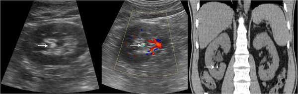
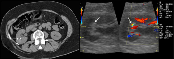
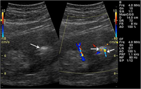
Purpose: The purpose of this study was to determine whether the color Doppler twinkling sign could be considered as an additional diagnostic feature of small renal lithiasis (_5mm).
Methods: 181 patients underwent CT scans performed for other pathologies; the images were also analyzed by a radiologists to identify the incidental presence of renal lithiasis equal to or smaller than 5 mm.These patients underwent an abdominal ultrasound examination, including grayscale analysis of the kidneys and color Doppler. Lithiasis were divided into three groups, on the basis of the diagnostic agreement provided by CT and gray scale results. Then, the twinkling sign sensitivity was assessed in the three groups.
Results: The twinkling sign was positive in 177 out of 206 lithiasis (86 %) visible on CT, while the grayscale was absolutely positive in 98 out of 206 lithiasis (47.6%) and doubtful positive in 71 out of 206 lithiasis (31%).The twinkling sign was positive in 100% of absolutely positive and doubtful positive lithiasis on bmode, and in 8 out of 31 lithiasis not visible on b-mode.
Conclusions: In the diagnosis of small renal lithiasis, integrating gray-scale with color Doppler may be the most suitable procedure, because the color-Doppler twinkling sign is able to confirm the doubtful diagnosis of renal lithiasis and to detect some lithiasis that are not visible on b-mode.
Figures
References
LinkOut - more resources
Full Text Sources
Other Literature Sources
