. 2013 Jul 15;5 Suppl 1(Suppl 1):S7.
doi: 10.1186/2036-7902-5-S1-S7. Epub 2013 Jul 15.

Intestinal Ischemia: US-CT findings correlations

Affiliations

Intestinal Ischemia: US-CT findings correlations

A Reginelli et al. Crit Ultrasound J. .

Abstract

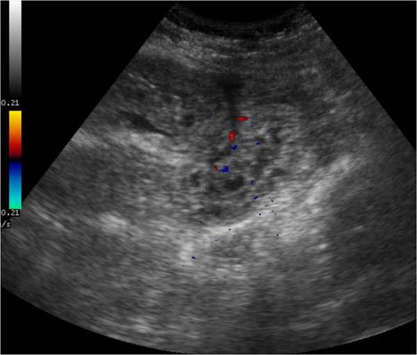
Background: Intestinal ischemia is an abdominal emergency that accounts for approximately 2% of gastrointestinal illnesses. It represents a complex of diseases caused by impaired blood perfusion to the small and/or large bowel including acute arterial mesenteric ischemia (AAMI), acute venous mesenteric ischemia (AVMI), non occlusive mesenteric ischemia (NOMI), ischemia/reperfusion injury (I/R), ischemic colitis (IC). In this study different study methods (US, CT) will be correlated in the detection of mesenteric ischemia imaging findings due to various etiologies.

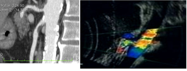
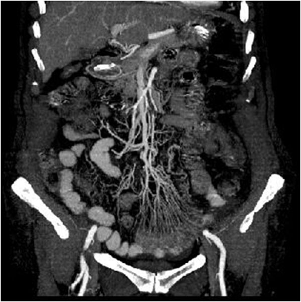
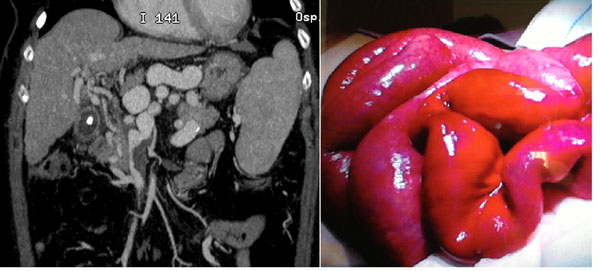
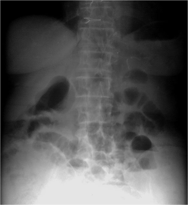
Methods: Basing on experience of our institutions, over 200 cases of mesenteric ischemia/infarction investigated with both US and CT were evaluated considering, in particular, the following findings: presence/absence of arterial/venous obstruction, bowel wall thickness and enhancement, presence/absence of spastic reflex ileus, hypotonic reflex ileus or paralitic ileus, mural and/or portal/mesenteric pneumatosis, abdominal free fluid, parenchymal ischemia/infarction (liver, kidney, spleen).

Results: To make an early diagnosis useful to ensure a correct therapeutic approach, it is very important to differentiate between occlusive (arterial,venous) and nonocclusive causes (NOMI). The typical findings of each forms of mesenteric ischemia are explained in the text.

Conclusion: At present, the reference diagnostic modality for intestinal ischaemia is contrast-enhanced CT. However, there are some disadvantages associated with these techniques, such as radiation exposure, potential nephrotoxicity and the risk of an allergic reaction to the contrast agents. Thus, not all patients with suspected bowel ischaemia can be subjected to these examinations. Despite its limitations, US could constitutes a good imaging method as first examination in acute settings of suspected mesenteric ischemia.


Figures

Figure 1
Figure 1
Acute arterial mesenteric ischemia Contrast-enhanced MDCT 2D reconstruction on sagittal plane and US Color Doppler features (b) shows thrombosis with impairment in the blood flow in the superior mesenteric artery (SMA)
Figure 2
Figure 2
Acute arterial mesenteric ischemia. Contrast-enhanced MDCT 2D reconstruction on coronal plane in early phase: the CT shows the presence of emboli or thrombi as filling defect in the lumen of the artery. If they are small and peripherally localized, the identification can be difficult. The loops of injured small bowel are contracted in consequence of spastic reflex ileus and intestinal wall shows lacking of/poor enhancement. The mesentery is bloodless, due to reduction in caliber of the vessels and apparently in number
Figure 3
Figure 3
Acute arterial mesenteric ischemia. Sonographic features show increased intraluminal secretions within the involved segments, the spasm of the bowel, the extraluminal fluid and the absent peristalsis
Figure 4
Figure 4
Acute venous mesenteric ischemia Contrast-enhanced MDCT 2D reconstruction on coronal plane in cases of superior mesenteric venous thrombosis in the SMV (a) confirmed at surgery (b).
Figure 5
Figure 5
Acute venous mesenteric ischemia Sonographic features show mural thickening with hyperechoic mucosal layers and hypoechoic submucosa attributable to edema of the affected bowel (a). In intermediate phase US examination may reveal increased intraluminal secretions and decreased peristalsis (b).
Figure 6
Figure 6
NOMI. Plain abdominal film shows in early phase: ischemia due to vasoconstriction of the splanchnic vessels leading to spastic reflex ileus
Figure 7
Figure 7
NOMI. US findings. US findings are in the early phase aspecific and poor indicative as thin layer of abdominal free fluid.

References

    1. Mazzei MA, Mazzei FG, Marrelli D, Imbriaco G, Guerrini S, Vindigni C, Civitelli S, Roviello F, Grassi R, Volterrani L. Computed tomographic evaluation of mesentery: diagnostic value in acute mesenteric ischemia. J Comput Assist Tomogr. 2012;5(1):1–7. doi: 10.1097/RCT.0b013e31823b4465. - DOI - PubMed
    1. Elder K, Lashner BA, Solaiman FAL. Clinical approach to colonic ischemia. Cleveland Clinic journal of medicine. 2009;5(7):401–409. doi: 10.3949/ccjm.76a.08089. - DOI - PubMed
    1. Sotiriadis J, Brandt LJ, Behin DS, Southern WN. Ischemic colitis has a worse prognosis when isolated to the right side of the colon. Am J Gastroenterol. 2007;5(10):2247–52. doi: 10.1111/j.1572-0241.2007.01341.x. - DOI - PMC - PubMed
    1. Wiesner W, Khurana B, Ji H, Ros PR. CT of acute bowel ischemia. Radiology. 2003;5:635–650. doi: 10.1148/radiol.2263011540. - DOI - PubMed
    1. Furukawa A, Kanasaki S, Kono N. et al. CT diagnosis of acute mesenteric ischemia from various causes. AJR. 2009;5:408–416. doi: 10.2214/AJR.08.1138. - DOI - PubMed