

US in the assessment of acute scrotum
- PMID: 23902859
- PMCID: PMC3711727
- DOI: 10.1186/2036-7902-5-S1-S8
US in the assessment of acute scrotum
Abstract
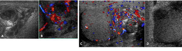
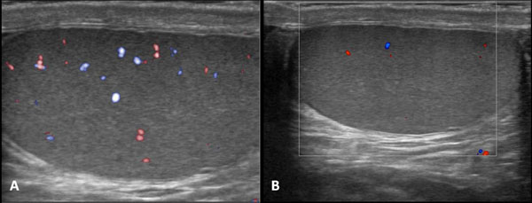
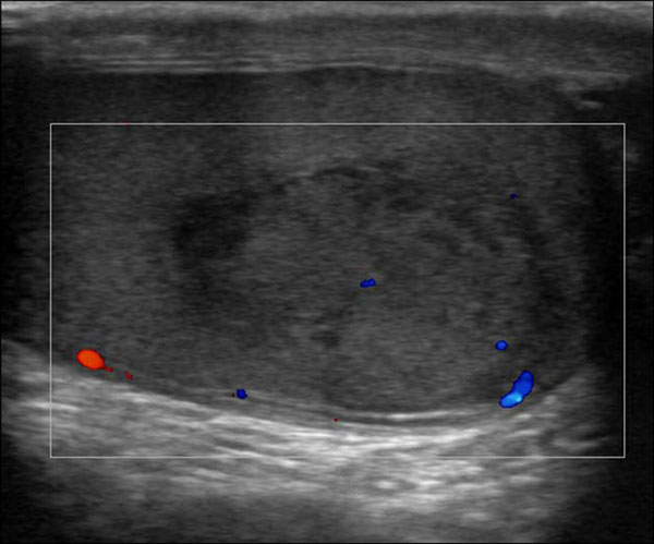
Background: The acute scrotum is a medical emergency . The acute scrotum is defined as scrotal pain, swelling, and redness of acute onset. Scrotal abnormalities can be divided into three groups , which are extra-testicular lesion, intra-testicular lesion and trauma. This is a retrospective analysis of 164 ultrasound examination performed in patient arriving in the emergency room for scrotal pain.The objective of this article is to familiarize the reader with the US features of the most common and some of the least common scrotal lesions.
Methods: Between January 2008 and January 2010, 164 patients aged few month and older with scrotal symptoms, who underwent scrotal ultrasonography (US), were retrospectively reviewed. The clinical presentation, outcome, and US results were analyzed. The presentation symptoms including scrotal pain, painless scrotal mass or swelling, and trauma.
Results: Of 164 patients, 125 (76%) presented with scrotal pain, 31 (19%) had painless scrotal mass or swelling and 8 (5%) had trauma. Of the 125 patients with scrotal pain, 72 had infection,10 had testicular torsion, 8 had testicular trauma, 18 had varicocele, 20 had hydrocele, 5 had cryptorchidism, 5 had scrotal sac and groin metastases, and 2 had unremarkable results. In the 8 patients who had history of scrotal trauma, US detected testicular rupture in 1 patients, scrotal haematomas in 2 patients .Of the 19 patients who presented with painless scrotal mass or swelling, 1 6 had extra-testicular lesions and 3 had intra-testicular lesions. All the extra-testicular lesions were benign. Of the 3 intra-testicular lesions, one was due to tuberculosis epididymo-orchitis, one was non-Hodgkin's lymphoma, and one was metastasis from liposarcoma
Conclusions: US provides excellent anatomic detail; when color Doppler and Power Doppler imaging are added, testicular perfusion can be assessed.
Figures
References
-
- McAndrew HF, Pemberton R, Kikiros CS, Gollow I. The incidence and investigation of acute scrotal problems in children. Pediatr Surg Int. 2002;5:435–437. - PubMed
-
- Nelson CP, Williams JF, Bloom DA. The cremasteric reflex: a useful but imperfect sign in testicular torsion. J Pediatr Surg. 2003;5:1248–1293. - PubMed
-
- Dogra VS, Gottlieb RH, Oka M. et al.Sonography of the scrotum. Radiology. 2003;5(1):18–36. - PubMed
LinkOut - more resources
Full Text Sources
Other Literature Sources
