. 2014 Jan;85(1):72-81.
doi: 10.1038/ki.2013.286. Epub 2013 Jul 31.

TNF-mediated damage to glomerular endothelium is an important determinant of acute kidney injury in sepsis

Affiliations

TNF-mediated damage to glomerular endothelium is an important determinant of acute kidney injury in sepsis

Chang Xu et al. Kidney Int. 2014 Jan.

Abstract

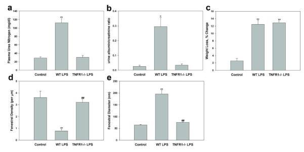
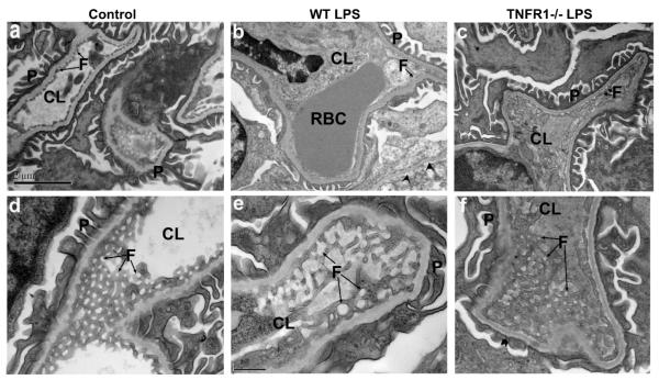
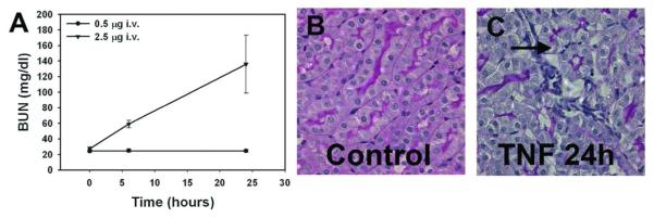
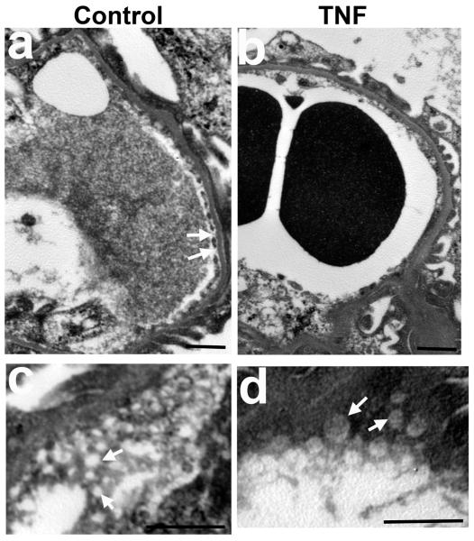
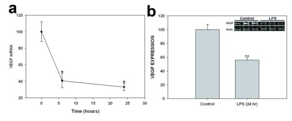
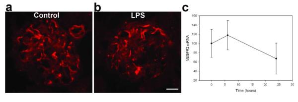
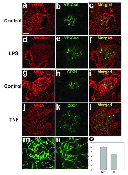
Severe sepsis is often accompanied by acute kidney injury (AKI) and albuminuria. Here we studied whether the AKI and albuminuria associated with lipopolysaccharide (LPS) treatment in mice reflects impairment of the glomerular endothelium with its associated endothelial surface layer. LPS treatment decreased the abundance of endothelial surface layer heparan sulfate proteoglycans and sialic acid, and led to albuminuria likely reflecting altered glomerular filtration permselectivity. LPS treatment decreased the glomerular filtration rate (GFR), while also causing significant ultrastructural alterations in the glomerular endothelium. The density of glomerular endothelial cell fenestrae was 5-fold lower, whereas the average fenestrae diameter was 3-fold higher in LPS-treated than in control mice. The effects of LPS on the glomerular endothelial surface layer, endothelial cell fenestrae, GFR, and albuminuria were diminished in TNF receptor 1 (TNFR1) knockout mice, suggesting that these LPS effects are mediated by TNF-α activation of TNFR1. Indeed, intravenous administration of TNF decreased GFR and led to loss of glomerular endothelial cell fenestrae, increased fenestrae diameter, and damage to the glomerular endothelial surface layer. LPS treatment decreased kidney expression of vascular endothelial growth factor (VEGF). Thus, our findings confirm the important role of glomerular endothelial injury, possibly by a decreased VEGF level, in the development and progression of AKI and albuminuria in the LPS model of sepsis in the mouse.


Figures

Figure 1
Figure 1
Endotoxemia induces TNFR1-dependent AKI and increases urine concentration of albumin, in association with decreased fenestral density and increased fenestral diameter of glomerular capillary endothelium. Effect of LPS (10 mg/kg for 24h) on (a) plasma BUN concentration, (b) urine albumin/creatinine ratio, (c) weight loss, (d) fenestral density, and fenestral diameter in wild type and Tnfr1−/− mice. Control, wild type treated with normal saline; WT LPS, wild type treated with LPS; TNFR1−/− LPS, Tnfr1−/− mice treated with LPS. Values are mean ± s.e.m. for 3-5 animals (a-c) 60-70 capillary loops (d) and 33-67 fenestrae (e). **P<0.01 vs. control. ##P<0.01 vs. wild-type mice injected with LPS.
Figure 2
Figure 2
Transmission electron microscopic ultrastructure of glomerular endothelial cells in glomeruli of 8-wk old wild-type control mice (a, d) or wild-type mice injected with LPS (10 mg/kg for 24h) (b, e) or Tnfr1−/− mice injected with LPS (10 mg/kg for 24h; c f). (a-c) low magnification electron micrographs. Control, wild type treated with normal saline; WT LPS, wild type treated with LPS; TNFR1−/− LPS, Tnfr1−/− mice treated with LPS. Scale bar, 2 μm. (d-e) high magnification electron micrographs, with en face view of the fenestrae. Scale bar, 0.5 μm. Arrows indicate endothelial fenestrae. Arrowheads in (b) indicate endothelial cell detachment. CL, capillary lumen; F, fenestrate; P, podocyte foot processes; RBC, red blood cell.
Figure 3
Figure 3
Intravenous injection of TNF-α recapitulates a form of ARF similar to that produced by LPS injection, with BUN elevation (A), pathological renal injury (B), and renal influx of neutrophils (C). Vacuolization and disruption of tubules is seen, with areas of leukocyte infiltration (arrow). Low dose intravenous TNF-α did not elevate BUN. BUN elevation required TNF-α levels similar to those achieved after LPS injection (3-10 ng/ml).
Figure 4
Figure 4
Transmission electron microscopic ultrastructure of glomerular endothelial cells in glomeruli of wild-type mice 24 h after intravenous administration of saline (control, a and c) or 2.5 μg of TNF-α (b and d). (a, b) Loss of endothelial fenestrae in the glomerular capillaries in TNF-treated mice compared to controls. (c, d) high magnification electron micrographs, with en face view of the fenestrae, showing enlarged glomerular endothelial fenestrae in TNF-treated mice. Scale bar, 1 μm. Arrows indicate endothelial fenestrae.
Figure 5
Figure 5
Effect of LPS on VEGF expression in kidney. (a) Time-dependent effect of LPS (10 mg/kg, 6h, 24h, 48h) on renal VEGF mRNA expression, as normalized to 18S mRNA expression. (g) Effect of LPS (10 mg/kg for 24h) on VEGF protein expression, as normalized to expression of β-actin protein. Top inset: Representative VEGF immunoblot of kidney lysate. Values are means ± s.e.m. for 4-6 animals. *P<0.05 vs. control. ** P<0.01 vs. control.
Figure 6
Figure 6
Effect of LPS on VEGFR2 expression in kidney. (a-b) Indirect immunofluorescence photomicrographs of frozen kidney cortex sections from wild-type control mice (a) and wild-type mice treated 24 h with 10/ mg/kg LPS (b), incubated with anti-VEGFR2 antibody. LPS did not change VEGFR2 expression in glomeruli. c) Time-dependent effect of LPS (10 mg/kg, 6h, 24h, 48h) on renal VEGFR2 mRNA expression, as normalized to 18S mRNA expression. Values are means ± s.e.m. for 4-6 animals. ANOVA shows no significant difference between the LPS and control groups. Scale bar 20 μm.
Figure 7
Figure 7
Effect of LPS on glomerular ESL. (a-l) Glomerular ESL is shown by Alexa Fluor 594-WGA staining and co-staining for the endothelial junctional proteins VE-cadherin and CD31 in wild-type mice treated 24 h with 10 mg/kg LPS (d-f) and their matched controls (a-c), or in mice treated 24 h with 2.5 μg TNF intravenously (j-l) and their matched controls (g-i). Glomerular ESL is also shown by staining PGS containing HS GAG chains in wild-type control mice (m) and in wild-type mice 24 hrs after treatment with 10 mg/kg LPS (n). (o) Fluorescence intensity of glomerular WGA staining. Data (normalized to staining intensity of control mice) are expressed as mean ± s.e.m. **p<0.01 vs wild-type control. Scale bar 20 μm.
Figure 8
Figure 8
Effect of LPS on glomerular heparanase levels and expression. Indirect immunofluorescence photomicrographs of frozen kidney sections from wild-type control mice (a-c) and from wild-type mice treated 24 hrs with LPS (10 mg/kg), incubated with antibodies against Heparanase-1 (red; a, d) and against nephrin (green; b, e). (i) Top inset: Representative immunoblot of Heparanase-1 protein expression in kidney. Bar graph quantifying kidney heparanase-1 expression from immunoblot densitometry. Data normalized to “control” band are expressed as mean ± s.e.m. **p<0.01 vs wild-type control. Scale bar 20 μm.

Comment in

References

    1. Oppert M, Engel C, Brunkhorst FM, et al. Acute renal failure in patients with severe sepsis and septic shock--a significant independent risk factor for mortality: results from the German Prevalence Study. Nephrol Dial Transplant. 2008;23:904–909. - PubMed
    1. Zarjou A, Agarwal A. Sepsis and acute kidney injury. J Am Soc Nephrol. 2011;22:999–1006. - PubMed
    1. Muntner P, Warnock DG. Acute kidney injury in sepsis: questions answered, but others remain. Kidney Int. 2010;77:485–487. - PubMed
    1. Parrillo JE. The cardiovascular pathophysiology of sepsis. Annu Rev Med. 1989;40:469–485. - PubMed
    1. Czabanka M, Peter C, Martin E, et al. Microcirculatory endothelial dysfunction during endotoxemia--insights into pathophysiology, pathologic mechanisms and clinical relevance. Curr Vasc Pharmacol. 2007;5:266–275. - PubMed

Publication types

Substances