. 2013 Sep 12;501(7466):237-41.
doi: 10.1038/nature12427. Epub 2013 Jul 31.

Completion of the entire hepatitis C virus life cycle in genetically humanized mice

Affiliations

Completion of the entire hepatitis C virus life cycle in genetically humanized mice

Marcus Dorner et al. Nature. .

Abstract

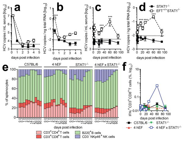
More than 130 million people worldwide chronically infected with hepatitis C virus (HCV) are at risk of developing severe liver disease. Antiviral treatments are only partially effective against HCV infection, and a vaccine is not available. Development of more efficient therapies has been hampered by the lack of a small animal model. Building on the observation that CD81 and occludin (OCLN) comprise the minimal set of human factors required to render mouse cells permissive to HCV entry, we previously showed that transient expression of these two human genes is sufficient to allow viral uptake into fully immunocompetent inbred mice. Here we demonstrate that transgenic mice stably expressing human CD81 and OCLN also support HCV entry, but innate and adaptive immune responses restrict HCV infection in vivo. Blunting antiviral immunity in genetically humanized mice infected with HCV results in measurable viraemia over several weeks. In mice lacking the essential cellular co-factor cyclophilin A (CypA), HCV RNA replication is markedly diminished, providing genetic evidence that this process is faithfully recapitulated. Using a cell-based fluorescent reporter activated by the NS3-4A protease we visualize HCV infection in single hepatocytes in vivo. Persistently infected mice produce de novo infectious particles, which can be inhibited with directly acting antiviral drug treatment, thereby providing evidence for the completion of the entire HCV life cycle in inbred mice. This genetically humanized mouse model opens new opportunities to dissect genetically HCV infection in vivo and provides an important preclinical platform for testing and prioritizing drug candidates and may also have utility for evaluating vaccine efficacy.


Conflict of interest statement

Competing financial interests. The following conflicts of interest are managed under University policy: C.M.R. has equity in Apath, LLC, which holds commercial licenses for the Huh-7.5 cell line, HCV cell culture system, the use of OCLN to construct HCV animal models and the fluorescent cell-based reporter system to detect HCV infection.

Figures

Figure 1
Figure 1. Transgenic expression of human CD81 and OCLN renders mice permissive to HCV entry
(a) Longitudinal bioluminescence imaging of Rosa26-Fluc mice expressing either hCD81, hOCLN, hSCARB1 and hCLDN1, hCD81 and hOCLN or all four HCV entry factors (4hEF). (b) Quantification of viral uptake into murine hepatocytes expressing hCD81 and OCLN or all four human entry factors determined by flow cytometry 72 h post infection with BiCre-Jc1. (c) Blocking of HCV infection in vivo by either blocking antibodies against CD81 (JS-81) or neutralizing antibodies against HCV E1E2 (AR4A). Rosa26-Fluc 4hEF mice were injected with the indicated amounts of antibodies 24 hours and 4 hours prior to infection with BiCre-Jc1. (d, e) Longitudinal quantification of HCV RNA by RT-qPCR in (d) serum and (e) liver of either wt or mice expressing all four HCV entry factors. (f, g) expression of the interferon-stimulated genes (f) ifi27, ifi44, mx1, pkr, (g) viperin, 2’oas and ip10 in the liver following infection with BiCre-Jc1 in wt or 4hEF mice. (h) Serum levels of IFNγ in wt or 4hEF mice infected with BiCre-Jc1. (i) Analysis of liver-infiltrating IFNγ-secreting NKp46-positive NK cells in BiCre-Jc1-infected wild-type or 4hEF mice. Data shown are mean ± SD of 4 independent experiments. For panels (e-g) four mice were used per time-point. *P < 0.05.
Figure 2
Figure 2. Blunting of antiviral immune responses in mice expressing HCV entry factors augments HCV RNA replication
(a–d) Bioluminescence kinetic of BiCre-Jc1 infected Rosa26-Fluc mice expressing all four HCV entry factors and entry-factor negative controls (a) with fully intact innate immune system or impaired in (b) PKR, MAVS, (c) IRF1, IRF3, IRF7, IRF9, (d) IFNαβR or STAT1. (e) HCV RNA levels in the serum of 4hEF mice or 4hEF mice deficient in STAT1, IRF1, IRF3, IRF7, MAVS or IRF9 6 weeks post infection with BiCre-Jc1.
Figure 3
Figure 3. Visualization and genetic and pharmacological interference with HCV infection
(a, b) Quantification of murine hepatocytes actively replicating HCV in wild-type, 4hEF mice and 4hEF STAT1−/− mice as measured by the HCV NS3-4a dependent cleavage of the TagBFPnlsMAVS transgenic reporter construct by ImageStream X analysis. (c, d) Longitudinal HCV RNA levels in 4hEF STAT1−/− mice, (c) 4hEF STAT1−/− mice lacking PPIA or (d) 4hEF STAT1−/− mice treated with an HCV NS5A inhibitor (BMS-790052) for 20 days. Data shown are mean ± SD of n=10–18 mice from 2 independent experiments. **P < 0.01.
Figure 4
Figure 4. HCV infection in 4hEF STAT1−/− mice leads to immune activation
(a–d) HCV RNA copies in (a, c) serum and (b, d) liver of 4hEF STAT1−/− mice and STAT1−/− mice (a, b) early or (c, d) late during infection with Con1/Jc1. (e) Relative frequencies of the indicated lymphocyte subsets in spleen of wild-type, 4hEF, STAT1−/− or STAT1−/− 4hEF mice isolated at the indicated time-points post infection with Con1/Jc1. (f) Analysis of liver-infiltrating IFNγ-producing CD3+CD8+ T cells of wild-type, 4hEF, STAT1−/− and 4hEF STAT1−/− mice following infection with Con1/Jc1. ***P < 0.001.
Figure 5
Figure 5. Evidence for production of infectious particles
(a) STAT1−/−, IRF1−/−, IRF3−/−, IRF7−/−, IRF9−/− and MAVS−/− mice expressing all four human HCV entry factors were infected with BiCre-Jc1. Sera were collected 6 weeks following infection and were used to infect naïve Huh-7.5 cells. NS5A staining was performed 72 h post infection and the frequency of HCV antigen bearing cells quantified by flow cytometry. (b) HCV infectious particles released into the serum of 4hEF STAT1−/− mice, 4hEF STAT1−/− mice lacking PPIA or 4hEF STAT1−/− mice treated with BMS-790052 for 20 days as determined by limiting dilution assay. * = not detectable.

Comment in

References

    1. Ploss A, et al. Human occludin is a hepatitis C virus entry factor required for infection of mouse cells. Nature. 2009;457:882–886. doi: 10.1038/nature07684. nature07684 [pii] - DOI - PMC - PubMed
    1. Dorner M, et al. A genetically humanized mouse model for hepatitis C virus infection. Nature. 2011;474:208–211. doi: 10.1038/nature10168. nature10168 [pii] - DOI - PMC - PubMed
    1. Barth H, et al. Cellular binding of hepatitis C virus envelope glycoprotein E2 requires cell surface heparan sulfate. J Biol Chem. 2003;278:41003–41012. - PubMed
    1. Agnello V, Abel G, Elfahal M, Knight GB, Zhang QX. Hepatitis C virus and other Flaviviridae viruses enter cells via low density lipoprotein receptor. Proc Natl Acad Sci USA. 1999;96:12766–12771. - PMC - PubMed
    1. Scarselli E, et al. The human scavenger receptor class B type I is a novel candidate receptor for the hepatitis C virus. EMBO Journal. 2002;21:5017–5025. - PMC - PubMed

Publication types

MeSH terms