. 2013 Jul;46(7):634-42.
doi: 10.1590/1414-431X20132647. Epub 2013 Jul 22.

A spheroid-based 3-D culture model for pancreatic cancer drug testing, using the acid phosphatase assay

Affiliations

A spheroid-based 3-D culture model for pancreatic cancer drug testing, using the acid phosphatase assay

Z Wen et al. Braz J Med Biol Res. 2013 Jul.

Abstract

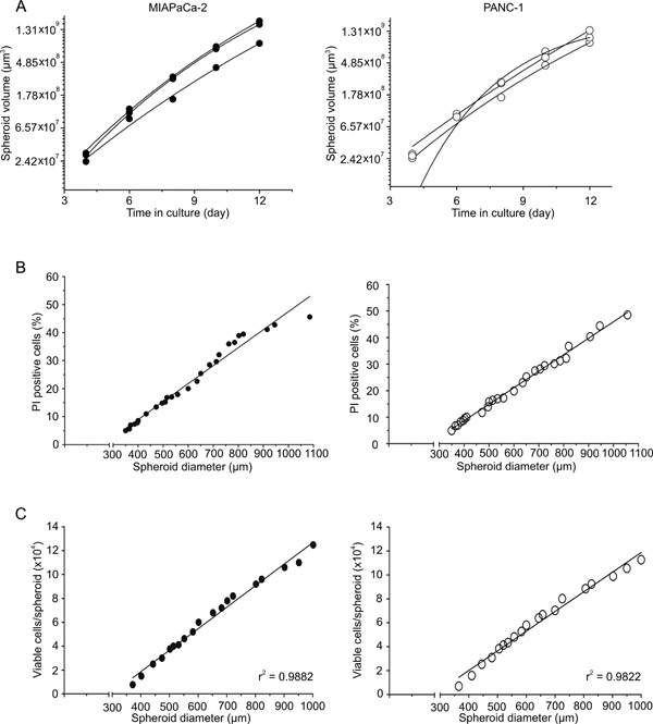
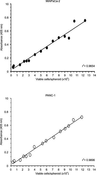
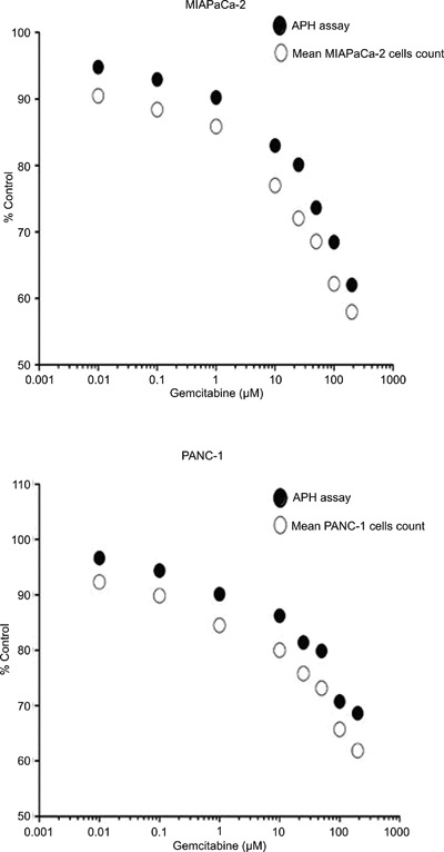
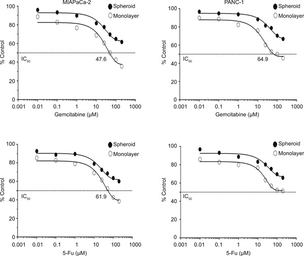
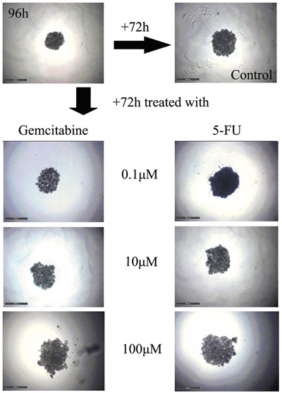
Current therapy for pancreatic cancer is multimodal, involving surgery and chemotherapy. However, development of pancreatic cancer therapies requires a thorough evaluation of drug efficacy in vitro before animal testing and subsequent clinical trials. Compared to two-dimensional culture of cell monolayer, three-dimensional (3-D) models more closely mimic native tissues, since the tumor microenvironment established in 3-D models often plays a significant role in cancer progression and cellular responses to the drugs. Accumulating evidence has highlighted the benefits of 3-D in vitro models of various cancers. In the present study, we have developed a spheroid-based, 3-D culture of pancreatic cancer cell lines MIAPaCa-2 and PANC-1 for pancreatic drug testing, using the acid phosphatase assay. Drug efficacy testing showed that spheroids had much higher drug resistance than monolayers. This model, which is characteristically reproducible and easy and offers rapid handling, is the preferred choice for filling the gap between monolayer cell cultures and in vivo models in the process of drug development and testing for pancreatic cancer.


Figures

Figure 1
Figure 1. General characteristics of spheroid growth. A, Volume of MIAPaCa-2 and PANC-1 spheroids as a function of time in culture, with an initiation interval of 4 days and a seeding density of 1.2×10³ MIAPaCa-2 cells and 1.0×10³ PANC-1 cells per well. Data points are mean spheroid volumes for 8 to 16 spheroids. B, Proportion of propidium iodide (PI)-positive cells in MIAPaCa-2 and PANC-1 spheroids as a function of the average spheroid diameter determined by flow cytometry following dissociation of 15 to 30 spheroids. C, Viable cells in MIAPaCa-2 and PANC-1 spheroids as a function of the average spheroid diameter. Data are reported as average cell numbers determined from 3 aliquots of 15 to 30 spheroids.
Figure 2
Figure 2. Linearity of the acid phosphatase (APH) assay in MIAPaCa-2 and PANC-1 spheroids. APH colorimetric measurement in MIAPaCa-2 and PANC-1 spheroids (mean, n≥8 spheroids) as a function of the average number of viable cells per spheroid. Three aliquots of 20 spheroids were measured for each data point.
Figure 3
Figure 3. Acid phosphatase (APH) activity reflects cell viability in MIAPaCa-2 and PANC-1 spheroids after treatment. Comparison of APH activity and live cell counts following dissociation in MIAPaCa-2 and PANC-1 spheroids after treatment with different concentrations of gemcitabine for 72 h. Drug efficacy was documented relative to the respective untreated controls. Data are reported as means.
Figure 4
Figure 4. Application of the acid phosphatase (APH) assay to determine drug effects in MIAPaCa-2 and PANC-1 spheroid cultures. APH activity/cell viability in MIAPaCa-2 and PANC-1 monolayer and spheroid cultures after 72 h of treatment with gemcitabine and 5-fluorouracil (5-FU). Data are reported as means of ≥3 individual experiments each with eight spheroids treated and measured per condition. IC50 values were calculated to investigate the difference of drug efficacy in 2-D vs 3-D culture and in these two pancreatic cancer cell lines.
Figure 5
Figure 5. Spheroid integrity following treatment with gemcitabine and 5-fluorouracil (5-FU). Phase contrast images of MIAPaCa-2 spheroids at the initiation of drug treatment and after a 72-h treatment interval with 0.1, 10, and 100 µM (Bar: 500 µm).

References

    1. Neoptolemos JP. Adjuvant treatment of pancreatic cancer. Eur J Cancer. 2011;47(Suppl 3):S378–S380. doi: 10.1016/S0959-8049(11)70210-6. - DOI - PubMed
    1. Griffith LG, Swartz MA. Capturing complex 3D tissue physiology in vitro . Nat Rev Mol Cell Biol. 2006;7:211–224. doi: 10.1038/nrm1858. - DOI - PubMed
    1. Mikos AG, Herring SW, Ochareon P, Elisseeff J, Lu HH, Kandel R, et al. Engineering complex tissues. Tissue Eng. 2006;12:3307–3339. doi: 10.1089/ten.2006.12.3307. - DOI - PMC - PubMed
    1. Burdett E, Kasper FK, Mikos AG, Ludwig JA. Engineering tumors: a tissue engineering perspective in cancer biology. Tissue Eng Part B Rev. 2010;16:351–359. doi: 10.1089/ten.teb.2009.0676. - DOI - PubMed
    1. Sutherland RM, Durand RE. Radiation response of multicell spheroids - an in vitro tumour model. Curr Top Radiat Res Q. 1976;11:87–139. - PubMed

MeSH terms