. 2013 Oct;87(20):10980-96.
doi: 10.1128/JVI.01646-13. Epub 2013 Jul 31.

Identification of a region in the stalk domain of the nipah virus receptor binding protein that is critical for fusion activation

Affiliations

Identification of a region in the stalk domain of the nipah virus receptor binding protein that is critical for fusion activation

Aparna Talekar et al. J Virol. 2013 Oct.

Abstract

Paramyxoviruses, including the emerging lethal human Nipah virus (NiV) and the avian Newcastle disease virus (NDV), enter host cells through fusion of the viral and target cell membranes. For paramyxoviruses, membrane fusion is the result of the concerted action of two viral envelope glycoproteins: a receptor binding protein and a fusion protein (F). The NiV receptor binding protein (G) attaches to ephrin B2 or B3 on host cells, whereas the corresponding hemagglutinin-neuraminidase (HN) attachment protein of NDV interacts with sialic acid moieties on target cells through two regions of its globular domain. Receptor-bound G or HN via its stalk domain triggers F to undergo the conformational changes that render it competent to mediate fusion of the viral and cellular membranes. We show that chimeric proteins containing the NDV HN receptor binding regions and the NiV G stalk domain require a specific sequence at the connection between the head and the stalk to activate NiV F for fusion. Our findings are consistent with a general mechanism of paramyxovirus fusion activation in which the stalk domain of the receptor binding protein is responsible for F activation and a specific connecting region between the receptor binding globular head and the fusion-activating stalk domain is required for transmitting the fusion signal.


Figures
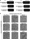
Fig 1
Fig 1
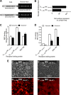
Chimeric proteins containing the NiV G stalk domain and the NDV globular can activate NiV F-mediated fusion. (A) Schematic diagram of chimera NiV-NDV. The stalk region was derived from residues 1 to 180 or from residues 1 to 186 of NiV G, and the globular head was derived from either residues 110 to 571, 120 to 571, or 124 to 571 of NDV HN. (B) Plaque size was evaluated by staining with anti-F antibodies.
Fig 2
Fig 2
Chimeric proteins containing the NiV G stalk domain and the NDV globular can activate NiV/NDV F-mediated cell-cell fusion. Cell-to-cell fusion promoted by the chimeric proteins (shown in Fig. 1A) coexpressed with NiV F was assessed by syncytium formation.
Fig 3
Fig 3
Chimeric proteins containing the NiV G stalk domain and the NDV globular head are efficiently expressed and can activate NiV F-mediated fusion. (A) Schematic diagram of chimera NiV-NDV. The stalk region is derived from residues 1 to 180 or residues 1 to 186 of NiV G, and the globular head is derived from residues 124 to 571 of NDV HN. (B) FACS analysis of cell surface expression from cells transfected with the chimeric proteins shown in Fig. 1A. The results are presented as percentages of NDV HN cell surface expression. (C) Receptor binding in the absence (□) or presence (■) of 2 mM zanamivir. (D) Neuraminidase activity of the receptor binding proteins, expressed in relative fluorescence intensity units (RFU) in the absence (□) or presence (■) of 2 mM zanamivir. The values in panels B, C, and D are means ± the standard deviations (SD) of results from samples assessed in triplicate and are representative of the experiment repeated at least three times. (E) Cell-to-cell fusion promoted by the chimeric proteins coexpressed with NiV F is observed in the top panel by syncytium formation using visible microscopy and in the bottom panel by redistribution of RFP (red fluorescent protein) using fluorescence microscopy.
Fig 4
Fig 4
Requirement for specific residues at residues 181 to 186 of the NiV G stalk for fusion activity of chimeric binding proteins. (A) Schematic diagram of alanine scanning mutagenesis of chimeric protein G1-186-HN124-571 constructs. (B) FACS analysis of cell surface expression from cells transfected with the chimeric proteins shown in panel A. The results are presented as percentages of NDV HN cell surface expression. (C) Receptor binding in the absence (□) or presence (■) of 2 mM zanamivir. (D) Neuraminidase activity of the receptor binding proteins, expressed in relative fluorescence intensity units (RFU) in the absence (□) or presence (■) of 2 mM zanamivir. The values in panels B, C, and D are means ± the SD of results from samples assessed in triplicate and are representative of the experiment repeated at least three times.
Fig 5
Fig 5
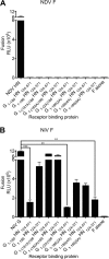
NiV G stalk residues 181 to 182 are required for activation of NiV F. Cell-to-cell fusion mediated by the chimeric NiV-NDV proteins with NDV F (A) or with NiV F (B) compared to the NDV HN/F (A) or NiV G/F (B) proteins. Fusion is measured by a β-Gal complementation assay. The values are means (± the standard errors) of results from four experiments. **, P < 0.05; ***, P < 0.005 (one-way analysis of variance, Dunn's multiple-comparison test).
Fig 6
Fig 6
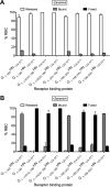
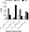
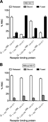
Engagement of site II of the receptor binding protein is required for activation of NiV F. 293T cells coexpressing NiV F with the NiV-NDV chimeric binding protein were allowed to bind to receptor-bearing RBCs at 4°C in the absence (A) or presence (B) of zanamivir. Zanamivir was added to activate NDV HN site II. Unbound RBCs were then washed, and standard medium without (A) or with (B) zanamivir, for the activation of binding site II, was added at 37°C for 120 min. The values on the y axis reflect quantification of RBCs that were (i) released (□), (ii) bound (▩), or (iii) fused (■). Note that there is no fusion (■) in panel A and that there are no released cells (□) in panel B. The values are means (± the standard errors) of results from three experiments.
Fig 7
Fig 7
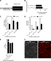
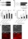
NiV G stalk residues 181 to 182 are sufficient for the activation of NiV F. (A) Schematic diagram of the G1-182-HN124-571 chimeric protein. (B) FACS analysis of cell surface expression from cells transfected with the chimeric proteins shown in Fig. 6A. The results are presented as percentages of NDV HN cell surface expression. (C) Receptor binding in the absence (□) or presence (■) of 2 mM zanamivir. (D) Neuraminidase activity of the receptor binding proteins, expressed in relative fluorescence intensity units (RFU) in the absence (□) or presence (■) of 2 mM zanamivir. The values are means ± the SD of results from samples assessed in triplicate and are representative of the experiment repeated at least three times. (E) Cell-to-cell fusion of the chimeric protein coexpressed with NiV F. Cell-to-cell fusion was measured by a β-Gal complementation assay. The values in panels B, C, D, and E are means (± the standard errors) of results from three experiments. (F) Cell-to-cell fusion promoted by the chimeric protein expressed with NiV F is observed in the left panel by syncytium formation using visible microscopy and in the right panel by redistribution of RFP (red fluorescent protein) using fluorescence microscopy.
Fig 8
Fig 8
The connecting region from NiV G stalk cannot be substituted by the NDV HN 110-124 or 120-124 stalk region. 293T cells coexpressing NiV F with the NiV-NDV chimeric binding protein were allowed to bind to receptor-bearing RBCs at 4°C in the presence of zanamivir. Zanamivir was added to activate NDV HN site II. Unbound RBCs were then washed, and standard medium with zanamivir, for the activation of binding site II, was added at 37°C for 120 min. The values on the y axis reflect quantification of RBCs that were (i) released (□), (ii) bound (▩), or (iii) fused (■). The values are means (± the standard errors) of results from two representative experiments.
Fig 9
Fig 9
Amino acids 181 and 182 regulate an early stage in F activation. 293T cells expressing the NiV-NDV chimeric binding protein alone (B) or coexpressing with NiV F (A) were allowed to bind to receptor-bearing RBCs at 4°C in the presence of zanamivir. Unbound RBCs were then removed, and medium with zanamivir, to activate binding site II, was added, followed by incubation at 37°C for 90 min. After incubation, the cells were washed, and medium at pH 6.5 was added. The cells were incubated at 37°C for 75 min. Values on the y axis reflect quantification of RBCs that were (i) released (□), (ii) bound (▩), or (iii) fused (■). Note that there is no fusion (■) in panel B, and there are no bound cells (▩) in panel A. The values are means (± the standard errors) of results from three experiments.
Fig 10
Fig 10
Requirement for specific residues at residues 181 and 182 of the NiV G stalk for binding and fusion promoting activity of NiV G. (A) Schematic diagram of alanine scanning mutagenesis of NiV G constructs. (B) FACS analysis of cell surface expression from cells transfected with the chimeric proteins shown in panel A. The results are presented as percentages of NiV G cell surface expression. (C) Receptor binding activity of the NiV G proteins to ephrin B2 (□) or ephrin B1 (■). (D) Ephrin B2 binding versus cell surface expression. (E) Cell-to-cell fusion mediated by NiV F coexpressed with the NiV G proteins in panel A. Fusion is measured by a β-Gal complementation assay. The values in panels B, C and D are means (± the standard errors) of results from four experiments. (F) Fusion measured by β-Gal complementation assay versus cell surface expression. (G) FACS analysis of cell surface expression from cells transfected with different levels of wt NiV G cDNA. The results are presented as percentages of NiV G cell surface expression at the highest cDNA concentration. (H) Fusion measured by β-Gal complementation under the same cell surface expression. (I) Ephrin B2 binding versus fusion measured by β-Gal complementation assay. **, P < 0.05; ****, P < 0.001 (one-way analysis of variance, Dunn's multiple comparison test).
Fig 11
Fig 11
Cysteine 123 in the NDV HN globular head alters neuraminidase and fusion promotion. (A) Schematic diagram of chimeric protein G1-186-HN123-571 constructs. (B) FACS analysis of cell surface expression from cells transfected with the chimeric proteins shown in panel A. The results are presented as percentages of NDV HN cell surface expression. (C) Receptor binding in the absence (□) or presence (■) of 2 mM zanamivir. (D) Neuraminidase activity of the receptor binding proteins, expressed in relative fluorescence intensity units (RFU) in the absence (□) or presence (■) of 2 mM zanamivir. The values in panels B, C, and D are means ± the SD of results from samples assessed in triplicate and are representative of the experiment repeated at least three times. (E) Cell-to-cell fusion promoted by the chimeric proteins expressed with NiV F was observed in the top panel by syncytium formation using visible microscopy and in the bottom panel by redistribution of RFP (red fluorescent protein) using fluorescence microscopy.
Fig 12
Fig 12
Cysteine 123 in the NDV HN globular head decreases the rate of F activation. 293T cells coexpressing NiV F and chimeric glycoproteins G1-186-HN123C-571 (A and B) or G1-186-HN123W-571 (C and D) were allowed to bind to RBCs at 4°C with (B and D) or without (A and C) zanamivir. Unbound RBCs were then washed, and standard medium with (B and D) or without (A and C) zanamivir was added at 37°C for up to 120 min. The values on the y axis reflect quantification of RBCs that were (i) released (●, dotted line), (ii) bound (■, dashed line), or (iii) fused (▲, solid line). The values are means (± the SD) of results from triplicate samples and are representative of the experiment repeated at least three times.
Fig 13
Fig 13
NiV G stalk domain determines the oligomerization state of the chimeric binding proteins. Monolayers of cells coexpressing NiV F and either the indicated chimeric glycoproteins, NDV HN, or NiV G were incubated in a medium supplemented with 35S-labeled amino acids. The cells were lysed, and the envelope glycoproteins were immunoprecipitated and subjected to SDS-PAGE under nonreducing (A and B) or reducing (C and D) conditions. Representative autoradiography shows the oligomeric state (A) and the level of protein expression (C) of the receptor binding glycoproteins, immunoprecipitated with anti-NDV HN antibodies. Note that monoclonal antibodies against NiV G were used in the lane marked NiV G. In panels B and D, the same samples were immunoprecipitated with anti-NiV F antibodies showing similar F expression (B) and processing (D) regardless of the coexpressed viral glycoproteins.
Fig 14
Fig 14
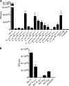
Chimeric envelope glycoproteins and the various NiV G mutant proteins on pseudotyped virions mediate infection. Monolayers of cells were infected with VSV-ΔG-NiV F pseudotyped virions with the indicated chimeric (A) or NiV G mutant (B) envelope glycoproteins. At 24 h postinfection, the PFU were determined as described in Materials and Methods. The values on the y axis reflect quantification of PFU/ml. The values are means (± the SD) of results from triplicate samples.
Fig 15
Fig 15
cis-Complementation of the F-triggering activity of the fusion-deficient chimeric proteins by wt NiV G. 293T cells coexpressing NiV G and F with the NiV-NDV chimeric binding protein were allowed to bind to receptor-bearing RBCs at 4°C in the absence (A) or presence (B) of zanamivir. Zanamivir was added to activate the NDV HN site II. Unbound RBCs were then washed, and standard medium without (A) or with (B) zanamivir, for the activation of binding site II, was added at 37°C for 120 min. The values on the y axis reflect quantification of RBCs that were (i) released (□), (ii) bound (▩), or (iii) fused (■). Note that there is no fusion (■) in panel A and that there are no released cells (□) in panel B. The values are means (± the standard errors) of results from two experiments.
Fig 16
Fig 16
Model of the NDV receptor binding domain (residues 123 to 571) and a portion of the NiV G stalk (residues 148 to 186). The stalk was modeled from the NDV structure (PDB ID 3T1E) based on sequence homology. The chimera model, like NDV HN, is a tetrameric complex formed by a dimer of dimers. The four subunits of the chimeric binding protein are colored differently and the LVGLPN region from residues 181 to 186 from one subunit (orange one) is shown as a peptide in an all-atom stick representation. Two Cys123 residues (ball representation) within NDV dimer are in close proximity for intermolecule disulfide bridge.

References

    1. Eckert DM, Kim PS. 2001. Mechanisms of viral membrane fusion and its inhibition. Annu. Rev. Biochem. 70:777–810. - PubMed
    1. White JM, Delos SE, Brecher M, Schornberg K. 2008. Structures and mechanisms of viral membrane fusion proteins: multiple variations on a common theme. Crit. Rev. Biochem. Mol. Biol. 43:189–219. - PMC - PubMed
    1. Harrison SC. 2008. Viral membrane fusion. Nat. Struct. Mol. Biol. 15:690–698. - PMC - PubMed
    1. Weissenhorn W, Hinz A, Gaudin Y. 2007. Virus membrane fusion. FEBS Lett. 581:2150–2155. - PMC - PubMed
    1. Sapir A, Avinoam O, Podbilewicz B, Chernomordik LV. 2008. Viral and developmental cell fusion mechanisms: conservation and divergence. Dev. Cell 14:11–21. - PMC - PubMed

Publication types

Substances

LinkOut - more resources