. 2013 Oct;87(19):10855-73.
doi: 10.1128/JVI.01535-13. Epub 2013 Jul 31.

Isolate-specific differences in the conformational dynamics and antigenicity of HIV-1 gp120

Affiliations

Isolate-specific differences in the conformational dynamics and antigenicity of HIV-1 gp120

Thaddeus M Davenport et al. J Virol. 2013 Oct.

Abstract

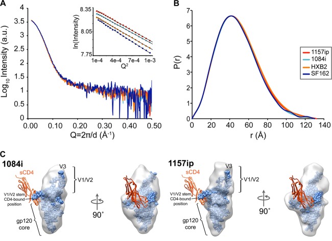
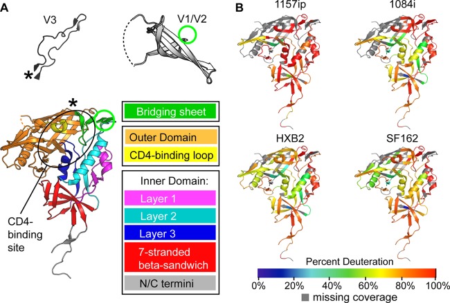
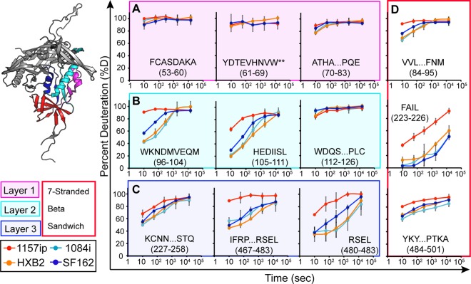
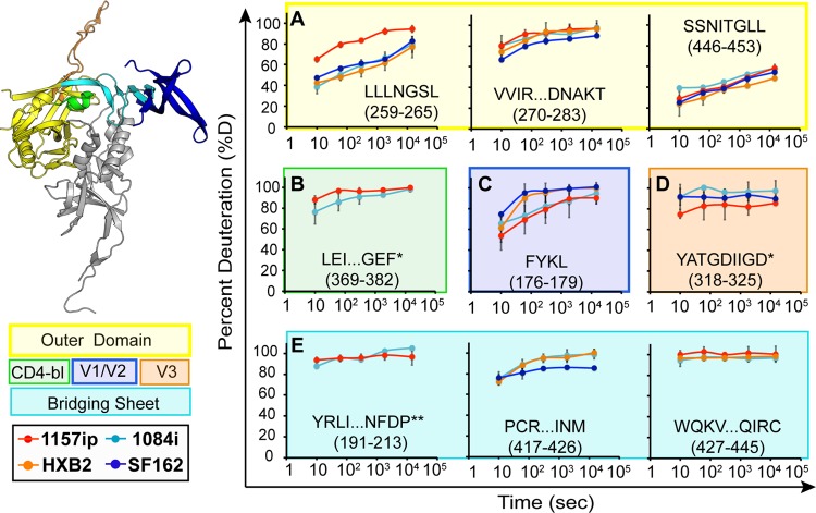
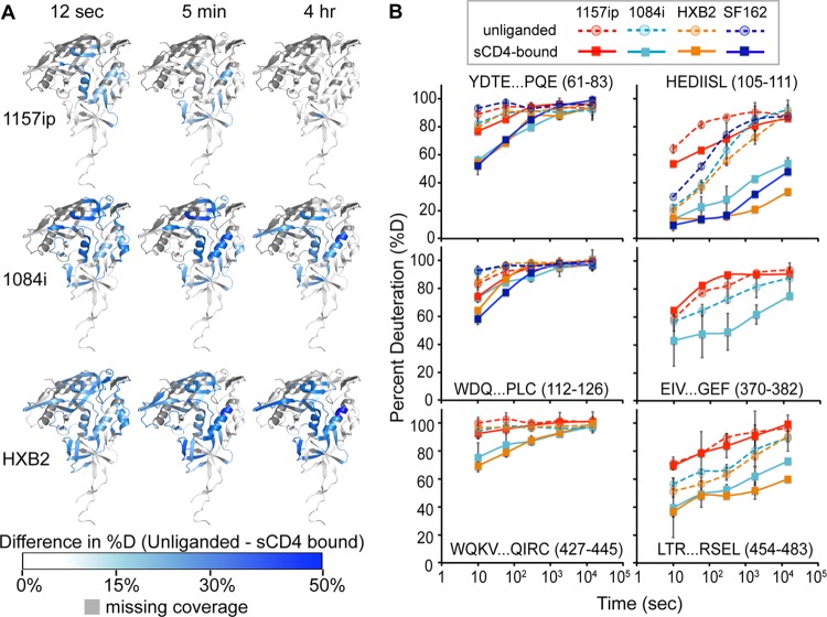
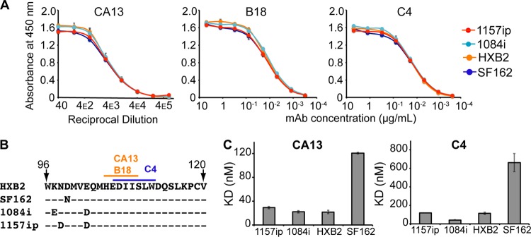
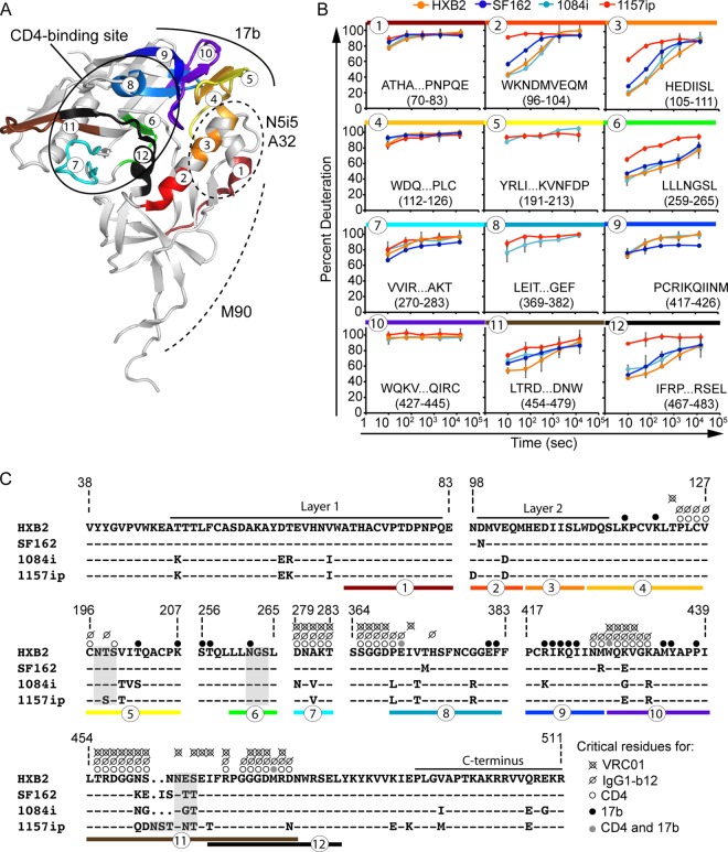
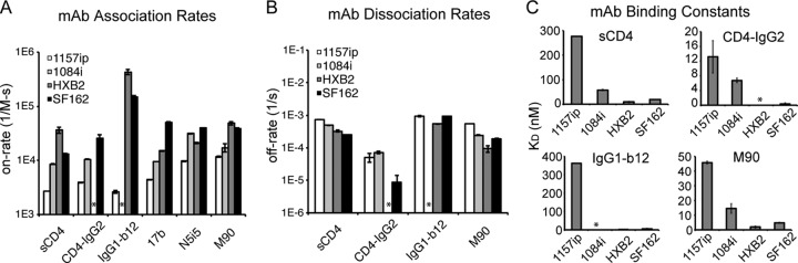
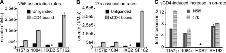
The HIV-1 envelope glycoprotein (Env) mediates viral entry into host cells and is the sole target of neutralizing antibodies. Much of the sequence diversity in the HIV-1 genome is concentrated within Env, particularly within its gp120 surface subunit. While dramatic functional diversity exists among HIV-1 Env isolates-observable even in the context of monomeric gp120 proteins as differences in antigenicity and immunogenicity-we have little understanding of the structural features that distinguish Env isolates and lead to isolate-specific functional differences, as crystal structures of truncated gp120 "core" proteins from diverse isolates reveal a high level of structural conservation. Because gp120 proteins are used as prospective vaccine immunogens, it is critical to understand the structural factors that influence their reactivity with antibodies. Here, we studied four full-length, glycosylated gp120 monomers from diverse HIV-1 isolates by using small-angle X-ray scattering (SAXS) to probe the overall subunit morphology and hydrogen/deuterium-exchange with mass spectrometry (HDX-MS) to characterize the local structural order of each gp120. We observed that while the overall subunit architecture was similar among isolates by SAXS, dramatic isolate-specific differences in the conformational stability of gp120 were evident by HDX-MS. These differences persisted even with the CD4 receptor bound. Furthermore, surface plasmon resonance (SPR) and enzyme-linked immunosorbance assays (ELISAs) showed that disorder was associated with poorer recognition by antibodies targeting conserved conformational epitopes. These data provide additional insight into the structural determinants of gp120 antigenicity and suggest that conformational dynamics should be considered in the selection and design of optimized Env immunogens.


Figures

Fig 1
Fig 1
Similar architecture of gp120 across isolates observed by SAXS. (A and B) SAXS curves (A) and pairwise distance distribution [P(r)] plots (B) show that the four full-length gp120s are similar in shape and size. Guinier plots (A, inset) indicate that the gp120 specimens did not exhibit aggregation or interparticle effects, which would have complicated the analysis. a.u., arbitrary units. (C) DAMMIN shape reconstructions of unliganded 1084i (left) and 1157ip (right) full-length gp120 (gray envelopes), with the gp120 extended core from 3JWD.pdb and containing the V3 loop from 2B4C.pdb (blue spheres) docked into the SAXS shape envelope. The V1/V2 stem in the CD4-bound conformation does not fit into the unliganded SAXS density. Instead, a lobe of mass atop the gp120 core is attributed to V1/V2 proximal to the likely V3 position in unliganded full-length gp120 (40). Two-domain sCD4 was not present in the SAXS experiment; however, it is shown as a red ribbon to aid in the interpretation of the SAXS envelopes.
Fig 2
Fig 2
Summary of isolate-specific differences in unliganded gp120 conformational dynamics. (A) The organization of monomeric gp120 into inner, outer, and bridging sheet domains is indicated by colors on the gp120 core with N- and C-terminal extensions (PDB ID 3JWD). The inner domain is further divided into mobile layers 1, 2, and 3 (magenta, cyan, and blue, respectively) that protrude from the 7-stranded β-sandwich (red) (22). Variable loops V1/V2 and V3 (from PDB 3U4E and PDB 2B4C, respectively) are included for reference. The positions of these elements in the context of the gp120 core are indicated by an asterisk for V3 and a green circle for V1/V2. The approximate location of the CD4 binding site is indicated with a black oval. The structures shown here and throughout the text reflect that of gp120 in a receptor-bound conformation, because no structure is currently available for full-length, unliganded gp120. These structures may not necessarily reflect the unliganded structure of full-length gp120 in solution. (B) Heat maps of HDX-MS data for unliganded gp120 reveal qualitative isolate-specific differences in structural dynamics. Colors mapped onto the gp120 core (PDB ID 3JWD) indicate the percent deuteration of peptides after 1 min of incubation in a deuterated buffer; warm colors correspond to high levels of deuterium uptake (dynamic regions), and cool colors correspond to low levels of deuteration (ordered or “protected” regions). Regions where peptide information is missing are indicated in gray.
Fig 3
Fig 3
Deuterium exchange profiles of homologous inner domain peptides. Each graph shows the deuterium uptake (percent deuteration) over time for peptides throughout the gp120 inner domain, which are either identical or homologous among the four gp120s. Each line in the graph reflects the deuterium uptake for that peptide in the context of a different isolate, as indicated in the figure legend. The peptide sequence and amino acid position (HXB2 numbering) are indicated for each graph. (A to C) Peptides from layers 1, 2, and 3 are color coded in the ribbon diagram, and deuteration uptake plots are grouped within the magenta, cyan, and blue boxes, respectively. (D) Peptides from the 7-stranded β-sandwich are bounded by a red box. A comparison of the deuterium uptake curves for a given peptide from multiple isolates revealed differences in stability, for example, the WKNDMVEQM peptide is more dynamic in 1157ip than in the other isolates. **, deuteration data obtained by subtraction of overlapping peptides. Error bars reflect standard deviations, calculated as described in Materials and Methods.
Fig 4
Fig 4
Deuterium exchange profiles for the homologous outer domain, bridging sheet, and variable loop peptides. Each graph shows deuterium uptake (percent deuteration) over time for peptides in the gp120 outer domain (A), CD4-binding loop (CD4-bl) (B), variable loops V2 (C) and V3 (D), and bridging sheets (E). Each line on the graphs corresponds to deuterium uptake for that peptide in the context of a different isolate, as indicated. The peptide sequence and amino acid position (HXB2 numbering) are indicated for each peptide. Each graph is color coded according to the position of the peptide in the gp120 structure, as indicated in the ribbon diagram and symbol key to the left. *, the listed peptide sequence is from an isolate other than HXB2, because peptide coverage was missing for HXB2; **, deuteration data were obtained by subtraction of overlapping peptides. Error bars reflect standard deviations, calculated as described in Materials and Methods.
Fig 5
Fig 5
Isolate-specific differences in sCD4-induced stabilization of gp120. (A) Difference heat maps show the spatial profile of sCD4-induced stabilization of gp120. The colors mapped onto the 3JWD structure indicate the quantitative differences in percent deuteration between unliganded and CD4-bound full-length gp120s. Darker blue areas correspond to greater CD4-induced stabilization. Missing peptide coverage is shown in gray. Difference heat maps for the 12-s, 5-min, and 4-h time points are shown. (B) Deuterium uptake plots (percent deuteration versus time) for identical or homologous peptides that are stabilized upon sCD4 binding. The peptide sequence and amino acid position (HXB2 numbering) are indicated for each peptide. The traces are color coded by isolates, as indicated in the symbol key. Stabilization as a result of sCD4 binding was observed as a downward shift in the deuterium uptake curve. Error bars reflect standard deviations, calculated as described in Materials and Methods.
Fig 6
Fig 6
Linear epitope-specific antibody binding to a region of gp120 differentially stabilized across isolates. (A) Linear epitope-specific antibody (CA13, B18, or C4) binding to gp120 proteins measured by the ELISA. The curves are color coded by Env isolate as defined in the symbol key. Concentrations of monoclonal antibodies (mAb) are shown on the x axis for C4 and B18, and the reciprocal dilution of antibody supernatant is shown on the x axis for CA13. Curves are representative of at least two independent experiments; error bars indicate standard deviations from duplicate measurements. (B) The primary sequence recognized by antibodies CA13, B18, and C4 is absolutely conserved among the four gp120s. A dash indicates that the residue is conserved in a given isolate, and amino acid differences are indicated. This region is differentially stabilized in the four gp120s, based on HDX-MS (Fig. 3B) (C) Summary of SPR-derived binding constants for CA13 and C4 binding to the four gp120s. Raw and fitted SPR curves are shown in Fig. S20 in the supplemental material. The B18-gp120 SPR binding curves did not fit well to a 1:1 binding model; therefore, estimates of affinity are not available, although qualitative isolate-specific differences in binding to B18 were similar to those observed for CA13 and C4 (see Fig. S20). Error bars indicate standard deviations, calculated as described in Materials and Methods.
Fig 7
Fig 7
Summary of isolate-specific differences in conformational dynamics and primary sequence for peptides involved in conformation-dependent antibody epitopes. (A) Colors and numbers mapped onto the 3JWD crystal structure indicate peptides within conformation-dependent antibody epitopes that can be tracked by HDX-MS. The structure is annotated to indicate the CD4 binding site (targeted by sCD4, IgG1-b12, and VRC01) as well as N5i5, A32, M90, and 17b epitopes. (B) Deuteration profiles (percent deuteration versus time) for peptides within conformation-dependent antibody epitopes. Plots are color coded and numbered as for panel A. (C) Amino acid alignment of the four isolates for regions of gp120 involved in conformation-dependent antibody binding. A dash indicates that the residue is conserved in a given isolate relative to HXB2; a period indicates an insertion in a separate sequence. N5i5 and A32 recognize a similar epitope involving layers 1 and 2 (61). Layer 1 and the C terminus are thought to be involved in the M90 epitope (63, 86). Critical residues for CD4, IgG1-b12, VRC01, and 17b binding are indicated with dots, as shown in the symbol key, and are based on references , , and . Peptides are indicated with colors and numbers as for panels A and B.
Fig 8
Fig 8
Conformation-dependent antibody binding to gp120s as determined by ELISA. Conformation-dependent antibodies specific for different regions of gp120 were tested: CD4 binding site ligands (CD4-IgG2, IgG1-b12, and VRC01), conformational inner domain antibodies (M90, N5i5, and A32), and the coreceptor binding site antibody (17b). Each graph corresponds to a different antibody binding to the four gp120s. The binding curves are color coded based on HIV isolate as shown in the symbol key. Antibody binding levels (measured as the absorbance at 450 nm) versus antibody concentrations are shown. Curves are representative of at least two independent experiments; error bars indicate standard deviations from duplicate measurements.
Fig 9
Fig 9
Summary of conformation-dependent antibody binding to gp120 as measured by SPR. (A) Rate of association of the four gp120 proteins with immobilized sCD4 or captured antibodies. Because of slow dissociation rates for 17b and N5i5, only on-rates could be derived for these antibodies. mAb, monoclonal antibody. (B) Rates of dissociation of the four gp120s from immobilized sCD4 or captured antibody ligands. Reliable off-rates could not be determined for 17b or N5i5. (C) Summary of binding constants for gp120 binding to immobilized sCD4 or captured antibody ligands. Large KD values reflect weaker binding interactions. Raw data and fitted curves for gp120 binding to conformation-dependent ligands are presented in the supplemental material. *, the experiment could not be reliably performed or the signal could not be fit with a 1:1 binding model. Error bars indicate standard deviations, calculated as described in Materials and Methods.
Fig 10
Fig 10
Changes in CD4i antibody binding to gp120 in the presence of sCD4 by SPR. (A) On-rates of N5i5 binding to the four gp120s in the presence and absence of sCD4. (B) On-rates of 17b binding to the four gp120s in the presence and absence of sCD4. (C) Change in rates of association (ka) of 17b and N5i5 with the gp120s as a result of sCD4 binding. Data are presented as the fold increase relative to the rate of association with unliganded gp120. *, the experiment could not be reliably performed. Error bars indicate standard deviations, calculated as described in Materials and Methods.

References

    1. Allan JS, Coligan JE, Barin F, McLane MF, Sodroski JG, Rosen CA, Haseltine WA, Lee TH, Essex M. 1985. Major glycoprotein antigens that induce antibodies in AIDS patients are encoded by HTLV-III. Science 228:1091–1094 - PubMed
    1. Lasky LA, Groopman JE, Fennie CW, Benz PM, Capon DJ, Dowbenko DJ, Nakamura GR, Nunes WM, Renz ME, Berman PW. 1986. Neutralization of the AIDS retrovirus by antibodies to a recombinant envelope glycoprotein. Science 233:209–212 - PubMed
    1. Barin F, McLane MF, Allan JS, Lee TH, Groopman JE, Essex M. 1985. Virus envelope protein of HTLV-III represents major target antigen for antibodies in AIDS patients. Science 228:1094–1096 - PubMed
    1. Hoxie JA. 2010. Toward an antibody-based HIV-1 vaccine. Annu. Rev. Med. 61:135–152 - PubMed
    1. Wei X, Decker JM, Wang S, Hui H, Kappes JC, Wu X, Salazar-Gonzalez JF, Salazar MG, Kilby JM, Saag MS, Komarova NL, Nowak MA, Hahn BH, Kwong PD, Shaw GM. 2003. Antibody neutralization and escape by HIV-1. Nature 422:307–312 - PubMed

Publication types

MeSH terms