. 2013 Aug;108(5):554-62.
doi: 10.1590/s0074-02762013000500004.

Study of vaccinia and cowpox viruses' replication in Rac1-N17 dominant-negative cells

Affiliations

Study of vaccinia and cowpox viruses' replication in Rac1-N17 dominant-negative cells

Ana Paula Carneiro Salgado et al. Mem Inst Oswaldo Cruz. 2013 Aug.

Abstract

Interfering with cellular signal transduction pathways is a common strategy used by many viruses to create a propitious intracellular environment for an efficient replication. Our group has been studying cellular signalling pathways activated by the orthopoxviruses Vaccinia (VACV) and Cowpox (CPXV) and their significance to viral replication. In the present study our aim was to investigate whether the GTPase Rac1 was an upstream signal that led to the activation of MEK/ERK1/2, JNK1/2 or Akt pathways upon VACV or CPXV' infections. Therefore, we generated stable murine fibroblasts exhibiting negative dominance to Rac1-N17 to evaluate viral growth and the phosphorylation status of ERK1/2, JNK1/2 and Akt. Our results demonstrated that VACV replication, but not CPXV, was affected in dominant-negative (DN) Rac1-N17 cell lines in which viral yield was reduced in about 10-fold. Viral late gene expression, but not early, was also reduced. Furthermore, our data showed that Akt phosphorylation was diminished upon VACV infection in DN Rac1-N17 cells, suggesting that Rac1 participates in the phosphoinositide-3 kinase pathway leading to the activation of Akt. In conclusion, our results indicate that while Rac1 indeed plays a role in VACV biology, perhaps another GTPase may be involved in CPXV replication.


Figures

Fig. 1
Fig. 1. murine fibroblasts displaying negative dominance for Rac1-N17 grow at similar levels as the wild-type cells. A31 wild-type cells (white bars), dominant-negative (DN) Rac1-N17 cells (clones 3 or 6, dark grey and black bars, respectively) and cells carrying an empty vector pcDNA3 (light grey bars) were cultured in 35 mm dishes starting at 1 x 10⁵ cells in 7.5% serum. At the times of 24 h, 48 h and 72 h cells were counted using a Neubauer chamber to calculate cellular growth. At least three independent experiments were performed with similar results, but the most representative curve is shown.
Fig. 2
Fig. 2. Vaccinia virus (VACV) but not Cowpox virus (CPXV) replication is impaired in Rac1-N17 dominant-negative (DN) cells. Viral growths were evaluated in either A31 wild-type cells or DN Rac1-N17 (clones 3 or 6). Cells were serum starved for 12 h prior infection with either VACV (A) or CPXV (B) at a multiplicity of infection of 10; at the time points of 3 h post-infection (hpi), 6 hpi, 12 hpi, 24 hpi, 36 hpi and 48 hpi cells were harvested to measure viral yields. At least three independent experiments were performed with similar results (C). Plaque phenotype of VACV (top) or CPXV (bottom) in either A31or DN Rac1-N17 cells. Confluent cell monolayers were serum starved for 12 h followed by infection with VACV or CPXV at 100 plaque forming units (PFU). Cells were stained with 10% formaldehyde/0.3% crystal violet solution at 48 hpi.
Fig. 3
Fig. 3. analysis of viral gene expression in A31 Rac1-N17 dominant-negative (DN) cells upon Vaccinia virus (VACV) and Cowpox virus (CPXV) infection. A31 (A-C, G, H) and DN Rac1-N17 (D-F, I, J) cells were serum starved for 12 h prior infection with VACV or CPXV at a multiplicity of infection (MOI) of 10. At the various time points shown, cell lysates were harvested and analysed by Western blotting using antibodies raised against the viral proteins CrmA (SPI-2) (A, D) or H3L (B, E, G, I). Anti-total ERK1/2 antibody (C, F) or anti-β-actin antibody (H, J) were used as internal controls for protein loading. Molecular masses (in kDa) are indicated on the left. hpi: hours post-infection.
Fig. 4
Fig. 4. ERK1/2 activation by Vaccinia virus (VACV) infection is Rac1-independent. A31 and dominant-negative (DN) Rac1-N17 cells were serum starved for 12 h and then infected with VACV at a multiplicity of infection of 10. At 4 h post-infection (hpi), cell lysates were collected and the phosphorylation status of ERK1/2 was analysed by Western blotting. Anti-total ERK1/2 antibody was used as an internal control for protein loading. Molecular masses (in kDa) are indicated on the left.
Fig. 5
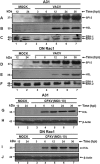
Fig. 5. Akt phosphorylation is affected in A31 Rac1-N17 dominant-negative (DN) cells only upon Vaccinia virus (VACV) infection. A31 (A, C) and DN Rac1-N17 (B, D) cells were serum starved for 12 h prior infection with VACV or Cowpox virus (CPXV) at a multiplicity of infection (MOI) of 10. At the various time points shown, cell lysates were collected and the phosphorylation status of Akt was analysed by Western blotting. Anti-β-actin antibody was used as an internal control for protein loading. Molecular masses (in kDa) are indicated on the left. hpi: hours post-infection.
Fig. 6
Fig. 6. examination of JNK1/2 activation during Vaccinia virus (VACV) and Cowpox virus (CPXV) infection. A31 and dominant-negative (DN) Rac1-N17 cells were serum starved for 12 h and then infected with VACV or CPXV at a multiplicity of infection (MOI) of 10. At the various time points indicated (for VACV) or 12 h post-infection (hpi) (for CPXV), cell lysates were collected to investigate JNK1/2 phosphorylation status (A, C, E) by Western blotting. Anti-β-actin antibody was used as an internal control for protein loading (B, D, F). Molecular masses (in kDa) are indicated on the left.

References

    1. Abrahão JS, Silva-Fernandes AT, Lima LS, Campos RK, Guedes MI, Cota MM, Assis FL, Borges IA, Souza-Júnior MF, Lobato ZI, Bonjardim CA, Ferreira PC, Trindade GS, Kroon EG. Vaccinia virus infection in monkeys, Brazilian Amazon. Emerg Infect Dis. 2010;16:976–979. - PMC - PubMed
    1. Aksamitiene E, Kiyatkin A, Kholodenko BN. Cross-talk between mitogenic Ras/MAPK and survival PI3K/Akt pathways: a fine balance. Biochem Soc Trans. 2012;40:139–146. - PubMed
    1. Andrade AA, Silva PN, Pereira AC, Sousa LP de, Ferreira PC, Gazzinelli RT, Kroon EG, Ropert C, Bonjardim CA. The vaccinia virus-stimulated mitogen-activated protein kinase (MAPK) pathway is required for virus multiplication. Biochem J. 2004;381:437–446. - PMC - PubMed
    1. Bishop A, Hall A. Rho GTPases and their effector proteins. Biochem J. 2000;348:241–255. - PMC - PubMed
    1. Campos MAS, Kroon EG. Critical period for irreversible block of vaccinia virus replication. Rev Microbiol. 1993;24:104–110.

Publication types

Substances